. 2006 Jul;17(7):3108-21.
doi: 10.1091/mbc.e05-09-0896. Epub 2006 Apr 26.

Multiple sequence elements facilitate Chp Rho GTPase subcellular location, membrane association, and transforming activity

Affiliations

Multiple sequence elements facilitate Chp Rho GTPase subcellular location, membrane association, and transforming activity

Emily J Chenette et al. Mol Biol Cell. 2006 Jul.

Abstract

Cdc42 homologous protein (Chp) is a member of the Rho family of small GTPases and shares significant sequence and functional similarity with Cdc42. However, unlike classical Rho GTPases, we recently found that Chp depends on palmitoylation, rather than prenylation, for association with cellular membranes. Because palmitoylation alone is typically not sufficient to promote membrane association, we evaluated the possibility that other carboxy-terminal residues facilitate Chp subcellular association with membranes. We found that Chp membrane association and transforming activity was dependent on the integrity of a stretch of basic amino acids in the carboxy terminus of Chp and that the basic amino acids were not simply part of a palmitoyl acyltransferase recognition motif. We also determined that the 11 carboxy-terminal residues alone were sufficient to promote Chp plasma and endomembrane association. Interestingly, stimulation with tumor necrosis factor-alpha activated only endomembrane-associated Chp. Finally, we found that Chp membrane association was not disrupted by Rho guanine nucleotide dissociation inhibitory proteins, which are negative regulators of Cdc42 membrane association and biological activity. In summary, the unique carboxy-terminal sequence elements that promote Chp subcellular location and function expand the complexity of mechanisms by which the cellular functions of Rho GTPases are regulated.


Figures
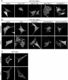
Figure 1.
Figure 1.
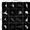
TNF-α–stimulated activation of endomembrane-associated Chp. (A) YFP-PAK-PBD colocalizes with activated Rac and Cdc42 small GTPases. NIH 3T3 cells were transiently transfected with 100 ng of YFP-PAK-PBD and 1 μg of the indicated pcDNA 3.1 construct [vector, Rac1, Rac1(17N), Rac1(61L), or Cdc42(61L)]. YFP fluorescence in live cells was visualized 20 h after transfection using a Zeiss 510 LSM confocal microscope, and 0.4-μm Z-slices were captured. Images shown are representative of at least two independent experiments, with >30 cells examined in each assay. YFP-PAK-PBD localized to the cytoplasm in cells that express vector, Rac1, or dominant negative Rac1(17N). Expression of constitutively active Rac1(61L) recruited YFP-PAK-PBD to membrane ruffles (arrows), whereas Cdc42(61L) recruited PAK-PBD to Golgi and plasma membranes (arrows). (B) Constitutively activated Chp recruits YFP-PAK-PBD to distinct cellular domains. NIH 3T3 cells were transiently transfected with 100 ng of YFP-PAK PBD and 1 μg of the indicated pcDNA3 construct [vector, Chp, Chp(40V), ΔN-Chp(40V), or ΔN/C-Chp(40V)]. YFP fluorescence in live cells was visualized 20 h after transfection using a Zeiss 510 LSM confocal microscope, and 0.4-dm Z-slices were captured. Images shown are representative of at least two independent experiments, with <30 cells examined in each assay. YFP-PAK-PBD is localized to the cytoplasm in cells that express vector, wild-type Chp, Chp(40V), and ΔN/C-Chp(40V). Expression of ΔN-Chp(40V) recruited PAKPBD to discrete and punctate intercellular domains (arrow), reminiscent of Chp localization. Treatment of cells that expressed vector or ΔN/ΔC-Chp(40V) with 10 ng/ml TNF-α for 15 min did not alter cytoplasmic localization of PAK-PBD. Wild-type Chp and Chp(40V) recruited YFP-PAK-PBD to endomembrane structures following TNF-a treatment. YFP-PAK-PBD retained its endomembrane localization in cells that expressed ΔN-Chp(40V) after TNF-α treatment. (C) TNF-α signaling does not cause redistribution of wild-type GFP-Chp. NIH 3T3 cells were transiently transfected with 1 μg of pEGFP-Chp and left untreated or stimulated with 10 ng/ml TNF-α. Images were captured as described above 15 min after treatment.
Figure 2.
Figure 2.
TNF-α causes prolonged Chp activation at the endomembrane. NIH 3T3 cells were transiently transfected with 1 μg of the indicated pcDNA construct [vector, Chp, ΔN-Chp(40V), or Rac1] together with 100 ng of YFP-PAK-PBD. Twenty hours after transfection, cells were then either incubated with growth medium (untreated) or growth medium supplemented with 10 ng/ml TNF-α. YFP fluorescence in live cells was visualized at the indicated times by using a Zeiss 510 LSM confocal microscope, and 0.4-μm Z-slices were captured. Images shown are representative of at least two independent experiments, with >30 cells examined in each assay. Arrows indicate Rac1-mediated recruitment of YFP-PAK-PBD to membrane structures (arrows) with concomitant clearing of cytoplasm.
Figure 3.
Figure 3.
Conserved residues in the carboxy termini of Chp and Wrch-1. (A) Sequence comparison of orthologues of Chp and Wrch-1 reveals the presence of conserved residues that may also play an important role in the membrane-targeting function of the Chp carboxy terminus. Gray boxes indicate amino acids that were mutated to aid in understanding of Chp membrane targeting. Arrows delineate basic amino acids that are conserved between Chp orthologues and homologues. Asterisk indicates Arg residue that is incompletely conserved between Chp homologues. Mm, Mus musculus (mouse); Rn, Rattus norvegicus (rat); Hs, Homo sapiens (human); Cd, Canis domesticus (dog); Pt, Pan troglodyte (chimp); Gg, Gallus gallus (chicken); Dr, Danio rerio (zebrafish). (B) Missense mutations were engineered in the carboxy terminus of Chp to evaluate the importance of basic amino acids as well as a conserved Leu, Ser, and Trp residues at positions 224, 225, and 229, respectively, in Chp localization. The terminal Val [Chp(236Stop)] was also removed to determine whether, as with the CAAX motif, the distance of the Cys residue from the carboxy terminus is critical for proper membrane targeting and localization.
Figure 4.
Figure 4.
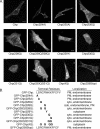
Chp membrane localization is dependent on basic amino acids. (A) NIH 3T3 cells were transiently transfected with 0.5 μg of the pEGFP expression vector encoding GFP fusion proteins of the indicated Chp proteins. Live cells were visualized 20 h after transfection using a Zeiss 510 LSM confocal microscope. Images shown are representative of at least two independent experiments, with >50 cells examined in each assay. Bars, 10 μm. (B) Summary of mutations engineered in the carboxy terminus of Chp and the effect of each mutation on subcellular localization of GFP-Chp. PM, plasma membrane; cyto, cytoplasm.
Figure 5.
Figure 5.
Basic residues are not part of the protein acyltransferase motif. 293T cells were transiently transfected with the indicated pEGFP constructs, lysed 48 h after transfection, and subjected to a biotin-BMCC labeling assay (Drisdel and Green, 2004). Nonpalmitoylated K-Ras was included as a negative control for this assay (top). An anti-GFP Western blot shows that equivalent concentration of GFP and GFP-tagged versions of Chp, Chp(229Y), Chp(230Q), Chp(231Q), Chp(4Q), K-Ras, and H-Ras protein was used in the assays (bottom). Data shown are representative of three independent experiments.
Figure 6.
Figure 6.
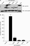
Basic residues are required for Chp transforming activity. Mass populations of NIH 3T3 cells that stably express the indicated Chp proteins were established and evaluated for expression of HA-tagged Chp by Western blot analyses with anti-HA antibody (A; top). Western blot analyses with anti-β-actin were done to verify equivalent total protein for each cell lysate (bottom). An intervening lane was removed between the mutant and wild-type Chp proteins in both blots, with crop boundaries as shown in the figure. (B) Mutant Chp proteins were analyzed for their ability to promote growth of NIH 3T3 cells in soft agar. The number of colonies was quantitated after 35 d. Data shown are representative of three independent experiments, with SE indicated by the bars.
Figure 7.
Figure 7.
The last 11 amino acids of Chp are necessary and sufficient for membrane targeting. (A) Truncation mutants were engineered to determine the minimal region of the carboxy terminus of Chp that was sufficient to target a heterologous protein (GFP) to cellular membranes. (B) NIH 3T3 cells were transiently transfected with 0.5 μg of the pEGFP expression vector encoding GFP fusion proteins that terminate with the indicated Chp carboxy-terminal sequences. GFP fluorescence in live cells was visualized 20 h after transfection using a Zeiss 510 LSM confocal microscope. Images are shown in duplicate and are representative of at least three independent experiments, with >50 cells examined in each assay.
Figure 8.
Figure 8.
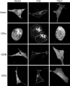
Chp membrane association is not regulated by RhoGDIs. NIH 3T3 cells were transiently transfected with 0.5 μg of the indicated GFP-tagged construct and 1 μg of pcDNA3.1 expression plasmid DNA encoding the indicated RhoGDI. Images shown are representative of at least three independent experiments, with >50 cells examined per assay. Live cells were visualized and photographed 20 h after transfection using a Zeiss 510 LSM confocal microscope.
Figure 9.
Figure 9.
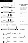
Rho GTPase carboxy-terminal membrane-targeting sequences. The greatest sequence divergence between otherwise closely-related Rho GTPases lies in the hypervariable (HV) motif at the carboxy terminus of the protein. Specific sequences in the HV motif signal for posttranslational modifications and direct localization of Rho GTPases to distinct membrane compartments. The majority (16 of 20) of human Rho family GTPases terminates with CAAX motifs that signal for modification by either farnesyl (F) or geranylgeranyl (G) isoprenoids, AAX proteolysis, and carboxymethylation (O-Me). These GTPases also possess a second membrane-targeting signal: dual palmitoylated cysteines (P), positively charged amino acid stretches (polybasic K/R), or a combination of both signals. Chp lacks a CAAX motif and is modified by palmitoylation of a single cysteine. At least two additional carboxy-terminal sequence elements (polybasic amino acids and an invariant tryptophan residue) are required for Chp subcellular membrane localization. RhoBTB proteins lack any known carboxy-terminal lipid modifications and instead terminate with tandem BR-C, ttk, and bab (BTB) domains.

References

    1. Allal C., Favre G., Couderc B., Salicio S., Sixou S., Hamilton A. D., Sebti S. M., Lajoie-Mazenc I., Pradines A. RhoA prenylation is required for promotion of cell growth and transformation and cytoskeleton organization but not for induction of serum response element transcription. J. Biol. Chem. 2000;275:31001–31008. - PubMed
    1. Aronheim A., Broder Y. C., Cohen A., Fritsch A., Belisle B., Abo A. Chp, a homologue of the GTPase Cdc42Hs, activates the JNK pathway and is implicated in reorganizing the actin cytoskeleton. Curr. Biol. 1998;8:1125–1128. - PubMed
    1. Ashby M. N., King D. S., Rine J. Endoproteolytic processing of a farnesylated peptide in vitro. Proc. Natl. Acad. Sci. USA. 1992;89:4613–4617. - PMC - PubMed
    1. Aspenstrom P., Fransson A., Saras J. Rho GTPases have diverse effects on the organization of the actin filament system. Biochem. J. 2004;377:327–337. - PMC - PubMed
    1. Berzat A. C., Buss J. E., Chenette E. J., Weinbaum C. A., Shutes A., Der C. J., Minden A., Cox A. D. Transforming activity of the Rho family GTPase, Wrch-1, a Wnt-regulated Cdc42 homolog, is dependent on a novel carboxyl-terminal palmitoylation motif. J. Biol. Chem. 2005;280:33055–33065. - PubMed

Publication types

MeSH terms

LinkOut - more resources