Comparative Study
. 2006 May;50(5):1689-95.
doi: 10.1128/AAC.50.5.1689-1695.2006.

Cellular accumulation and activity of quinolones in ciprofloxacin-resistant J774 macrophages

Affiliations
Comparative Study

Cellular accumulation and activity of quinolones in ciprofloxacin-resistant J774 macrophages

Jean-Michel Michot et al. Antimicrob Agents Chemother. 2006 May.

Abstract

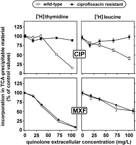
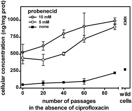
Ciprofloxacin is the substrate for a multidrug resistance-related protein (MRP)-like multidrug transporter in J774 mouse macrophages, which also modestly affects levofloxacin but only marginally affects garenoxacin and moxifloxacin (J.-M. Michot et al., Antimicrob. Agents Chemother. 49:2429-2437, 2005). Two clones of ciprofloxacin-resistant cells were obtained by a stepwise increase in drug concentration (from 34 to 51 to 68 mg/liter) in the culture fluid. Compared to wild-type cells, ciprofloxacin-resistant cells showed (i) a markedly reduced ciprofloxacin accumulation (12% of control) and (ii) a two- to threefold lower sensitivity to the enhancing effect exerted by MRP-inhibitors (probenecid and MK571) on ciprofloxacin accumulation or by ciprofloxacin itself. ATP-depletion brought ciprofloxacin accumulation to similarly high levels in both wild-type and ciprofloxacin-resistant cells. Garenoxacin and moxifloxacin accumulation remained unaffected, and levofloxacin showed an intermediate behavior. DNA and protein synthesis were not impaired in ciprofloxacin-resistant cells for ciprofloxacin concentrations up to 100 mg/liter (approximately 85 and 55% inhibition, respectively, in wild-type cells). In Listeria monocytogenes-infected ciprofloxacin-resistant cells, 12-fold higher extracellular concentrations of ciprofloxacin were needed to show a bacteriostatic effect in comparison with wild-type cells. The data suggest that the resistance mechanism is mediated by an overexpression and/or increased activity of the MRP-like ciprofloxacin transporter expressed at a basal level in wild-type J774 macrophages, which modulates both the intracellular pharmacokinetics and activity of ciprofloxacin.


Figures

FIG. 1.
FIG. 1.
Cellular concentration of quinolones each at an extracellular concentration of 17 mg/liter in wild-type (WT) and in ciprofloxacin-resistant (RS) macrophages after 2 h of incubation. All values are the means of three independent determinations ± standard deviations. CIP, ciprofloxacin; LVX, levofloxacin; GRN, garenoxacin; MXF, moxifloxacin.
FIG. 2.
FIG. 2.
Influence of efflux pump inhibitors on the cellular concentration of quinolones in wild-type and in ciprofloxacin-resistant macrophages. Cells were incubated for 2 h in the simultaneous presence of 17 mg/liter of each quinolone and increasing concentrations of each inhibitor. Results are expressed as percentages of the maximal values measured in each individual experiment. All values are the means of three independent determinations ± standard deviations (when not visible, error bars are smaller than the size of the symbol). CIP, ciprofloxacin; LVX, levofloxacin; GRN, garenoxacin; MXF, moxifloxacin.
FIG. 3.
FIG. 3.
Influence of the extracellular concentration of quinolones on their cellular concentrations in wild-type cells or ciprofloxacin-resistant cells measured after 2 h of incubation. All values are the means of three independent determinations ± standard deviations (when not visible, error bars are smaller than the size of the symbol). Details of the data for low extracellular concentrations of levofloxacin, garenoxacin, and moxifloxacin are shown in the insets. CIP, ciprofloxacin; LVX, levofloxacin; GRN, garenoxacin; MXF, moxifloxacin.
FIG. 4.
FIG. 4.
Incorporation of [3H]thymidine and [3H]leucine in trichloroacetic acid (TCA)-precipitable material of wild-type and ciprofloxacin-resistant macrophages. Cells were incubated during 24 h with increasing concentrations of ciprofloxacin (CIP) or moxifloxacin (MXF) and then exposed for 3 h to an 80 nM concentration radiolabeled thymidine or a 200 nM concentration of radiolabeled leucine. All values are expressed as a percentage of the control and are the means of three independent determinations ± standard deviations (when not visible, error bars are smaller than the size of the symbol).
FIG. 5.
FIG. 5.
Cellular concentration of ciprofloxacin in ciprofloxacin-resistant macrophages cultivated for an increasing number of passages in the absence of ciprofloxacin (each passage was made at a 3-day interval). Cells were incubated for 2 h with 17 mg/liter ciprofloxacin alone (filled squares) or with ciprofloxacin plus either 5 mM probenecid (open circles) or 10 mM probenecid (gray circles). Reference values for wild-type cells are shown on the right of the graph. All values are the means of three independent determinations ± standard deviations (when not visible, error bars are smaller than the size of the symbol).
FIG. 6.
FIG. 6.
Concentration killing curves of ciprofloxacin toward L. monocytogenes in wild-type (WT) cells or ciprofloxacin-resistant (RS) cells. In the right panel, cells were incubated with probenecid as indicated (the probenecid concentrations were selected to obtain a grossly similar content in ciprofloxacin based on data from Fig. 2). The abscissas show the extracellular concentrations of ciprofloxacin in multiples of its MIC in broth (2 mg/liter). The arrow points to the concentration corresponding to the maximum concentration (Cmax; peak concentration in serum, 4.3 mg/liter [9]) commonly observed in the serum of patients treated with ciprofloxacin. The ordinates show the changes in CFU (log10) per mg of cell protein as observed after 5 h of incubation in comparison with the original inocula (horizontal dotted lines). All values are the means of three independent determinations ± standard deviations (when not visible, error bars are smaller than the size of the symbol).

References

    1. Cao, C., T. H. Steinberg, H. C. Neu, D. Cohen, S. B. Horwitz, S. Hickman, and S. C. Silverstein. 1993. Probenecid-resistant J774 cell expression of enhanced organic anion transport by a mechanism distinct from multidrug resistance. Infect. Agents Dis. 2:193-200. - PubMed
    1. Carlier, M. B., B. Scorneaux, A. Zenebergh, J. F. Desnottes, and P. M. Tulkens. 1990. Cellular uptake, localization and activity of fluoroquinolones in uninfected and infected macrophages. J. Antimicrob. Chemother. 26(Suppl. B):27-39. - PubMed
    1. Deng, L., S. Tatebe, Y. C. Lin-Lee, T. Ishikawa, and M. T. Kuo. 2002. MDR and MRP gene families as cellular determinant factors for resistance to clinical anticancer agents. Cancer Treat. Res. 112:49-66. - PubMed
    1. Eriksson, E., A. Forsgren, and K. Riesbeck. 2003. Several gene programs are induced in ciprofloxacin-treated human lymphocytes as revealed by microarray analysis. J. Leukoc. Biol. 74:456-463. - PubMed
    1. Gollapudi, S., F. Thadepalli, C. H. Kim, and S. Gupta. 1995. Difloxacin reverses multidrug resistance in HL-60/AR cells that overexpress the multidrug resistance-related protein (MRP) gene. Oncol. Res. 7:213-225. - PubMed

Publication types

LinkOut - more resources