Comparative Study
. 2006 May;50(5):1788-97.
doi: 10.1128/AAC.50.5.1788-1797.2006.

Sodium antimony gluconate induces generation of reactive oxygen species and nitric oxide via phosphoinositide 3-kinase and mitogen-activated protein kinase activation in Leishmania donovani-infected macrophages

Affiliations
Comparative Study

Sodium antimony gluconate induces generation of reactive oxygen species and nitric oxide via phosphoinositide 3-kinase and mitogen-activated protein kinase activation in Leishmania donovani-infected macrophages

Jayati Mookerjee Basu et al. Antimicrob Agents Chemother. 2006 May.

Abstract

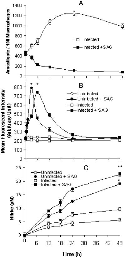
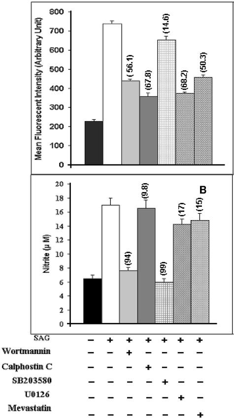
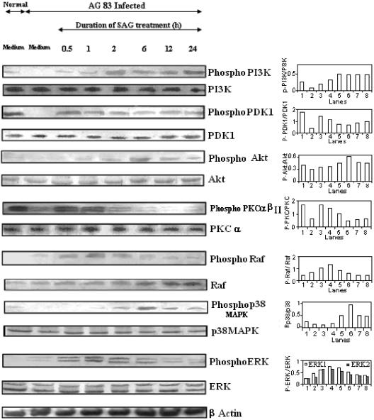
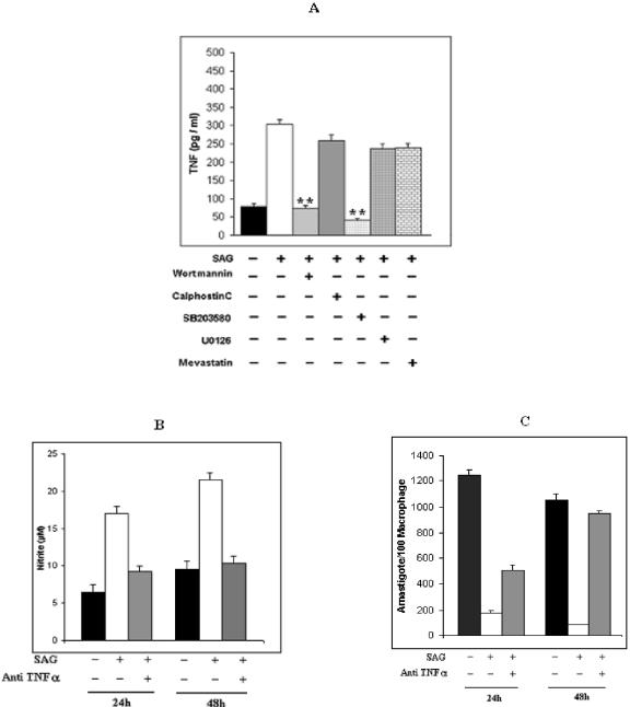
Pentavalent antimony complexes, such as sodium stibogluconate and sodium antimony gluconate (SAG), are still the first choice for chemotherapy against various forms of leishmaniasis, including visceral leishmaniasis, or kala-azar. Although the requirement of a somewhat functional immune system for the antileishmanial action of antimony was reported previously, the cellular and molecular mechanism of action of SAG was not clear. Herein, we show that SAG induces extracellular signal-regulated kinase 1 (ERK-1) and ERK-2 phosphorylation through phosphoinositide 3-kinase (PI3K), protein kinase C, and Ras activation and p38 mitogen-activated protein kinase (MAPK) phosphorylation through PI3K and Akt activation. ERK-1 and ERK-2 activation results in an increase in the production of reactive oxygen species (ROS) 3 to 6 h after SAG treatment, while p38 MAPK activation and subsequent tumor necrosis factor alpha release result in the production of nitric oxide (NO) 24 h after SAG treatment. Thus, this study has provided the first evidence that SAG treatment induces activation of some important components of the intracellular signaling pathway, which results in an early wave of ROS-dependent parasite killing and a stronger late wave of NO-dependent parasite killing. This opens up the possibility of this metalloid chelate being used in the treatment of various diseases either alone or in combination with other drugs and vaccines.


Figures

FIG. 1.
FIG. 1.
SAG-induced generation of ROS and NO in Mφs and killing of intracellular parasites. Uninfected Mφs and Mφs infected with L. donovani (I-Mφs) were either kept untreated or treated with SAG for different durations. (A) Intracellular parasite numbers were measured for I-Mφs in the absence or presence of SAG. (B) ROS generation was measured with DCFDA; uninfected Mφs and I-Mφs showed significant increases in ROS generation at 3 h and 6 h post-SAG treatment, respectively, compared with levels for corresponding untreated controls (*, P < 0.001). (C) Nitrite was measured using Griess reagent in cell-free culture supernatants of SAG-treated uninfected Mφs; I-Mφs showed significant increase in nitrite generation compared to untreated counterparts (*, P < 0.001; **, P < 0.005). All results are presented as means ± SEM of five independent experiments.
FIG. 2.
FIG. 2.
Effect of NAC (scavenger of ROS) and l-NMMA (competitive inhibitor of iNOS) on SAG-mediated intracellular parasite killing. (A) I-Mφs cultured on cover glasses were either kept untreated or treated with NAC or l-NMMA prior to SAG treatment. After indicated time points, cover glasses were stained with Giemsa stain and assessed for intracellular parasite number. Pretreatment with NAC and l-NMMA significantly inhibited SAG-mediated parasite killing (*, P < 0.005 for NAC; **, P < 0.001 for l-NMMA) compared to levels for corresponding SAG-treated controls. Results are presented as means ± SEM of four independent experiments. (B) Peritoneal Mφs isolated from iNOS−/− and wild-type C57BL/6 mice were cultured on cover glasses, infected with L. donovani, and either kept untreated or treated with SAG for 24 h and 48 h, following which their intracellular parasite numbers were assessed. SAG-mediated intracellular L. donovani killing was significantly inhibited in infected Mφs of iNOS−/− mice (*, P < 0.005; **, P < 0.001). Results are presented as means ± SEM of three independent experiments.
FIG. 3.
FIG. 3.
Effect of inhibitors of PI3K (wortmannin), PKC (calphostin C), Ras (mevastatin), and ERK (U0126) on SAG-induced generation of ROS and that of inhibitor of p38 MAPK (SB203580) on SAG-induced generation of NO. I-Mφs were treated (+) with wortmannin, calphostin C, mevastatin, U0126, or SB203580 prior to SAG treatment. (Top) ROS levels were measured after 6 h of SAG treatment, and corresponding intracellular parasite numbers were also assessed. Pretreatment with wortmannin, calphostin C, mevastatin, or U0126 significantly inhibited SAG-mediated ROS generation as well as parasite killing (P < 0.005). (Bottom) Nitrite levels in cell-free culture supernatant were estimated after 24 h of SAG treatment, and corresponding intracellular parasite numbers were also assessed. Pretreatment with wortmannin or SB203580 significantly inhibited SAG-mediated NO generation as well as parasite killing (P < 0.001). For both panels, numbers in parentheses show percentages of inhibition of SAG-mediated intracellular parasite killing compared to levels for respective SAG-treated controls. Results are presented as means ± SEM of five independent experiments. As mentioned in Materials and Methods, the inhibitors did not exhibit any cytotoxicity at the doses used in this and subsequent experiments.
FIG. 4.
FIG. 4.
SAG treatment induces phosphorylation of PI3K, PDK1, Akt, PKC α/βII, Raf, ERK, and p38 MAPK. I-Mφs were either kept untreated or treated with SAG for different durations; cell lysates were prepared, run in 10% polyacrylamide gels, and immunoblotted with MAbs against whole and phosphorylated forms (labeled Phospho or P) of PI3K, PKC α/βII (phosphorylated form of α/βII and whole α only), PDK1, Raf, ERK-1/ERK-2, p38 MAPK, and Akt, with β-actin as an internal control. The phosphorylation status of each of the above molecules was expressed as the densitometric ratio of the phosphorylated form versus the expression control. Representative data of three similar experiments are presented. AG83, MHOM/IN/1983/AG83.
FIG. 5.
FIG. 5.
PI3K phosphorylation due to SAG treatment induces phosphorylation of PDK1, PKC α/βII, Raf, and ERK-1/ERK-2 as well as that of Akt and p38 MAPK. I-Mφs were treated with an inhibitor of PI3K (wortmannin), PKC (calphostin C), or Ras (mevastatin) for 1 h prior to SAG treatment, and the phosphorylation status of PDK1 and PKC α/βII at 30 min of SAG treatment, Raf and ERK at 1 h of SAG treatment, and Akt and p38 MAPK at 6 h of SAG treatment (SAG-induced phosphorylation status of each of the above-mentioned signaling molecules was maximal at these time points) was determined by immunoblotting. Phosphorylation status of each of the above-mentioned molecules was expressed as the densitometric ratio of the phosphorylated form (labeled Phospho or P) versus the internal control (β-actin). Representative data of three similar experiments are presented here.
FIG. 6.
FIG. 6.
SAG induces TNF-α production from L. donovani-infected Mφs through participation of PI3K and p38 MAPK. (A) I-Mφs were either kept untreated (−) or treated (+) with wortmannin, calphostin C, mevastatin, U0126, or SB203580 prior to SAG treatment; cell-free culture supernatants were collected after 18 h and assayed for TNF-α by sandwich enzyme-linked immunosorbent assay. Pretreatment with wortmannin or SB203580 significantly inhibited SAG-mediated TNF-α generation (**, P < 0.001). (B) In some experiments, I-Mφ was either kept untreated or treated with SAG in the presence or absence of neutralizing anti-TNF-α MAb, and resulting levels of nitrite accumulated in cell-free culture supernatants after 24 h of SAG treatment were measured. (C) Corresponding intracellular parasite numbers were also assessed. Neutralization of TNF-α significantly inhibited SAG-mediated NO generation (P < 0.005) as well as intracellular L. donovani killing (P < 0.005 at 24 h). Results presented are means ± SEM of four independent experiments.

References

    1. Alexander, J., K. C. Carter, N. Al-Fasi, A. Satoskar, and F. Brombacher. 2000. Endogenous IL-4 is necessary for effective drug therapy against visceral leishmaniasis. Eur. J. Immunol. 30:2935-2943. - PubMed
    1. Amer, A. O., and M. S. Swanson. 2002. Phagosome of one's own: a microbial guide to life in the macrophage. Curr. Opin. Microbiol. 5:56-61. - PubMed
    1. Awasthi, A., R. Mathur, A. Khan, B. N. Joshi, N. Jain, S. Sawant, R. Boppana, D. Mitra, and B. Saha. 2003. CD40 signaling is impaired in L. major-infected macrophages and is rescued by a p38MAPK activator establishing a host-protective memory T cell response. J. Exp. Med. 197:1037-1043. - PMC - PubMed
    1. Berman, J. D., and D. J. Wyler. 1980. An in vitro model for investigation of chemotherapeutic agents in leishmaniasis. J. Infect. Dis. 142:83-86. - PubMed
    1. Blanchette, J., N. Racette, R. Faure, K. A. Siminovitch, and M. Olivier. 1999. Leishmania-induced increases in activation of macrophage SHP-1 tyrosine phosphatase are associated with impaired IFN-gamma-triggered JAK2 activation. Eur. J. Immunol. 29:3737-3744. - PubMed

Publication types

MeSH terms

Substances

LinkOut - more resources