. 2006 May;15(5):976-86.
doi: 10.1110/ps.051957006.

3-Nitrotyrosine as a spectroscopic probe for investigating protein protein interactions

Affiliations

3-Nitrotyrosine as a spectroscopic probe for investigating protein protein interactions

Vincenzo De Filippis et al. Protein Sci. 2006 May.

Abstract

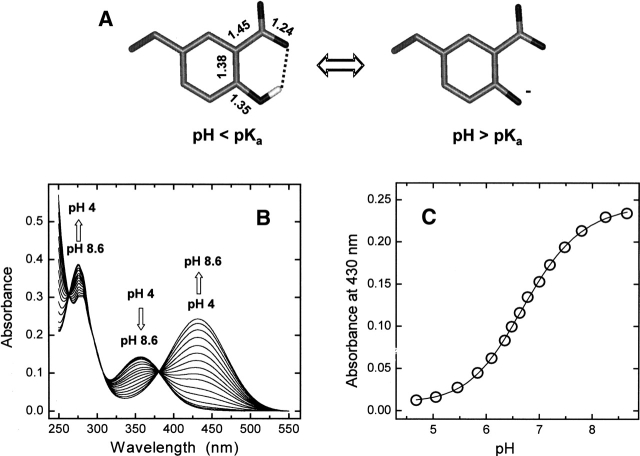
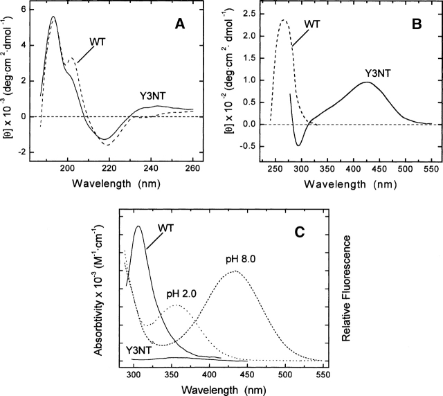
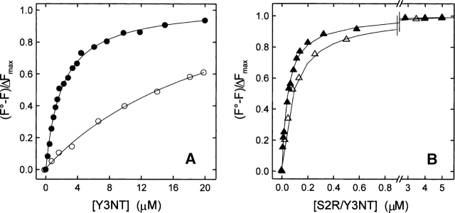
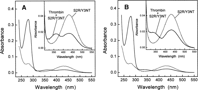
3-Nitrotyrosine (NT) is approximately 10(3)-fold more acidic than Tyr, and its absorption properties are strongly pH-dependent. NT absorbs radiation in the wavelength range where Tyr and Trp emit fluorescence (300-450 nm), and it is essentially nonfluorescent. Therefore, NT may function as an energy acceptor in resonance energy transfer (FRET) studies for investigating ligand protein interactions. Here, the potentialities of NT were tested on the hirudin thrombin system, a well-characterized protease inhibitor pair of key pharmacological importance. We synthesized two analogs of the N-terminal domain (residues 1-47) of hirudin: Y3NT, in which Tyr3 was replaced by NT, and S2R/Y3NT, containing the substitutions Ser2 --> Arg and Tyr3 --> NT. The binding of these analogs to thrombin was investigated at pH 8 by FRET and UV/Vis-absorption spectroscopy. Upon hirudin binding, the fluorescence of thrombin was reduced by approximately 50%, due to the energy transfer occurring between the Trp residues of the enzyme (i.e., the donors) and the single NT of the inhibitor (i.e., the acceptor). The changes in the absorption spectra of the enzyme inhibitor complex indicate that the phenate moiety of NT in the free state becomes protonated to phenol in the thrombin-bound form. Our results indicate that the incorporation of NT can be effectively used to detect protein protein interactions with sensitivity in the low nanomolar range, to uncover subtle structural features at the ligand protein interface, and to obtain reliable Kd values for structure activity relationship studies. Furthermore, advances in chemical and genetic methods, useful for incorporating noncoded amino acids into proteins, highlight the broad applicability of NT in biotechnology and pharmacological screening.


Figures

Figure 1.
Figure 1.
(A) Structure of 3-nitrotyrosine. Bond distances (Å) are taken from the crystallographic structure of free NT (Mostad and Natarajan 1990). (B,C) Spectrophotometric titration of Y3NT by UV/Vis absorption spectroscopy. Absorption spectra (B) and plot of the absorbance values at 430 nm of Y3NT (2 mL, 48 μM) as a function of pH (C). Measurements were carried out in 2 mM citrate-borate-phosphate buffer, at the indicated pH. The data points were fitted to Equation 1, yielding a pKa value for NT of 6.74 ± 0.02.
Figure 2.
Figure 2.
Conformational characterization of wild-type hirudin fragment 1 47 (WT) and Y3NT analog. Far-UV (A) and near-UV (B) CD spectra of hirudin analogs were taken at protein concentrations of 40 and 200 μM in the far- and the near-UV region, respectively. (C) Superimposition of the fluorescence spectra (continuous lines) of WT and Y3NT with the absorption spectra (dashed/dotted lines) of Y3NT at pH 2.0 and 8.0. All measurements were carried out at 25° in 5 mM Tris-HCl buffer (pH 8.0), containing 0.1% (w/v) PEG 8000 and 0.2 M NaCl.
Figure 3.
Figure 3.
Binding of Y3NT and S2R/Y3NT to thrombin, monitored by Trp-to-NT fluorescence energy transfer. (A) Fluorescence spectra of thrombin alone (50 nM) and in the presence of Y3NT (10 μM). For comparison, the spectrum of the free inhibitor Y3NT (10 μM) is also reported. (B) Change in the fluorescence of thrombin as a function of S2R/Y3NT concentration, under fast (▴-▴, 0.2 M NaCl) and slow (△-△, 0.2 M ChCl) conditions. (C) Superimposition of the fluorescence spectrum of thrombin (continuous line) with the absorption spectra of Y3NT at pH 2 and 8.0 (dashed/dotted lines). (D) Change in thrombin fluorescence as a function of Y3NT concentration (•). As a control, the fluorescence intensity of thrombin in the presence of free NT (○) is reported. The signal of Y3NT alone (△) is also included. All measurements were carried out at 25°C by exciting the protein samples at 295 nm in 5 mM Tris-HCl buffer (pH 8.0), containing 0.1% (w/v) PEG 8000 and 0.2 M salt, as indicated, and recording the fluorescence signal at 342 nm.
Figure 4.
Figure 4.
Determination of the dissociation constant (Kd) of the complexes formed by the synthetic analogs Y3NT (A) and S2R/Y3NT (B) with thrombin, under fast (filled symbols) and slow (hollow symbols) conditions. Fluorescence data were obtained as detailed in Materials and Methods (see also Fig. 3) and expressed as (F)/ΔFmax. Continuous lines represent the best fit of the data points to Equation 2, which allowed us to obtain the Kd values reported in Table 1.
Figure 5.
Figure 5.
Schematic representation of the interaction of the N-terminal tripeptide of Y3NT with thrombin. The inhibitor is color-coded (carbon, gray; nitrogen, blue; oxygen, red), while the relevant residues of thrombin in the S2 (Tyr60a and Trp60d) and S3 site (Trp215, Leu99, and Ile174) are shown in magenta. Tyr3′ of hirudin fills the apolar S3 site of thrombin, Val1′ contacts the S2 site, and Ser2′ covers but does not penetrate the S1 site. The structure of Y3NT was modeled on the structure of hirudin thrombin complex, crystallized under conditions stabilizing the fast form (i.e., sodium acetate) (4HTC.pdb) (Rydel et al. 1991). The structural water molecules (w432, B-factor 36 Å2, occupancy 1.0; w606, B-factor 22 Å2, occupancy 0.52; w672, B-factor 49 Å2, occupancy 0.65) are indicated by green spheres, and lie approximately in the geometrical plane of NT. The nitro-group was easily accommodated at the enzyme inhibitor interface without bump by keeping the orientation of Tyr3 (χ1 = −62° and χ2 = −56°) unchanged, together with the rest of the hirudin thrombin structure. The bond-lengths and angles of NT are derived from the crystal structure of the free amino acid (Mostad and Natarajan 1990). Relevant NT thrombin distances, in the 2.5 3.5 Å range, are indicated by dashed lines. Of note, w606, which in the wild-type hirudin thrombin structure connects Tyr3′ of the inhibitor to Tyr60a of the enzyme, is well suited as a hydrogen bond donor to stabilize the NT ring system (see text).
Figure 6.
Figure 6.
Binding of the synthetic analog S2R/Y3NT to the fast (A) or the slow (B) form of thrombin, monitored by UV/Vis absorption spectroscopy. Spectra of the inhibitor (8.6 μM) were taken at 25° in 5 mM Tris-HCl buffer (pH 8.0), containing 0.1% PEG 8000 and 0.2 M NaCl for the fast form, or 0.2 M ChCl for the slow form, in the absence (---) and presence (___) of thrombin (4.1 μM). For clarity, the insets show the spectra in the wavelength range 300 550 nm.

References

    1. Abraham M.H., Du C.M., Platts J.A. 2000. Lipophilicity of the nitrophenols J. Org. Chem. 65: 7114–7118. - PubMed
    1. Albericio F. 2004. Developments in peptides and amide synthesis Curr. Opin. Chem. Biol. 8: 211–221. - PubMed
    1. Bell R., Stevens W.K., Jia Z., Samis J., Coté H.C.F., MacGillivray R.T.A., Nesheim M.E. 2000. Fluorescence properties and functional roles of tryptophan residues 60d, 96, 148, and 215 of thrombin J. Biol. Chem. 275: 29513–29520. - PubMed
    1. Bode W., Turk D., Karshikov A. 1992. The refined 1.9-Å X-ray crystal structure of D-Phe-Pro-Arg chloromethylketone-inhibited human α-thrombin: Structure analysis, overall structure, electrostatic properties, detailed active-site geometry, and structure-function relationships Protein Sci. 1: 426–471. - PMC - PubMed
    1. Copeland R.A. In Enzymes: A practical introduction to structure, mechanism, and data analysis . 2000. 2d ed J. Wiley & Sons, New York.

Publication types