. 2006 Jun 7;25(11):2287-96.
doi: 10.1038/sj.emboj.7601126. Epub 2006 Apr 27.

Bcl-2 changes conformation to inhibit Bax oligomerization

Affiliations

Bcl-2 changes conformation to inhibit Bax oligomerization

Paulina J Dlugosz et al. EMBO J. .

Abstract

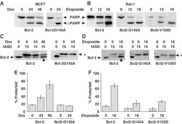
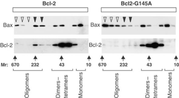
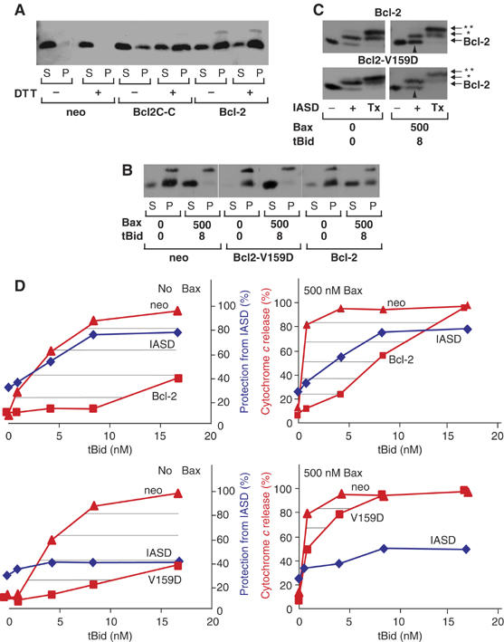
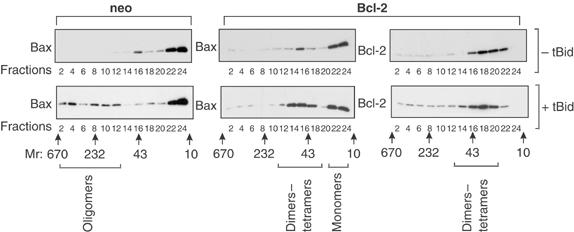
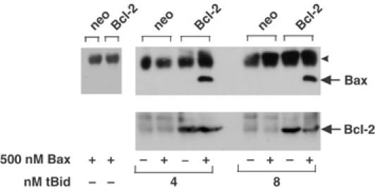
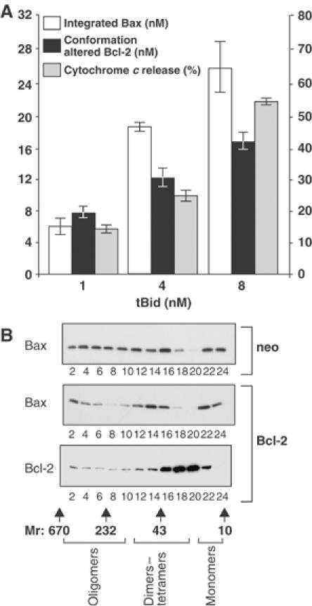
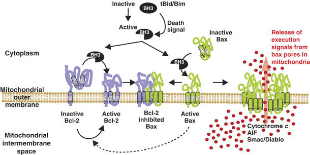
Bcl-2 inhibits apoptosis by regulating the release of cytochrome c and other proteins from mitochondria. Oligomerization of Bax promotes cell death by permeabilizing the outer mitochondrial membrane. In transfected cells and isolated mitochondria, Bcl-2, but not the inactive point mutants Bcl-2-G145A and Bcl-2-V159D, undergoes a conformation change in the mitochondrial membrane in response to apoptotic agonists such as tBid and Bax. A mutant Bcl-2 with two cysteines introduced at positions predicted to result in a disulfide bond that would inhibit the mobility of alpha5-alpha6 helices (Bcl-2-S105C/E152C) was only active in a reducing environment. Thus, Bcl-2 must change the conformation to inhibit tBid-induced oligomerization of integral membrane Bax monomers and small oligomers. The conformationally changed Bcl-2 sequesters the integral membrane form of Bax. If Bax is in excess, apoptosis resumes as Bcl-2 is consumed by the conformational change and in complexes with Bax. Thus, Bcl-2 functions as an inhibitor of mitochondrial permeabilization by changing conformation in the mitochondrial membrane to bind membrane-inserted Bax monomers and prevent productive oligomerization of Bax.


Figures

Figure 1
Figure 1
In two different cell lines inactive Bcl-2 mutants do not change topology at the membrane during apoptosis. MCF7 human breast cancer (A, C, E) and Rat-1 fibroblast (B, D, F) cell lines were transfected with vector control (neo), or expression plasmids encoding Bcl-2 or the inactive mutants Bcl-2-G145A or Bcl-2-V159D, as indicated below the panels. (A, B) Bcl-2-G145A and Bcl-2-V159D do not inhibit apoptosis. Cell lines were exposed to 10 μM doxorubicin (Dox) or 6 μM etoposide for the time indicated in hours above the blots. Apoptosis measured by cleavage of the caspase substrate PARP (ΔPARP) was assessed by immunoblotting of cell lysates. (CF) Cysteine 158 of Bcl-2 but not the inactive mutants is protected from IASD in drug-treated cells. After drug treatment for the time indicated (hours), membrane fractions were incubated with IASD for 0 (control reactions) or 15 min to label proteins with exposed cysteine residues. After separation by SDS–PAGE, labeling was assessed by immunoblotting with antibodies to Bcl-2. (C, D) The migration position of unmodified Bcl-2 is indicated to the left of the panels. Bcl-2 modified by IASD is indicated by an asterisk (*) to the right of the panels. (E, F) Protection of cysteine 158 from IASD labeling for Bcl-2, Bcl-2-G145A and Bcl-2-V159D determined by densitometry from at least three independent experiments for each drug treatment (indicated below the histograms), for samples from (E) MCF7 cells or (F) Rat-1 cells. As reported previously, the limited linear range of the blotting procedure and the small migration difference between labeled and unlabeled proteins limits the range that can be measured to 10–80% protection (Kim et al, 2004). The error bars indicate s.d.
Figure 2
Figure 2
Bcl-2 but not Bcl-2-G145A inhibits oligomerization of Bax. Rat-1 cells expressing either Bcl-2 or Bcl-2-G145A were treated with etoposide for 12 h, and oligomerization of Bax and Bcl-2 were measured by gel filtration chromatography of whole-cell membranes solubilized in CHAPS, followed by immunoblotting of fractions for either Bax or Bcl-2, as indicated. Large oligomers (>250 kDa) are indicated by open arrowheads and are seen primarily in fractions from cells expressing Bcl-2-G145A. A smaller oligomer of approximately 230 kDa seen reproducibly in cells expressing Bcl-2 is indicated by closed arrowheads. Control experiments with untreated cells established elution positions for dimers of Bcl-2 (30–40 kDa fractions) and Bax monomers (15 kDa) as indicated below the panels. The elution positions of molecular weight standards (Mr in kDa) are shown below the panels.
Figure 3
Figure 3
(A) Change in membrane topology of Bcl-2 is required to prevent release of cytochome c from mitochondria treated with recombinant Bax and tBid. The heavy membrane fraction of vector control (neo), Bcl-2-, Bcl-2C-C- or Bcl-2-V159D- (V159D) expressing Rat-1 fibroblasts was exposed to Bax and tBid, as indicated. (A, B) Cytochrome c release was assayed by pelleting the mitochondria and visualizing the cytochrome c (cyt c) in the supernatant (S) and pellet (P) fractions after SDS–PAGE by immunoblotting. X-crossreacting mitochondrial protein. In (A), the heavy membrane fractions were incubated in 1 mM DTT throughout isolation and incubated with tBid (1 nM) and Bax (100 nM). In other panels, the concentration (nM) of tBid and Bax are indicated. (C) Bcl-2 and Bcl-2-V159D were assayed for conformational change in membranes from Rat-1 fibroblasts by IASD labeling as described in Figure 1. In Bcl-2 (upper panel), cysteine 158 was protected; therefore, migration of more than half of the Bcl-2 is not changed after incubation with IASD (upward arrowhead). Much less Bcl-2-V159D (lower panel) was protected from IASD (upward arrowhead). Bands corresponding to IASD labeling of cysteine 158 are indicated by *. Tx indicates an additional control in which Triton X-100 (1%) was added to solubilize the membranes. Labeling of both cysteine 158 and the constitutively membrane embedded cysteine in helix 9 (migration position indicated by **) confirms that protection from labeling was due to integration in the lipid bilayer. (D) Protection (%) of cysteine 158 from IASD labeling (blue diamonds) in heavy membranes from cells expressing Bcl-2 (upper panels) or Bcl-2-V159D (lower panels) compared to the extent of cytochrome c release from mitochondria from the same cells (red, squares indicated Bcl-2 or V159D), and from vector control cells (red, triangles). Immunoblots were quantified by densitometry (n>3) and averaged. The area hatched by horizontal lines indicate protection from apoptosis by Bcl-2 (upper panels) or Bcl-2V-159D (lower panels) for reactions without added Bax (left) or containing 500 nM Bax (right). The amount of tBid added to the reactions is indicated. One of three complete data sets for cytochrome c release at different Bax and tBid concentrations is shown in Supplementary Figure 3.
Figure 4
Figure 4
Bcl-2 prevents tBid-induced oligomerization of Bax. The heavy membrane fraction of vector control (neo) or Bcl-2-expressing Rat-1 fibroblasts was incubated with 500 nM Bax without or with tBid, upper and lower panels, respectively. Oligomerization of Bax and Bcl-2 was measured by gel filtration chromatography of the heavy membrane fraction solubilized in 2% CHAPS, followed by immunoblotting of alternate fractions (indicated below the panels) for Bax or Bcl-2 as indicated. In the absence of tBid, Bax elutes primarily as monomers. Although a very small amount of dimers was detected, large oligomers were not observed (upper panels). Oligomerization of Bax induced by tBid resulted in large oligomers (fractions 2–12) in mitochondria from control cells (neo), but was limited to dimers–tetramers in mitochondria from cells expressing Bcl-2 (fractions 14–18, lower middle panel). Bcl-2 (right panels) was found in small complexes (fractions 14–22), regardless of the presence of tBid. However, the small increase in apparent molecular weight of Bcl-2 complexes when tBid was added was very reproducible. The elution positions of molecular weight standards, and the fractions in which different sized complexes of Bax and/or Bcl-2 were detected, are indicated below the blots.
Figure 5
Figure 5
Bcl-2 binds to tBid-activated Bax. Heavy membrane fractions of vector control (neo) or Bcl-2-expressing Rat-1 fibroblasts were incubated with Bax and/or tBid, as indicated. The membranes were solubilized in CHAPS and immunoprecipitated with sheep anti-Bcl-2 antibody, then analyzed by immunoblotting with monoclonal anti-Bax antibody (upper panels) or with sheep anti-Bcl-2 antibody (lower panels). The single arrowhead indicates sheep immunoglobulin light chain, and the migration of Bax and Bcl-2 is indicated at the right.
Figure 6
Figure 6
Bcl-2 conformational change correlates with the amount of integrated Bax. (A) Bax (500 nM) and the indicated amount of tBid were added to reactions containing heavy membranes from Bcl-2-expressing cells (final concentration of Bcl-2, 20 nM). Protection of cysteine 158 of Bcl-2 from labeling by IASD measured by immunoblotting indicates the amount of conformationally changed Bcl-2 (solid bars). The amount of Bax integrated into mitochondria was assayed by alkaline extraction (open bars). When the amount of integrated Bax exceeds the amount of conformationally changed Bcl-2, cytochrome c release begins (gray bars) but remains less than that for mitochondria from vector-transfected control cells (>90%). The error bars indicate s.e.m.'s for three independent experiments, each performed in triplicate. (B) Large oligomers of Bax detected by gel filtration chromatography when 8 nM tBid and 500 nM Bax were added to mitochondria from control cells (top panel) were reduced but clearly detectable in Bcl-2-expressing cells (center panel). Bcl-2 is excluded from large oligomers of Bax detected by gel filtration chromatography (bottom panel). The elution position of molecular weight standards on the gel filtration column, and the fractions in which different sized complexes of Bax and/or Bcl-2 are detected, are shown below the blot.
Figure 7
Figure 7
Conformationally changed Bcl-2 inhibits membrane-bound activated Bax. In dividing cells, Bcl-2 is constitutively bound to intracellular membranes by a carboxyl-terminal tail-anchor sequence including α-helix 9. This form of Bcl-2 is inactive for preventing Bax oligomerization, but is probably a homodimer with other antiapoptosis functions, and Bax is an inactive monomer located in the cytoplasm or loosely bound to mitochondria. Death signals activate BH3-only proteins (tBid, Bim), which cause changes in the membrane conformation of both Bax and Bcl-2: Bax translocates and inserts into organellar membrane such that α-helices 5, 6 and 9 are embedded in the bilayer (Annis et al, 2005), and Bcl-2 changes conformation such that cysteine 158 (α-helices 5 and 6) are inserted into the membrane (Figures 1, 3, 6 and Roberts et al, manuscript in preparation). In the absence of conformationally changed Bcl-2, membrane-integrated Bax monomers form large oligomers that permeabilize membranes and release proapoptotic factors. Oligomerization of membrane-embedded Bax (monomers or small oligomers) is inhibited by only those Bcl-2 molecules that have changed conformation. It is possible that when activated by a BH3 protein that does not bind Bcl-2, Bax may induce the conformation change in Bcl-2 (dotted line). The precise stoichiometry of binding of polytopic Bcl-2 to membrane-associated Bax is unknown, but inhibited complexes elute as if they are less than 100 kDa. Binding of Bcl-2 to Bax in nonproductive oligomers appears irreversible, and prevents Bcl-2 from inhibiting other Bax molecules. Thus, when excess Bax molecules are activated, large oligomers form and permeabilize the outer mitochondrial membrane releasing proapoptotic molecules. The number of Bax molecules required to form an oligomer that permeabilizes membranes is not known.

References

    1. Adams JM, Cory S (1998) The Bcl-2 protein family: arbiters of cell survival. Science 281: 1322–1326 - PubMed
    1. Annis MG, Soucie EL, Dlogosz PJ, Cruz-Acado JA, Penn LZ, Leber B, Andrews DW (2005) Bax forms multispanning momomers that oligomerize to permeabilize membranes during apoptosis. EMBO J 24: 2096–2103 - PMC - PubMed
    1. Asoh S, Ohtsu T, Ohta S (2000) The super anti-apoptotic factor Bcl-xFNK constructed by disturbing intramolecular polar interactions in rat Bcl-xL. J Biol Chem 275: 37240–37425 - PubMed
    1. Eskes R, Desagher S, Antonsson B, Martinou JC (2000) Bid induces the oligomerization and insertion of Bax into the outer mitochondrial membrane. Mol Cell Biol 20: 929–935 - PMC - PubMed
    1. Hsu YT, Youle RJ (1997) Nonionic detergents induce dimerization among members of the Bcl-2 family. J Biol Chem 272: 13829–132834 - PubMed

Publication types

MeSH terms

Substances

LinkOut - more resources