. 2006 May 17;25(10):2253-62.
doi: 10.1038/sj.emboj.7601117. Epub 2006 Apr 27.

A Drosophila model of oculopharyngeal muscular dystrophy reveals intrinsic toxicity of PABPN1

Affiliations

A Drosophila model of oculopharyngeal muscular dystrophy reveals intrinsic toxicity of PABPN1

Aymeric Chartier et al. EMBO J. .

Abstract

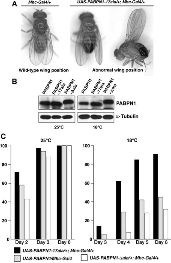
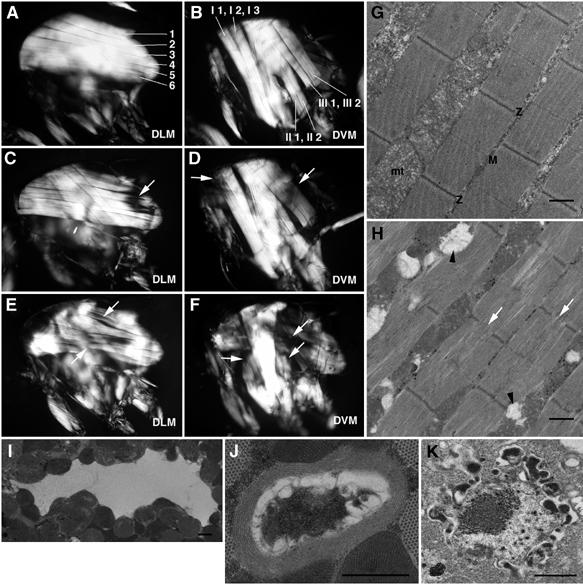
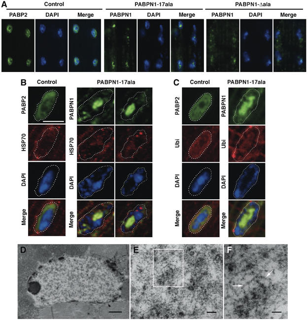
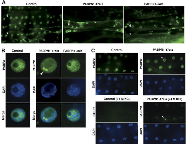
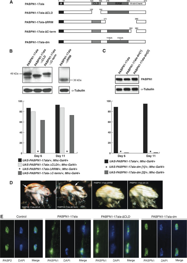
Oculopharyngeal muscular dystrophy (OPMD) is an adult-onset syndrome characterized by progressive degeneration of particular muscles. OPMD is caused by short GCG repeat expansions within the gene encoding the nuclear poly(A)-binding protein 1 (PABPN1) that extend an N-terminal polyalanine tract in the protein. Mutant PABPN1 aggregates as nuclear inclusions in OMPD patient muscles. We have created a Drosophila model of OPMD that recapitulates the features of the human disorder: progressive muscle degeneration, with muscle defects proportional to the number of alanines in the tract, and formation of PABPN1 nuclear inclusions. Strikingly, the polyalanine tract is not absolutely required for muscle degeneration, whereas another domain of PABPN1, the RNA-binding domain and its function in RNA binding are required. This demonstrates that OPMD does not result from polyalanine toxicity, but from an intrinsic property of PABPN1. We also identify several suppressors of the OPMD phenotype. This establishes our OPMD Drosophila model as a powerful in vivo test to understand the disease process and develop novel therapeutic strategies.


Figures

Figure 1
Figure 1
Muscular expression of mammalian PABPN1 induces a progressive wing position phenotype in adults. (A) Abnormal wing position phenotypes (wings down or up, right panel) observed when PABPN1, PABPN1-17ala or PABPN1-Δala is expressed in muscles with the Mhc-Gal4 driver; wild-type wing position in the control Mhc-Gal4/+ (left panel). (B) Quantitation of PABPN1 levels by Western blot. Protein extracts are from 0.25 thoraxes of UAS-PABPN1/Mhc-Gal4, UAS-PABPN1-17ala/+; Mhc-Gal4/+, UAS-PABPN1-Δala/+; Mhc-Gal4/+ adult males at day 1 raised at 25°C (left panel) or 18°C (right panel). α-Tubulin was used as a loading control. Comparable levels of PABPN1 are produced in these three selected transgenic lines. (C) Quantitative analysis of the abnormal wing position phenotype of UAS-PABPN1/Mhc-Gal4, UAS-PABPN1-17ala/+; Mhc-Gal4/+ and UAS-PABPN1-Δala/+; Mhc-Gal4/+ individuals raised at 25 or 18°C. The percentages of adults with abnormal wing position were calculated based on 200 flies from two independent crosses. Control Mhc-Gal4/+ flies did not show abnormal wing position at 25 or 18°C (0% abnormal wing position, n>200).
Figure 2
Figure 2
Muscular expression of PABPN1-17ala induces progressive muscle degeneration. (A–F) Indirect flight muscles (IFMs) in the adult thorax observed under polarized light. (A, B) Control fly with focus on dorso-longitudinal muscles (DLMs, six muscles 1–6) (A) and dorso-ventral muscles (DVMs, seven muscles I1, I2, I3 out of focus, II1, II2, III1, III2) (B). Anterior is to the left and dorsal is up. (C–F) UAS-PABPN1-17ala/+; Mhc-Gal4/+ adult raised at 25°C, at day 6 (C, D) and day 16 (E, F). Muscle defects are clearly visible at day 6, with thinner muscles (white arrows, C, D). At day 16, these defects are strongly enhanced, with very thin or missing muscles (white arrows, E, F). (G, H) Ultrastructure of IFMs visualized by electron microscopy. (G) Longitudinal section through IFM in a control fly showing sarcomeric structure of myofibrils. Z-bands (Z) separate adjacent sarcomeres showing central M-bands (M). mt: mitochondria. (H–K) UAS-PABPN1-17ala/+; Mhc-Gal4/+ adult at day 6, raised at 25°C. (H) Muscle degeneration is characterized by disintegration of mitochondria and their replacement by vacuoles (black arrowheads), and disruption of myofibril integrity with the dissociation of the myosin–actin network and broken Z-bands (white arrows). Similar defects are observed when PABPN1 or PABPN1-Δala is expressed in muscles at 25°C. (I, J) Examples of vacuoles observed in degenerating muscles. (J) Rimmed vacuole surrounded by a ring of membranous structures that closely resembles rimmed vacuoles characteristic of OPMD patient biopsies. (K) Apoptotic nucleus showing a disintegrating nuclear membrane. Scale bars : 1 μm.
Figure 3
Figure 3
Characterization of nuclear inclusions induced by expression of PABPN1-17ala in muscles. (A) Immunostaining of wild-type IFMs with anti-PABP2 (control), and of IFMs from UAS-PABPN1-17ala/+; Mhc-Gal4/+ (PABPN1-17ala) and UAS-PABPN1-Δala/+; Mhc-Gal4/+ (PABPN1-Δala) adults raised at 25°C, at day 6, with anti-PABPN1. DNA is revealed with DAPI staining. The control shows the distribution of PABP2 throughout nuclei, whereas dense nuclear inclusions of PABPN1 are observed when PABPN-17ala is expressed. A diffuse nuclear staining is visible when PABPN1-Δala is expressed. A substantial level of PABPN1 is also present in the cytoplasm when PABPN1-17ala and PABPN1-Δala are expressed using Mhc-Gal4. (B, C) Presence of HSP70 and conjugated ubiquitin in PABPN1 nuclear inclusions. (B) Immunostaining of wild-type IFMs (control) with anti-PABP2 and anti-HSP70, and immunostaining of UAS-PABPN1-17ala/+; Mhc-Gal4/+ (PABPN1-17ala) IFMs with anti-PABPN1 and anti-HSP70. (C) Immunostaining of wild-type IFMs (control) with anti-PABP2 and anticonjugated ubiquitin (Ubi), and immunostaining of UAS-PABPN1-17ala/+; Mhc-Gal4/+ (PABPN1-17ala) IFMs with anti-PABPN1 and anticonjugated ubiquitin. DNA is visualized by DAPI staining. DNA is excluded from the dense PABPN1 nuclear inclusions that show anti-HSP70 and anticonjugated ubiquitin staining. Scale bar is identical for B-C: 4 μm. (D–F) Ultrastructure of nuclei in UAS-PABPN1-17ala/+; Mhc-Gal4/+ IFMs at day 6 and 25°C showing a nuclear inclusion. In this example (D), the nuclear inclusion almost fills the entire nucleus (clear zone); it is surrounded by chromatin (black) close to the nuclear membrane. (E) Higher magnification of a central region of the inclusion shown in (D). Tangled filaments are visible. (F) Higher magnification of the region within the rectangle in (E). Arrows indicate tubular filaments disposed in various directions. Scale bars: 1 μm in (D), 0.2 μm in (E) and 0.1 μm in (F).
Figure 4
Figure 4
The formation of nuclear inclusions depends on the presence of the alanine tract in PABPN1. (A) Immunostaining of third instar larval muscles 6 (top) and 7 (bottom) from wild-type larvae with anti-PABP2 (control) and from UAS-PABPN1-17ala/+; Mhc-Gal4/+ (PABPN1-17ala) and UAS-PABPN1-Δala/+; Mhc-Gal4/+ (PABPN1-Δala) larvae raised at 29°C, with anti-PABPN1. (B) Immunostaining of third instar larval muscles as in (A) showing higher magnification of nuclei. DNA is visualized by DAPI staining. After expression of PABPN1-17ala, dense nuclear inclusions are visible (arrowhead) and the nuclear structure is affected, with DNA at the periphery and excluded from the inclusions. Inclusions do not form, however, after expression of PABPN1-Δala and the nuclear structure does not appear affected. (C) PABPN1-17ala nuclear inclusions are resistant to KCl. Immunostaining of third instar larval muscles from wild-type larvae with anti-PABP2 (control) and from UAS-PABPN1-17ala/+; Mhc-Gal4/+ (PABPN1-17ala) larvae raised at 25°C, with anti-PABPN1. After 1 M KCl treatment, PABP2 or PABPN1-17ala that is diffuse in nuclei are soluble, but PABPN1-17ala within nuclear inclusions is not (bottom panels). Arrowheads indicate nuclear inclusions. Nuclei are visualized by DAPI staining.
Figure 5
Figure 5
The PABPN1 RNA-binding domain is required for muscle defects. (A) Schematic representation of the truncated and mutant versions of PABPN1-17ala. A, alanine tract; CLD, coiled-coil domain; RRM, RNP-type RNA-binding domain; R rich C-term, arginine-rich C-terminal domain. (B) Expression of the PABPN1-17ala-truncated forms, analyzed by Western blots, and quantitative analysis of the abnormal wing position phenotype. Protein extracts were from 0.25 thoraxes of UAS-PABPN1-17ala/+; Mhc-Gal4/+ (PABPN1-17ala), UAS-PABPN1-17ala-ΔCLD/+; Mhc-Gal4/+ (PABPN1-17ala-ΔCLD), UAS-PABPN1-ΔC-term/+; Mhc-Gal4/+ (PABPN1-17ala-ΔC-term) and UAS-PABPN1-17ala-ΔRRM/+; Mhc-Gal4/+ (PABPN1-17ala-ΔRRM) adult males at day 1. α-Tubulin was used as a loading control. Note that PABPN1-17ala-ΔRRM protein is substantially shorter and might be less reactive to the antibody than other PABPN1-17ala versions. Thus, expression level of UAS-PABPN1-17ala-ΔRRM transgene could not be assayed by Western blots and was analyzed by RT–PCR (Supplementary Figure 3). The percentages of adults with abnormal wing position were calculated based on 200 flies from two independent crosses at 18°C. *Indicates that 0% of flies shows an abnormal wing position phenotype. Expression of PABPN1-17ala-ΔRRM from four copies of the transgene in the presence of two copies of Mhc-Gal4 did not produce any abnormal wing position phenotype either. (C) Expression of PABPN1-17ala bearing a double point mutation (PABPN1-17ala-dm) in UAS-PABPN1-17ala-dm/+; Mhc-Gal4/+ individuals, analyzed by Western blots, and quantitative analysis of the abnormal wing position phenotype. Legend is as in (B). Results of two independent transgenic lines (1 and 2) are shown. (D) IFMs visualized under polarized light in (from left to right) UAS-PABPN1-17ala-ΔCLD/+; Mhc-Gal4/+, UAS-PABPN1-17ala-ΔC-term/+; Mhc-Gal4/+, UAS-PABPN1-17ala-ΔRRM/+; Mhc-Gla4/+, UAS-PABPN1-17ala-dm/+; Mhc-Gal4/+ individuals raised at 18°C, at day 16. Defects were visible after expression of PABPN1-17ala-ΔCLD or PABPN1-17ala-ΔC-term, whereas muscles appeared normal after expression of PABPN1-17ala-ΔRRM or PABPN1-17ala-dm. White arrows indicate regions where muscle fibers are affected. (E) Immunostaining of adult IFMs with anti-PABP2 for the wild-type control (left panels), or anti-PABPN1. DNA was revealed with DAPI. Expression of PABPN1-17ala or PABPN1-17ala-ΔCLD led to the formation of nuclear inclusions. In contrast, expression of PABPN1-17ala-dm did not induce the formation of nuclear inclusions, nor did it affect nuclear structure. The distribution of PABPN1-17ala-dm in nuclei is similar to that of PABP2 in wild-type nuclei.

Similar articles

Cited by

References

    1. Abu-Baker A, Messaed C, Laganiere J, Gaspar C, Brais B, Rouleau GA (2003) Involvement of the ubiquitin–proteasome pathway and molecular chaperones in oculopharyngeal muscular dystrophy. Hum Mol Genet 12: 2609–2623 - PubMed
    1. Arrasate M, Mitra S, Schweitzer ES, Segal MR, Finkbeiner S (2004) Inclusion body formation reduces levels of mutant huntingtin and the risk of neuronal death. Nature 431: 805–810 - PubMed
    1. Bao YP, Cook LJ, O'Donovan D, Uyama E, Rubinsztein DC (2002) Mammalian, yeast, bacterial, and chemical chaperones reduce aggregate formation and death in a cell model of oculopharyngeal muscular dystrophy. J Biol Chem 277: 12263–12269 - PubMed
    1. Bear DG, Fomproix N, Soop T, Bjorkroth B, Masich S, Daneholt B (2003) Nuclear poly(A)-binding protein PABPN1 is associated with RNA polymerase II during transcription and accompanies the released transcript to the nuclear pore. Exp Cell Res 286: 332–344 - PubMed
    1. Bellen HJ, Levis RW, Liao G, He Y, Carlson JW, Tsang G, Evans-Holm M, Hiesinger PR, Schulze KL, Rubin GM, Hoskins RA, Spradling AC (2004) The BDGP gene disruption project: single transposon insertions associated with 40% of Drosophila genes. Genetics 167: 761–781 - PMC - PubMed

Publication types

MeSH terms

Substances