. 2006 Jul 1;71(1):139-48.
doi: 10.1016/j.cardiores.2006.03.008. Epub 2006 Mar 16.

Monocyte-specific Bcl-2 expression attenuates inflammation and heart failure in monocyte chemoattractant protein-1 (MCP-1)-induced cardiomyopathy

Affiliations

Monocyte-specific Bcl-2 expression attenuates inflammation and heart failure in monocyte chemoattractant protein-1 (MCP-1)-induced cardiomyopathy

Jianli Niu et al. Cardiovasc Res. .

Abstract

Objective: Infiltrating inflammatory cells within the myocardium have been shown to be apoptotic, but the significance of apoptotic inflammatory cells to the development of cardiomyopathy remains undefined. Transgenic mice with cardiac-specific expression of MCP-1 exhibit extensive apoptosis of infiltrating mononuclear cells and develop heart failure. Here, we tested the hypothesis that in vivo selective inhibition of apoptosis of infiltrating mononuclear cells would preserve cardiac structure and function and improve survival in this murine model.

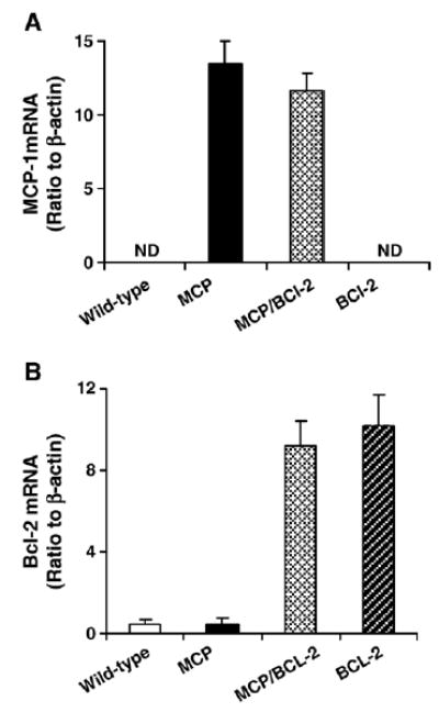
Methods: Mice with cardiac-specific expression of MCP-1 and monocyte-specific expression of Bcl-2 were generated by cross-breeding MCP-1 transgenic mice with hMRP8-Bcl-2 mice that over-express Bcl-2 in the monocytes. Structural and functional parameters and the inflammatory response of the heart were evaluated and compared between the wild-type and transgenic mice.

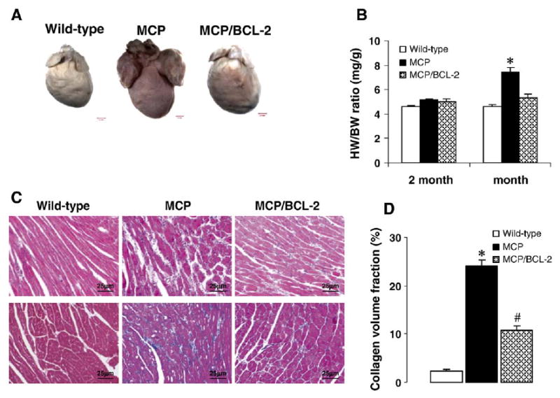
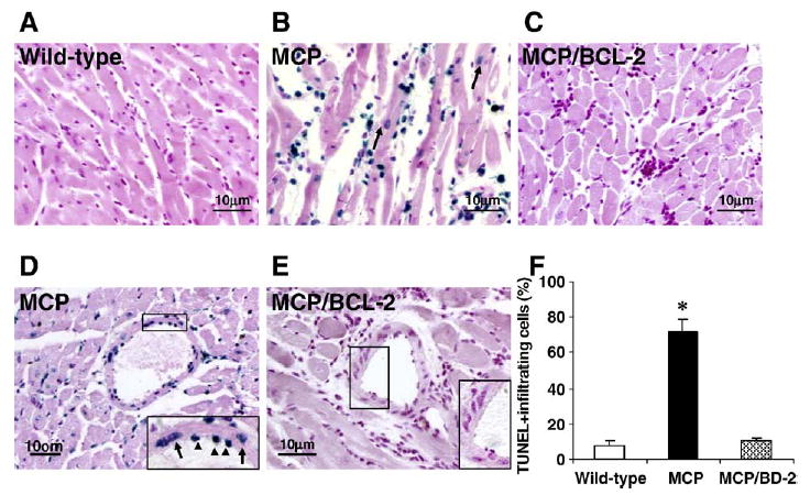
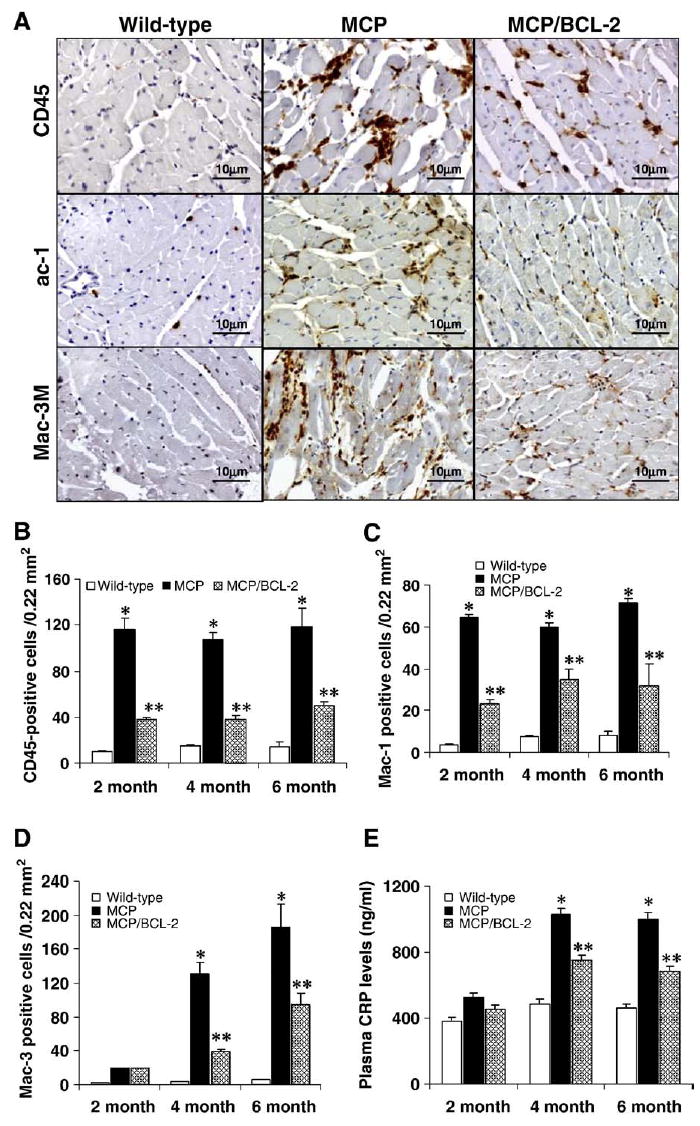
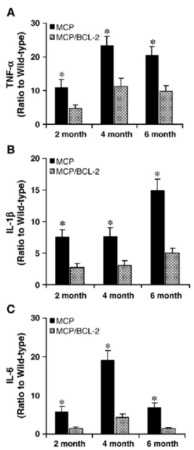
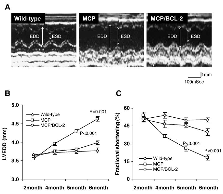
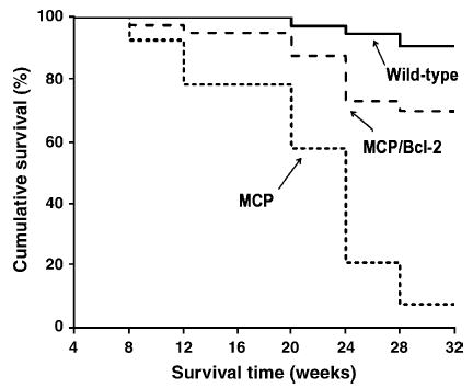
Results: Expression of Bcl-2 in monocytes results in superior preservation of myocardial structure, cardiac function and a significant prolongation of survival of MCP-1 transgenic mice. The beneficial effects of monocyte-specific Bcl-2 expression are associated with inhibition of apoptosis of infiltrating mononuclear cells, normalization of circulating C-reactive protein levels, attenuation of cellular infiltrates, macrophage activation and production of proinflammatory cytokines, tumor necrosis factor (TNF-alpha), interleukin (IL)-1 beta and IL-6 in the hearts.

Conclusions: These results demonstrate that apoptosis of infiltrating mononuclear cells plays a detrimental role in the development of heart failure in this murine model, suggesting that modulation of apoptosis of infiltrating mononuclear cells may be of clinical benefit in heart failure.


Figures

Fig. 1
Fig. 1
Real-time PCR demonstrates transgene mRNA expression in the hearts. Histograms showing the levels of MCP-1 (A) and Bcl-2 (B) mRNA expression in the hearts of wild-type and transgenic mice of 4 months of age (n =3 per group). Equal expression of transgene was found in mice expressing only one transgene versus mice expressing both transgenes. ND, not detectable.
Fig. 2
Fig. 2
Monocyte-specific Bcl-2 expression in MCP mice attenuates cardiac deterioration. (A) Representative photographs showing the whole hearts from the wild-type, MCP and MCP/Bcl-2 mouse at 6 months of age. The heart from MCP mouse is significantly larger and noticeably discolored when compared to the wild-type and MCP/Bcl-2 mouse. (B) Heart weight to body weight (HW/BW) ratio was depicted from 2 to 6 months of age. *P <0.001 versus MCP/Bcl-2 mice and wild-type mice, n =6 per group per time point. (C) Representative photomicrographs demonstrating morphology of heart sections from 6-month-old wild-type and transgenic mice stained with H&E (upper panel) and Masson’s trichrome (lower panel). Blue staining represents collagen deposition. (D) Collagen volume fraction in the hearts of 6-month-old wild-type, MCP and MCP/Bcl-2 mice. n =6 per group. *P <0.001 versus wild-type and MCP/Bcl-2 mice; #P <0.05 versus wild-type mice.
Fig. 3
Fig. 3
Bcl-2 expression in monocytes prevents apoptotic cell death in the hearts of MCP mice. (A–E) Representative photomicrographs demonstrating TUNEL staining of heart sections from 6-month-old wild-type, MCP and MCP/Bcl-2 mice. Blue staining indicates TUNEL-positive cells and the TUNEL-positive cardiac myocytes are indicated by arrows in B. The insert in D shows TUNEL-positive vascular endothelial (arrowheads) and smooth muscle cells (arrows) in the heart of MCP mouse. The insert in E shows no TUNEL-positive vascular cells in the heart of MCP/Bcl-2 mouse. (F) Histogram showing the quantitative analysis of TUNEL-positive infiltrating cells in the hearts of mice at 6 months of age. n =5 per group. *P <0.001 versus wild-type mice and MCP/Bcl-2 mice.
Fig. 4
Fig. 4
Bcl-2 expression in monocytes results in reduced inflammatory cells infiltration in the hearts of MCP mice. (A) Representative photomicrographs demonstrating immunohistochemical staining with anti-CD45, -Mac-1 and -Mac-3 antibodies in heart sections from 6-month-old wild-type and transgenic mice. Positive-stained cells were visualized with diaminobenzidine (brown). (B–D) Histograms showing the number of CD45-, Mac-1- and Mac-3-positive cells in the hearts of wild-type, MCP and MCP/Bcl-2 mice. *P <0.001 versus wild-type mice; #P <0.05 versus wild-type and MCP mice; n =6 per group per time point. (E) Circulating levels of CRP assayed by ELISA. n =6 per group per time point. *P <0.05 versus wild-type and MCP/Bcl-2 mice; #P <0.05 versus wild-type mice.
Fig. 5
Fig. 5
Bcl-2 expression in monocytes inhibits proinflammatory cytokines mRNA expression in the hearts of MCP mice. Relative levels of TNF-α (A), IL-1β (B) and IL-6 (C) mRNA expression in the hearts of wild-type and transgenic mice were assayed by real-time PCR. The expression of TNF-α, IL-1β and IL-6 in the hearts was expressed as a ratio relative to age-matched wild-type controls. Over-expression of Bcl-2 in monocytes resulted in decreased TNF-α, IL-1β and IL-6 mRNA expression in the hearts of MCP/Bcl-2 mice compared to age-matched MCP mice. *P <0.05, n =6 per group per time point.
Fig. 6
Fig. 6
Bcl-2 expression in monocytes prevents progression of left ventricle (LV) dilatation and preserves cardiac function. (A) Representative M-mode tracing showing changes in left ventricle dimensions at 6 months of age in wild-type and transgenic mice. EDD and ESD indicate LV end-diastolic dimension and end-systolic dimension, respectively. (B–C) LV end-diastolic dimension (LVEDD) and percent functional shortening (FS) were depicted from 2 to 6 months of age. MCP mice had progressive LV dilatation and cardiac dysfunction compared to wild-type and MCP/Bcl-2 mice, whereas MCP/Bcl-2 mice had slightly increased LVEDD and reduced FS, but were not significantly different from LVEDD and FS in wild-type mice, P <0.001 versus wild-type and MCP/Bcl-2 mice, n =6 per group per time point.
Fig. 7
Fig. 7
Kaplan–Meier survival analysis of wild-type (n =28), MCP (n =36) and MCP/Bcl-2 (n =32) mice showing improvement of survival profile of MCP/Bcl-2 mice compared with MCP mice ( P <0.001).

References

    1. Ikeda U. Inflammation and coronary artery disease. Curr Vasc Pharmacol. 2003;1:65–70. - PubMed
    1. Nian M, Lee P, Khaper N, Liu P. Inflammatory cytokines and postmyocardial infarction remodeling. Circ Res. 2004;94:1543–53. - PubMed
    1. Hansson GK. Inflammation, atherosclerosis, and coronary artery disease. N Engl J Med. 2005;352:1685–95. - PubMed
    1. Dawson J, Miltz W, Mir AK, Wiessner C. Targeting monocyte chemoattractant protein-1 signaling in disease. Expert Opin Ther Targets. 2003;7:35–48. - PubMed
    1. Sheikine Y, Hansson GK. Chemokines and atherosclerosis. Ann Med. 2004;36:98–118. - PubMed

Publication types

MeSH terms