

Influence of ageing and physical activity on vascular morphology in rat skeletal muscle
- PMID: 16644803
- PMCID: PMC1819439
- DOI: 10.1113/jphysiol.2006.108431
Influence of ageing and physical activity on vascular morphology in rat skeletal muscle
Abstract
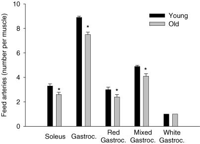
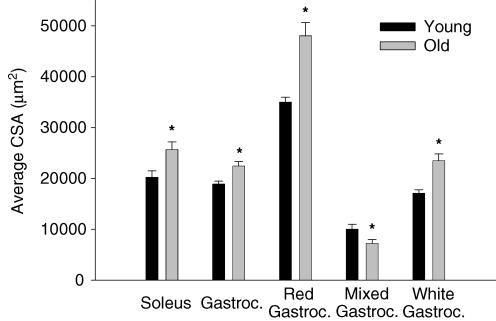
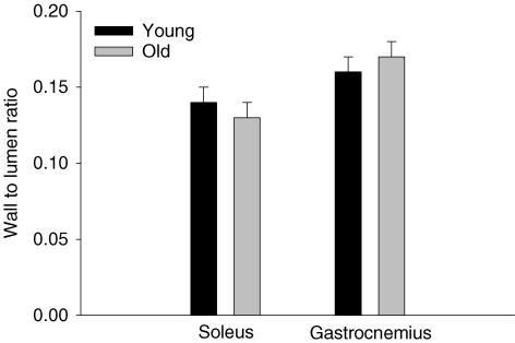
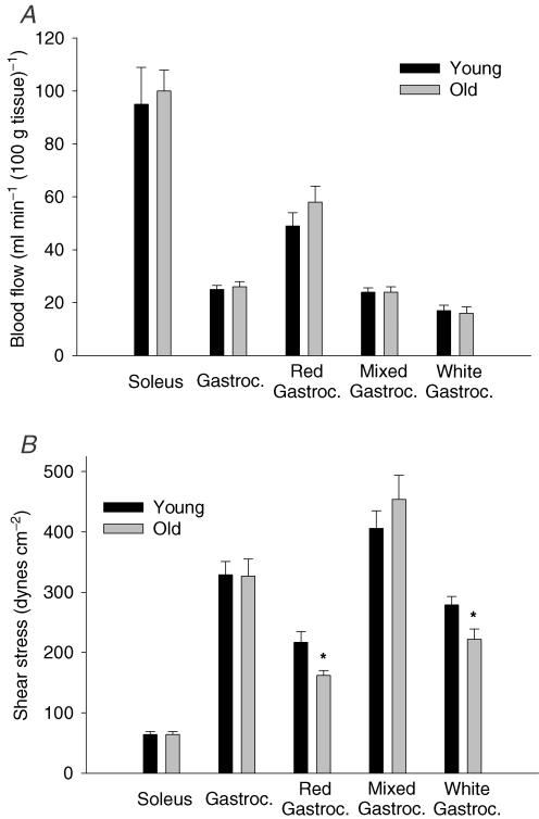
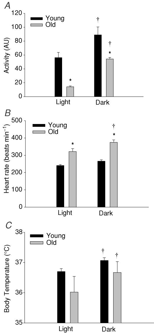
Key structural and functional properties of the skeletal muscle vasculature that underlie diminished vascular conductance with ageing remain obscure. The purpose of this investigation was to test the hypotheses that (1) reduced levels of spontaneous physical activity in old rats are associated with skeletal muscle vascular remodelling (e.g. arterial rarefaction), and (2) consequent to a vascular remodelling, calculated shear stress is maintained in feed arteries of aged muscle at levels commensurate with that in young. Activity during daily light and dark cycles (12-12 h) was measured at 30-s intervals for 2 weeks in young (6 months; n = 9) and old (24 months; n = 9) Fisher 344 rats via telemetry. Subsequently, the gastrocnemius complex and soleus muscles were excised and all feed arteries were counted, isolated, cannulated and maximally dilated for measurement of luminal diameter. Resting blood flow was also measured to estimate vessel wall shear-stress in the feed arteries perforating the soleus and gastrocnemius muscles. Overall, young rats were approximately 1.6 times more active during dark periods and approximately 4 times more active during light periods than old rats. In addition, young rats had approximately one additional feed artery perforating both the soleus (young, 3.3 +/- 0.2; old, 2.6 +/- 0.2 vessels; P < 0.05) and gastrocnemius (young, 8.8 +/- 0.1; old, 7.5 +/- 0.2 vessels, P < 0.05) muscles compared with old rats. However, average vessel wall shear stress at rest was similar between young and old rats (soleus: Y, 65 +/- 5; O, 64 +/- 5 dynes cm(-2); gastrocnemius: Y, 329 +/- 22; O, 327 +/- 27 dynes cm(-2)) resulting from a larger vessel diameter in arteries from old rats. In conclusion, lower activity levels of old rats likely contribute to resistance artery rarefaction and, consequently, this provides a plausible mechanism for the altered blood flow patterns observed during exercise in aged skeletal muscle.
Figures
References
-
- Armstrong RB, Delp MD, Goljan EF, Laughlin MH. Distribution of blood flow in muscles of miniature swine during exercise. J Appl Physiol. 1987;62:1285–1298. - PubMed
-
- Armstrong RB, Laughlin MH. Exercise blood flow patterns within and among rat muscles after training. Am J Physiol. 1984;246:H59–H68. - PubMed
-
- Armstrong RB, Marum P, Saubert CWT, Seeherman HJ, Taylor CR. Muscle fiber activity as a function of speed and gait. J Appl Physiol. 1977;43:672–677. - PubMed
-
- Asai K, Kudej RK, Shen YT, Yang GP, Takagi G, Kudej AB, et al. Peripheral vascular endothelial dysfunction and apoptosis in old monkeys. Arterioscler Thromb Vasc Biol. 2000;20:1493–1499. - PubMed
-
- Behnke BJ, Delp MD, Dougherty PJ, Musch TI, Poole DC. Effects of aging on microvascular oxygen pressures in rat skeletal muscle. Respir Physiol Neurobiol. 2005;146:259–268. - PubMed
Publication types
MeSH terms
Grants and funding
LinkOut - more resources
Full Text Sources
Medical