. 2006 Sep 1;108(5):1668-76.
doi: 10.1182/blood-2006-04-015586. Epub 2006 Apr 27.

Constitutive activation of Akt contributes to the pathogenesis and survival of mantle cell lymphoma

Affiliations

Constitutive activation of Akt contributes to the pathogenesis and survival of mantle cell lymphoma

Martina Rudelius et al. Blood. .

Abstract

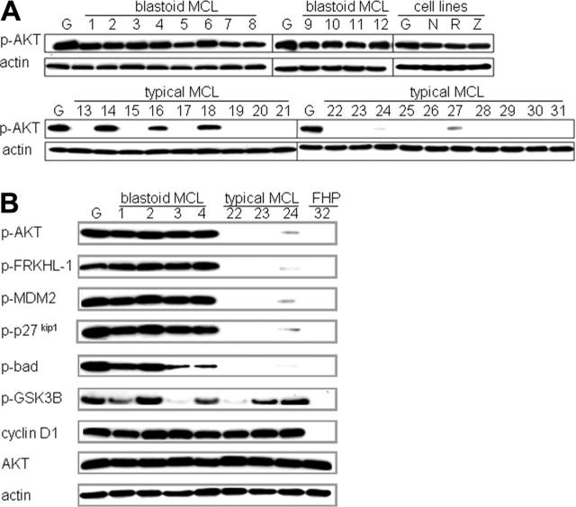
To determine whether the PI3K/Akt signaling pathway is involved in the pathogenesis of mantle cell lymphoma (MCL), we investigated the phosphorylation status of Akt and multiple downstream targets in primary MCL cases and cell lines. Akt was phosphorylated in 12 of 12 aggressive blastoid MCL variants and in 4 of 4 MCL cell lines. In contrast, phosphorylated Akt was present in only 5 of 16 typical MCL, 3 at comparable levels to the blastoid cases, and 2 at low levels. The presence of p-Akt was accompanied by the phosphorylation of p27(kip1), FRKHL-1, MDM2, Bad, mTOR, and p70S6K. Inhibition of the PI3K/Akt pathway in the MCL cell lines abrogated or reduced the phosphorylation of Akt, p27(kip1), FRKHL-1, MDM2, Bad, mTOR, GSK-3beta, IkappaB, and led to cell-cycle arrest and apoptosis. Six MCL cases (5 with activated Akt and 1 with inactive Akt) and 3 of 4 cell lines showed loss of PTEN expression. PIK3CA mutations were not detected. We conclude that constitutive activation of the PI3K/Akt pathway contributes to the pathogenesis of MCL and preferentially occurs in blastoid variants. One possible mechanism of activation is loss of PTEN expression. These data suggest that PI3K/Akt inhibitors may be effective in the treatment of Akt-activated MCL.


Figures

Figure 1.
Figure 1.
Akt is preferentially activated in the blastoid variant of MCL and in MCL cell lines and is associated with phosphorylation of several downstream targets. (A) Western blot analysis for activated p-Akt (Ser473) in MCL cases and cell lines (Granta 519 [G], NCEB-1 [N], REC-1 [R], Z138C [Z]). High levels of p-Akt were detected in 12 of 12 blastoid MCLs (cases 1-12) and in the 4 cell lines, whereas only 5 of 19 typical MCLs displayed p-Akt—3 at high levels (cases 14, 16, and 18) and 2 at low levels (cases 24 and 27). Actin is shown as loading control. (B) Western blot analyses of downstream Akt targets shown for representative MCL cases, the Granta 519 cell line, and a follicular hyperplasia control (FHP). Blastoid variants (cases 1-4) and the Granta 519 cell line show high levels of p-Akt and multiple phosphorylated downstream targets (p-FRKHL-1, p-MDM2, p-p27kip1, p-Bad), compared with typical MCL (cases 22-24). The low activation of Akt in case 24 results in modest phosphorylation of the downstream targets. Note that GSK-3β, a classic Akt target, is phosphorylated in both p-Akt+ and p-Akt-cases. The negative control follicular hyperplasia (FHP) shows no activation of the Akt pathway. Cyclin D1 is strongly positive in all MCL cases. Actin shows equal loading of protein for each lane.
Figure 2.
Figure 2.
Akt is constitutively activated in MCL cell lines. Serum-depletion experiments were performed with all 4 MCL cell lines. Cells were grown for 48 hours with (FBS +) or without serum (FBS -). Western blot analysis with p-Akt under serum starvation shows no significant decrease of p-Akt in MCL cell lines (shown are Granta 519, NCEB-1, and Z138C). In contrast, p-Akt levels in the Burkitt cell line BJAB are dramatically decreased under serum-starvation conditions. Total Akt levels are shown as loading control.
Figure 3.
Figure 3.
Activation of Akt and downstream targets are dependent upon PI3K activity. PI3K/Akt pathway inhibition studies were performed with all 4 MCL cell lines using 3 different inhibitors: LY294002, wortmannin, and Akt inhibitor (Calbiochem). Comparable results were obtained for each line, and results with Granta 519 using LY294002 or Akt inhibitor are shown in representative experiments. Akt inactivation occurs after 8 hours (8 h), followed by the abrogation of phosphorylation of Bad, FRKHL-1, MDM2, and p27kip1 after 24 hours (24 h). Translational control proteins mTOR, p70S6K, and S6K show a time-dependent decrease in phospho-protein levels. S6K and total Akt levels are shown as loading control.
Figure 4.
Figure 4.
Inhibition of the PI3K/Akt pathway decreases activation of the NF-κB pathway. PI3K/Akt pathway inhibition studies were performed with the Granta 519 and Z138C cell lines using 3 different inhibitors: LY294002, Akt inhibitor, and wortmannin. Comparable results were obtained for both lines, and results with Granta 519 using LY294002 or Akt inhibitor are shown as representative experiments. (A,C) Western blot analysis reveals Akt inactivation by 8 hours, and a time-dependent decrease in phosphorylated IκBα and p65. Total Akt and α-tubulin are shown as loading controls. (B,D) Western blot analysis of separated nuclear and cytoplasmic fractions reveals a gradual accumulation of the NF-κB p65 subunit in the cytoplasm following addition of LY294002 or Akt inhibitor and a corresponding reduction in nuclear NF-κB. α-tubulin is shown as loading control for cytoplasmic protein; oct-1 as loading control for nuclear protein.
Figure 5.
Figure 5.
Inhibition of the PI3K/Akt pathway induces cell-cycle arrest and apoptosis. PI3K/Akt pathway inhibition studies were performed with 2 MCL cell lines (Granta 519 and Z138C) and a non-Akt-activated control cell line (RAJI) using 3 different inhibitors: LY294002, Akt inhibitor, and wortmannin. Comparable results were obtained for each MCL cell line, and results with Granta 519 using LY294002 and Akt inhibitor are shown as representative experiments in comparison to RAJI. (A,C) Cell-cycle analysis by flow cytometry indicates G1-S phase arrest in the MCL cell line Granta 519 at 24 hours and an increase in apoptotic cells after 48 hours of treatment. In contrast, the non-Akt-activated control cell line RAJI shows no significant alterations under treatment. (B,D) Western blot analysis shows a corresponding activation of pro-caspase-3 as assessed by appearance of the cleaved caspase-3, the loss of phosphorylated Bad, and the subsequent accumulation of Bad-bcl-xL complexes after 24 hours (Western blot preceded by immunoprecipitation [IP] with bcl-xL). Bcl-xL is shown as a loading control for the IP; α-tubulin for the standard Western.
Figure 6.
Figure 6.
Inhibition of the PI3K/Akt pathway leads to up-regulation of p27kip1 and loss of cyclin D1 expression. PI3K/Akt pathway inhibition studies were performed with the 4 MCL cell lines and the non-Akt-activated control cell line RAJI using 3 different inhibitors: LY294002, wortmannin, and Akt inhibitor (Calbiochem). Comparable results were obtained for each MCL cell line, and results with Granta 519 using LY294002 or Akt inhibitor are shown as representative experiments. (A,C) Proliferation/viability as assessed by MTT test. Results are averages of 3 individual experiments and are displayed as percent absorbance of control cells (untreated) with Granta 519 (□), Z138C (formula image
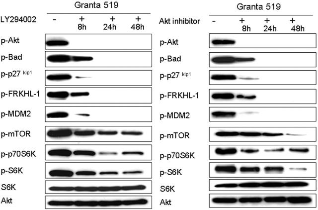
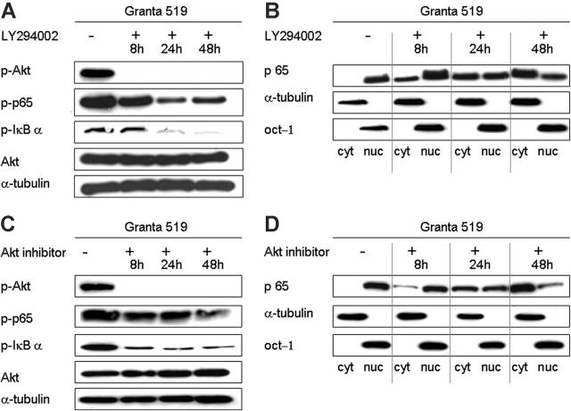
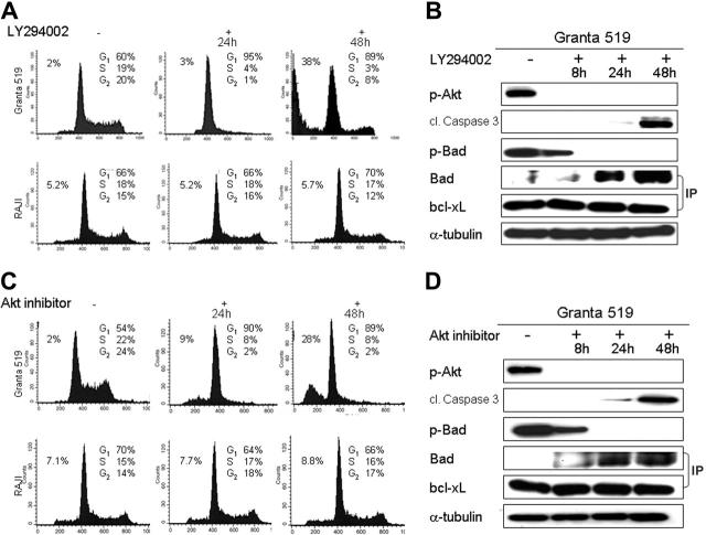
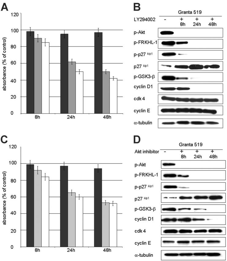
), and RAJI (▪). A significant reduction in absorbance to 50% of control values after 48 hours is seen in the MCL cell lines, whereas the non-Akt-activated control RAJI shows no significant reduction. (B,D) Western blot analysis with Granta 519 demonstrates Akt inactivation at 8 hours, followed by abrogation of phosphorylation of FRKHL-1, p27kip1, and GSK-3β by 24 hours. The cell-cycle inhibitor p27kip1 shows a gradual increase in expression levels over time, and cyclin D1 is dramatically down-regulated. In contrast, cdk4 and cyclin E remain constant. α-tubulin expression is shown as loading control.
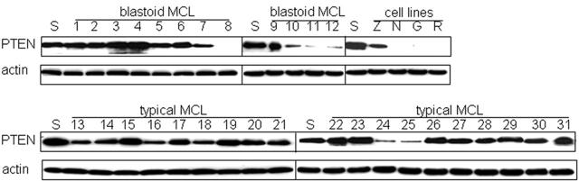
Figure 7.
Figure 7.
Loss of PTEN contributes to the activation of Akt in a subset of cases. Western blot analysis for PTEN was performed for all MCL cases and cell lines. SUDHL-1 (S) served as positive control. PTEN expression was decreased in 5 of 17 (29%) cases with activated Akt, including 4 high-expressing blastoid MCL (cases 8, 10, 11, 12) and 1 low-expressing typical MCL (case 24). In contrast, 1 of 14 cases without detectable Akt activation (case 25) showed decreased expression of PTEN. Actin is shown as loading control.

References

    1. Raffeld M, Jaffe ES. bcl-1, t(11;14), and mantle cell derived neoplasms. Blood. 1991;78: 259-263. - PubMed
    1. Meeker TC, Sellers W, Harvey R, et al. Cloning of the t(11;14)(q13;q32) translocation breakpoints from two human leukemia cell lines. Leukemia. 1991;5: 733-737. - PubMed
    1. Tsujimoto Y, Jaffe E, Cossman J, Gorham J, Nowell PC, Croce CM. Clustering of breakpoints on chromosome 11 in human B-cell neoplasms with the t(11;14) chromosome translocation. Nature. 1985;315: 340-343. - PubMed
    1. Bosch F, Lopez-Guillermo A, Campo E, et al. Mantle cell lymphoma: presenting features, response to therapy, and prognostic factors. Cancer. 1998;82: 567-575. - PubMed
    1. Argatoff LH, Connors JM, Klasa RJ, Horsman DE, Gascoyne RD. Mantle cell lymphoma: a clinicopathologic study of 80 cases. Blood. 1997;89: 2067-2078. - PubMed

Publication types

MeSH terms

Substances