. 2006 Sep;35(3):337-46.
doi: 10.1165/rcmb.2005-0474OC. Epub 2006 Apr 27.

Persistent effects induced by IL-13 in the lung

Affiliations

Persistent effects induced by IL-13 in the lung

Patricia C Fulkerson et al. Am J Respir Cell Mol Biol. 2006 Sep.

Abstract

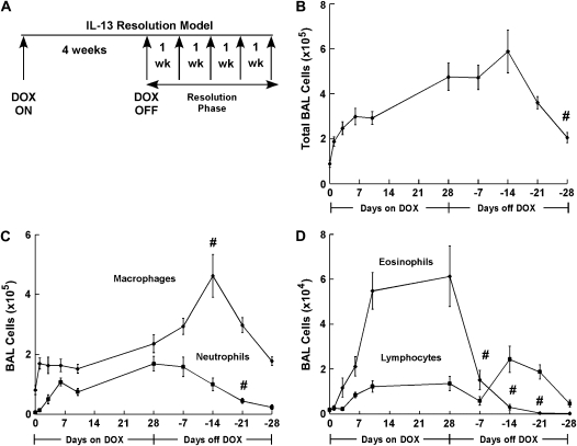
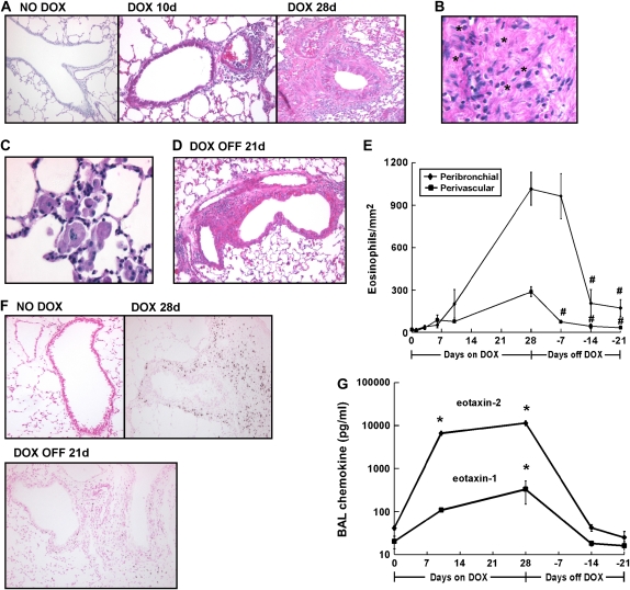
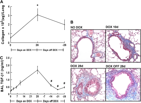
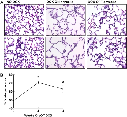
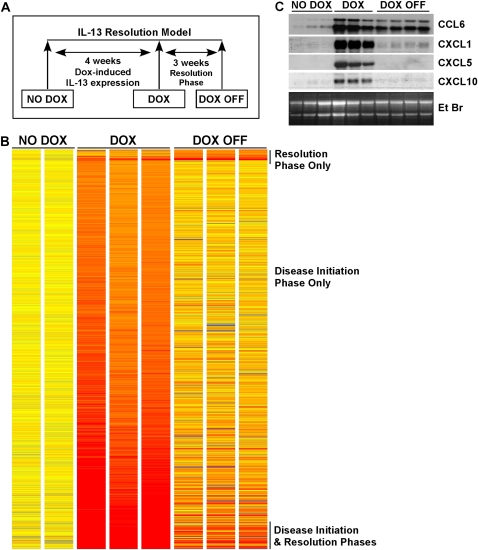
IL-13 overexpression in the lung induces inflammatory and remodeling responses that are prominent features of asthma. Whereas most studies have concentrated on the development of IL-13-induced disease, far fewer studies have focused on the reversibility of IL-13-induced pathologies. This is particularly important because current asthma therapy appears to be poor at reversing lung remodeling. In this manuscript, we used an externally regulatable transgenic system that targets expression of IL-13 to the lung with the aim of characterizing the reversibility process. After 4 wk of doxycycline (dox) exposure, IL-13 expression resulted in mixed inflammatory cell infiltration, mucus cell metaplasia, lung fibrosis, and airspace enlargement (emphysema). After withdrawal of dox, IL-13 protein levels were profoundly reduced by 7 d and below baseline by 14 d. During this time frame, the level of lung eosinophils returned to near normal, whereas macrophages, lymphocytes, and neutrophils remained markedly elevated. IL-13-induced mucus cell metaplasia significantly decreased (91%) 3 wk after withdrawal of dox, showing strong correlation with reduced eosinophil levels. In contrast, IL-13-induced lung fibrosis did not significantly decline 4 wk after dox withdrawal. Importantly, IL-13-induced emphysema persisted, but modestly declined 4 wk after dox. Examination of transcript expression profiles identified a subset of genes that remained increased weeks after transgene expression was no longer detected. Notably, numerous IL-13-induced cytokines and enzymes were reversible (IL-6 and cathepsins), whereas others were sustained (CCL6 and chitinases) after IL-13 withdrawal, respectively. Thus, several hallmark features of IL-13-induced lung pathology persist and are dissociated from eosinophilia after IL-13 overexpression ceases.


Figures

<b>Figure 1.</b>
Figure 1.
Inducible IL-13 expression in the lung. (A) Constructs used in the generation of CC10-iIL-13 mice. (B) Kinetics of IL-13 induction and cessation. Dual transgene positive mice were grown to adulthood on normal food pellets and then switched to dox-impregnated food. The levels of pulmonary IL-13 mRNA (B) were assessed at intervals before and after the addition of dox food. RNA was isolated from the lungs of CC10-iIL-13 mice fed dox-impregnated food for the indicated time periods. Each lane represents an individual mouse. In C, dual transgene positive mice received dox food for 28 d and then the dox food was removed. The levels of BALF IL-13 protein (C) were assessed at intervals before and after dox removal. Protein levels represent the mean ± SD of 4–6 mice at each time point.
<b>Figure 2.</b>
Figure 2.
IL-13 transgene expression results in persistent airway inflammation. (A) A schematic representation of the IL-13 resolution model is depicted. Adult mice are fed dox-impregnated food for 4 wk and then given normal food. Multiple parameters are evaluated weekly after dox withdrawal (Resolution Phase). (B) Total BALF cells from CC10-iIL-13 mice fed dox-impregnated food (Days on DOX) and after dox withdrawal (Days off DOX) for the indicated days (n = 6–9 mice/group). All data points were statistically different (P < 0.001) from non–dox-fed control mice after 1 d of dox administration. #P = 0.003 when compared with mice fed dox food for 28 d. (C) Macrophage and neutrophil composition of BALF from CC10-iIL-13 mice at indicated times in resolution model (n = 6–9 mice/group). All data points were statistically different (P < 0.02) than non–dox-fed mice after 1 d of dox administration for BAL macrophages and after 6 d for BAL neutrophils. #P = 0.01 when compared with mice fed dox-food for 28 d. (D) Eosinophil and lymphocyte composition of BALF from CC10-iIL-13 mice at indicated times in the resolution model (n = 6–9 mice/group). All points were significantly different (P < 0.002) from non–dox-fed control mice after 6 d of dox administration. #P = 0.03 when compared with mice fed dox food for 28 d.
<b>Figure 3.</b>
Figure 3.
IL-13 transgene expression results in sustained tissue inflammation. (A) H&E-stained lungs from CC10-iIL-13 mice fed normal (NO DOX) or dox-food (DOX) for the indicated times are shown. (B) Infiltrating leukocytes trapped in extracellular matrix are shown. *Eosinophils. (C) Enlarged macrophages within the airspace are shown. (D) Inflammation surrounding blood vessels and airways 3 wk after dox withdrawal (DOX OFF) is shown. (E) Kinetics of IL-13–induced eosinophil infiltration around blood vessels (perivascular, squares) and bronchioles (peribronchial, diamonds) is shown (n = 4–11 mice/group). All points were significantly different (P < 0.05) from non–dox-fed control mice after 10 d of dox administration. #P ⩽ 0.004 when compared with mice fed dox food for 28 d. (F) Eosinophils, detected by anti-MBP immunohistochemistry, are shown in lung tissue at different time points within the resolution model. (G) Eotaxin-1 and eotaxin-2 protein levels in the BALF of CC10-iIL-13 mice fed normal (NO DOX) or dox-food (DOX) for the indicated time are shown. Protein levels represent mean ± SD of 4–7 mice at each time point. *P < 0.0001 when compared with non–dox-fed control mice. Magnification in A–D, F: ×100–400.
<b>Figure 4.</b>
Figure 4.
Resolution of IL-13–induced mucus production. (A) Kinetics of mucus induction and resolution as shown in PAS-stained lungs from CC10-iIL-13 mice fed normal (NO DOX) or dox food (DOX) for the indicated times. Magnification: ×100. (B) Quantitation of PAS+ cells (mean ± SD) in airways of CC10-iIL-13 mice at indicated times in the resolution model (n = 4–6 mice/group). *P < 0.001 when compared with 10 d of dox administration.
<b>Figure 5.</b>
Figure 5.
Persistence of IL-13–induced collagen deposition. (A) Collagen content of the left lobe of CC10-iIL-13 mice using Sircol assay at indicated times in resolution model is shown (n = 2 experiments with a representative experiment shown). *P ⩽ 0.003 when compared with non–dox-fed control mice. (B) Localization of collagen in CC10-iIL-13 mice over time as shown by Masson's trichrome staining. (C) Levels of active TGF-β1 in BALF from CC10-iIL-13 mice at indicated times in resolution model are shown (4–8 mice/group). #P ⩽ 0.03 when compared with mice fed dox food for 28 d.
<b>Figure 6.</b>
Figure 6.
Persistence of IL-13–induced emphysema. (A) Induction of airspace enlargement as shown in H&E stained lungs from CC10-iIL-13 mice fed normal (NO DOX) or dox food (DOX) for the indicated number of weeks. Each panel is an individual mouse. Magnification: ×200. (B) Quantitation of percentage of fractional area of airspace (% fx airspace area) of CC10-iIL-13 mice at indicated times in the resolution model (n = 2 experiments, with 4–8 mice/group/experiment). A representative experiment is shown. *P < 0.0001 when compared with non–dox-fed control mice. #P = 0.002 when compared with mice fed dox food for 28 d.
<b>Figure 7.</b>
Figure 7.
Identification of IL-13–induced genes. (A) A schematic diagram of the IL-13 resolution model is depicted, indicating the three time points of analysis: before transgene induction (NO DOX), 4 wk with transgene expression (DOX), and 3 wk after transgene turned off (DOX OFF). (B) The 767 genes differentially expressed (P < 0.05) in the lungs of CC10-iIL-13 mice at indicated time points in the model compared with NO DOX control mice are shown; upregulated genes are represented in red and downregulated genes in blue. The magnitude of the gene changes is proportional to the darkness of the color. Each column represents an individual mouse and each line a gene. (C) Northern blot analysis confirmed sustained induction of CCL6 and CXCL1, but not CXCL5 or CXCL10 by IL-13 transgene expression. The ethidium bromide (EtBr)-stained gel is shown. Each lane represents an individual mouse.

Similar articles

Cited by

References

    1. Elias JA, Zhu Z, Chupp G, Homer RJ. Airway remodeling in asthma. J Clin Invest 1999;104:1001–1006. - PMC - PubMed
    1. Bousquet J, Chanez P, Lacoste JY, White R, Vic P, Godard P, Michel FB. Asthma: a disease remodeling the airways. Allergy 1992;47:3–11. - PubMed
    1. Wills-Karp M, Luyimbazi J, Xu X, Schofield B, Neben TY, Karp CL, Donaldson DD. Interleukin-13: central mediator of allergic asthma. Science 1998;282:2258–2261. - PubMed
    1. Kuperman DA, Huang X, Koth LL, Chang GH, Dolganov GM, Zhu Z, Elias JA, Sheppard D, Erle DJ. Direct effects of interleukin-13 on epithelial cells cause airway hyperreactivity and mucus overproduction in asthma. Nat Med 2002;8:885–889. - PubMed
    1. Zheng T, Zhu Z, Wang Z, Homer RJ, Ma B, Riese RJ Jr, Chapman HA Jr, Shapiro SD, Elias JA. Inducible targeting of IL-13 to the adult lung causes matrix metalloproteinase- and cathepsin-dependent emphysema. J Clin Invest 2000;106:1081–1093. - PMC - PubMed

Publication types