Review
. 2006 Aug;101(2):618-27.
doi: 10.1152/japplphysiol.00252.2006. Epub 2006 Apr 27.

Central pathways of pulmonary and lower airway vagal afferents

Affiliations
Review

Central pathways of pulmonary and lower airway vagal afferents

Leszek Kubin et al. J Appl Physiol (1985). 2006 Aug.

Abstract

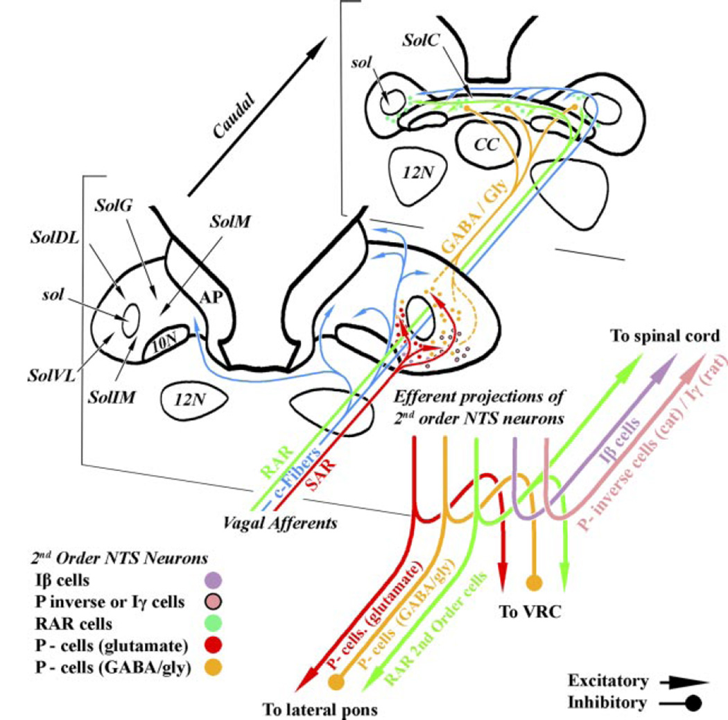
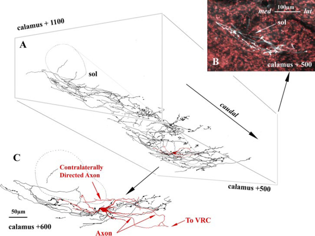
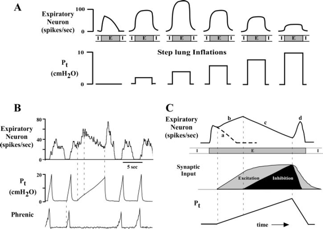
Lung sensory receptors with afferent fibers coursing in the vagus nerves are broadly divided into three groups: slowly (SAR) and rapidly (RAR) adapting stretch receptors and bronchopulmonary C fibers. Central terminations of each group are found in largely nonoverlapping regions of the caudal half of the nucleus of the solitary tract (NTS). Second order neurons in the pathways from these receptors innervate neurons located in respiratory-related regions of the medulla, pons, and spinal cord. The relative ease of selective activation of SARs, and to a lesser extent RARs, has allowed for more complete physiological and morphological characterization of the second and higher order neurons in these pathways than for C fibers. A subset of NTS neurons receiving afferent input from SARs (termed pump or P-cells) mediates the Breuer-Hering reflex and inhibits neurons receiving afferent input from RARs. P-cells and second order neurons in the RAR pathway also provide inputs to regions of the ventrolateral medulla involved in control of respiratory motor pattern, i.e., regions containing a predominance of bulbospinal premotor neurons, as well as regions containing respiratory rhythm-generating neurons. Axon collaterals from both P-cells and RAR interneurons, and likely from NTS interneurons in the C-fiber pathway, project to the parabrachial pontine region where they may contribute to plasticity in respiratory control and integration of respiratory control with other systems, including those that provide for voluntary control of breathing, sleep-wake behavior, and emotions.


Figures

Fig. 1
Fig. 1
Distribution within the nucleus of the solitary tract (NTS) of terminal regions for slowly and rapidly adapting stretch receptors (SARs and RARs, respectively) and bronchopulmonary C fibers, and major projections of their 2nd order neurons. The 3 afferent systems project to largely nonoverlapping areas. The diagram summarizes studies in cats in which individual vagal afferent fibers were recorded and their central projections determined by antidromic mapping (e.g., Refs. 33, 39, 89) and in rats or cats in which 2nd order neurons activated by these primary afferent fibers were recorded within the NTS (34, 45, 52, 53, 90, 109, 110). Of note in the rat, pump cells (P-cells) are not recorded dorsolateral to the solitary tract, but presumably homologous inhibitory P-cells located in SolVL project caudally to second order RAR neurons in the SolC (48). P-inverse cells (deflation-sensitive neurons) are rare in the cat but have frequently been found by Ezure and Tanaka (48) in more caudal portions of SolVL in rats. P-cells and P-inverse cells may be homologous. Abbreviations generally conform to Paxinos and Watson (117): 10N, dorsal motor nucleus of the vagus; 12N, hypoglossal nucleus; AP, area postrema; CC, central canal; sol, solitary tract; SolDL, dorsolateral subnucleus; SolG, gelatinous subnucleus; SolIM, intermediate subnucleus; SolM, medial subnucleus; SolVL, ventrolateral subnucleus; VRC, ventral respiratory column of the ventrolateral medulla.
Fig. 2
Fig. 2
Composite image of an intra-axonally labeled rat SAR axon superimposed on an intracellular labeled P-cell (from a separate experiment). A: oblique perspective of an SAR reconstructed over six 100-µm sections; a reconstructed P-cell (red) is inserted within the terminal field of the SAR axon at the approximate level it was located in vivo. B: pseudocolored image of the SAR axon (white) from a single caudal coronal plane of a Nissl (red) counterstained section. Note the involvement of SolIM and SolVL by the medial (med) to lateral (lat) excursion of the SAR. C: enlarged view of the P-cell and the SAR axonal arborization for a single coronal section at the same rostrocaudal level as in B. Note that the horizontal orientation of the P-cell dendrites matches the distribution of the SAR terminals and that the P-cell dendrites may receive terminations within both the SolIM and SolVL. Both the SAR axon and P-cell were filled with neurobiotin.
Fig. 3
Fig. 3
Biphasic responses of a caudal VRC expiratory bulbospinal neuron to lung inflations. A: diagram illustrating the responses of an expiratory neuron to a series of increasing lung inflations delivered during the expiratory (E) phase. Without inflation, the neuron has a decrementing pattern typical of canine expiratory bulbospinal neuron. Transpulmonary pressures (Pt) below ∼5 cmH2O produce a graded increase in neuronal discharge frequency, whereas Pt above this threshold level produce a graded decrease in activity. The threshold level closely approximates the Pt at functional residual capacity. B: the biphasic response of a canine E bulbospinal neuron is evoked within a single expiratory phase by a slow ramp inflation. In an anesthetized, paralyzed dog the onset of phrenic activity was used to synchronize inflations during control cycles, whereas the offset was used to trigger a slow ramp inflation during the E phase. In B, note the increase in expiratory neuron activity at Pt in the low range and the progressive linear decrease in unit discharge for Pt at higher pressures. When the inflation is abruptly terminated, unit activity increases. C: schematic of the neuronal response shown in B illustrating the response characteristics and the hypothesized mechanisms. The dashed line in part a of the curve indicates the intrinsic decrementing discharge pattern without inflation. In part b the excitatory portion of the inflation response shown by gray shading is hypothesized to be due to excitatory P-cell inputs. In part c of the response, the ongoing excitation appears to be masked by inflation-mediated inhibition. This may result from inhibitory P-cell inputs (shown by black shading). In part d, the more rapid decline in inhibition unmasks the excitation.

References

    1. Averill DB, Cameron WE, Berger AJ. Monosynaptic excitation of dorsal medullary respiratory neurons by slowly adapting pulmonary stretch receptors. J Neurophysiol. 1984;52:771–785. - PubMed
    1. Backman SB, Anders C, Ballantyne D, Röhrig N, Camerer H, Mifflin S, Jordan D, Dickhaus H, Spyer KM, Richter DW. Evidence for a monosynaptic connection between slowly adapting pulmonary stretch receptor afferents and inspiratory beta neurones. Pflügers Arch. 1984;402:129–136. - PubMed
    1. Bajic J, Zuperku EJ, Hopp FA. Processing of pulmonary afferent input patterns by respiratory I-beta neurons. Am J Physiol Regul Integr Comp Physiol. 1989;256:R379–R393. - PubMed
    1. Bajic J, Zuperku EJ, Tonkovic-Capin M, Hopp FA. Expiratory bulbospinal neurons of dogs I. Control of discharge patterns by pulmonary stretch receptors. Am J Physiol Regul Integr Comp Physiol. 1992;262:R1075–R1086. - PubMed
    1. Bajic J, Zuperku EJ, Tonkovic-Capin M, Hopp FA. Interaction between chemoreceptor and stretch receptor inputs at medullary respiratory neurons. Am J Physiol Regul Integr Comp Physiol. 1994;266:R1951–R1961. - PubMed

Publication types

LinkOut - more resources