Review
. 2006 May;8(2):141-51.
doi: 10.2353/jmoldx.2006.050083.

FISH analysis for the detection of lymphoma-associated chromosomal abnormalities in routine paraffin-embedded tissue

Affiliations
Review

FISH analysis for the detection of lymphoma-associated chromosomal abnormalities in routine paraffin-embedded tissue

Roland A Ventura et al. J Mol Diagn. 2006 May.

Abstract

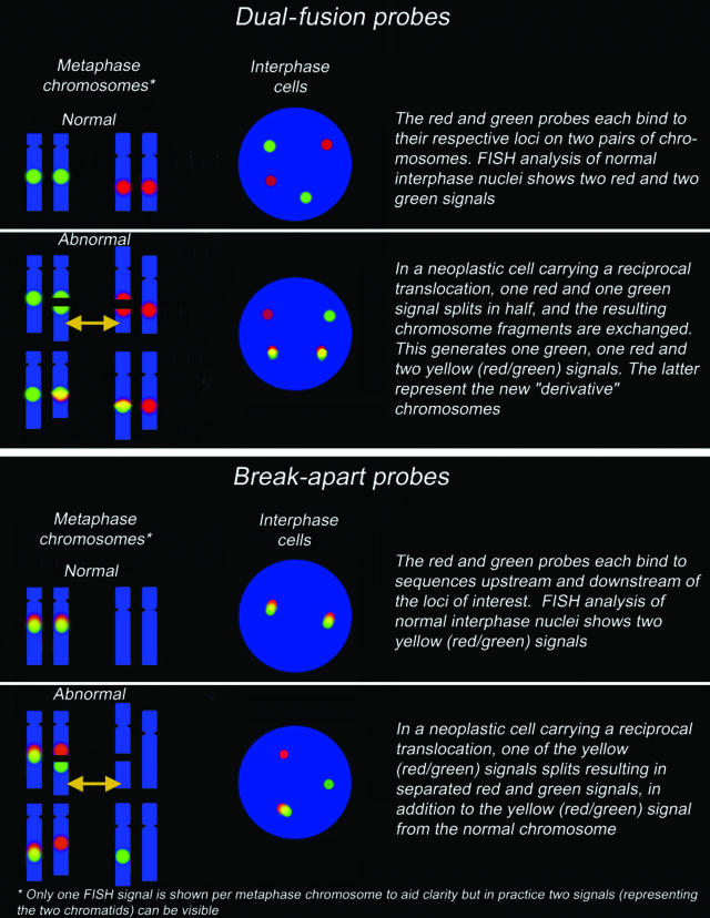
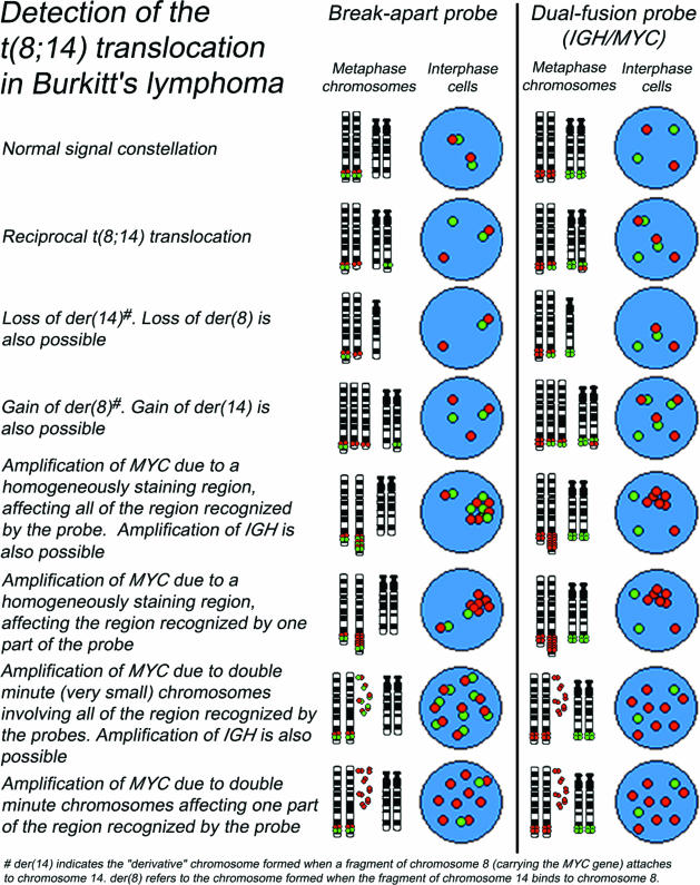
Over the last decade, fluorescence in situ hybridization (FISH) has become a firmly established technique in the diagnosis and assessment of lymphoid malignancies. However, this technique is not wide-ly used in the routine diagnostic evaluation of paraffin-embedded biopsies, most likely because of a perception that it is technically more demanding. There are also uncertainties regarding diagnostic thresholds and the way in which results should be interpreted. In this Review, we describe practical strategies for using FISH analysis to detect lymphoma-associated chromosomal abnormalities in routine paraffin-embedded lymphoma biopsies. Furthermore, we provide proposals on how FISH results should be interpreted (including how to calculate cutoff levels for FISH probes), recorded, and reported. An online appendix (available at http://jmd.amjpathol.org) details various simple, yet robust procedures for paraffin FISH analysis; it also provides additional information on the production of FISH probes, evaluating and reporting FISH results, sources for reagents and equipment, and troubleshooting. We hope that these suggestions will make FISH technology for the study of lymphoma biopsies more accessible to routine diagnostic and research laboratories.


Figures

Figure 1
Figure 1
Schematic representation of the characteristics of dual-fusion and break-apart probes. Depending on probe design (eg, the distance between the regions recognized) and the state of the genomic DNA at the time of fixation, a fused signal may appear either as a colocalized red and green signal or as a single yellow signal. When using break-apart probes, red/green signal pairs will occasionally appear to be slightly separated because of the secondary structure of the target DNA.
Figure 2
Figure 2
Examples of normal and common variant signal constellations for t(8;14)(q24;q32) using break-apart and dual-fusion probes. Similar variant patterns may be seen in other translocations.
Figure 3
Figure 3
A and B: Photomicrographs of FISH patterns obtained in paraffin-embedded tissue sections with IGHbreak-apart and dual-fusion probes for t(11;14) in mantle cell lymphoma. A: A neoplastic cell with a split IGH locus (one fused, single red, and single green signals) is indicated by the red arrow. The green arrow indicates a normal cell (two fused signals). B: The red arrow indicates a neoplastic cell containing single red and green signals (corresponding to the normal CCND1 and IGH loci, respectively) together with two fused signals (corresponding to the genes fused by the translocation). A normal cell, with two red and two green signals, is indicated by the green arrow. C: Single locus probe for the detection of amplifications of the REL gene (2p16) in Hodgkin’s disease. Green arrow, normal diploid cell with two copies of the REL gene. Red arrow, larger cell showing gains (six copies) of the REL gene. D: Loss of nuclear material due to tissue sectioning demonstrated by FISH analysis of normal squamous epithelium using CEPs for chromosomes X and Y (red and green signals, respectively). The red arrows indicate cells containing only a single signal. However, two nuclei retain both signals (green arrows).
Figure 4
Figure 4
Examples of FICTION and a variant FISH signal pattern. A: FICTION analysis of paraffin-embedded lymphoma tissue sections using an antibody against proliferating cells (MIB1, blue immunostaining) (see online appendix for protocol) and t(11;14) dual-fusion FISH probe without DAPI counterstain. Green arrow, MIB1-positive (proliferating) cell (immunostained blue) containing the IGH-CCND1 fusion, indicating the presence of a t(11;14) translocation (two fused, one red, and one green signal). The red arrow indicates a normal, ie, nonproliferating, cell. B: Mantle cell lymphoma showing a variant dual-fusion t(11;14) signal pattern. Red arrow, one red, one green, and three fused signals are present in contrast to the classic two fused, one red, and one green pattern. White arrows, variation of this pattern due to truncation artifacts and/or intraclonal heterogeneity. Green arrow, occasionally, two small probe signals corresponding to a single locus will appear slightly separated (dependent on the probe design, DNA secondary structure, and phase of the cell cycle). Care should be taken not to interpret these as separated signals as being indicative of an abnormality. Blue arrow, when the FISH signals in a neoplastic cell are grouped together, they might be scored as un-interpretable.

References

    1. Kearney L. The impact of the new fish technologies on the cytogenetics of haematological malignancies. Br J Haematol. 1999;104:648–658. - PubMed
    1. Gozzetti A, Le Beau MM. Fluorescence in situ hybridization: uses and limitations. Semin Hematol. 2000;37:320–333. - PubMed
    1. Spagnolo DV, Ellis DW, Juneja S, Leong AS, Miliauskas J, Norris DL, Turner J. The role of molecular studies in lymphoma diagnosis: a review. Pathology. 2004;36:19–44. - PubMed
    1. Belaud-Rotureau MA, Parrens M, Dubus P, Garroste JC, de Mascarel A, Merlio JP. A comparative analysis of FISH, RT-PCR, PCR, and immunohistochemistry for the diagnosis of mantle cell lymphomas. Mod Pathol. 2002;15:517–525. - PubMed
    1. Cataldo KA, Jalal SM, Law ME, Ansell SM, Inwards DJ, Fine M, Arber DA, Pulford KA, Strickler JG. Detection of t(2;5) in anaplastic large cell lymphoma: comparison of immunohistochemical studies, FISH, and RT-PCR in paraffin-embedded tissue. Am J Surg Pathol. 1999;23:1386–1392. - PubMed

Publication types