

Review
doi: 10.1002/art.21813.
Molecular mechanisms of cartilage destruction: mechanics, inflammatory mediators, and aging collide
- PMID: 16645963
- PMCID: PMC1774815
- DOI: 10.1002/art.21813
Item in Clipboard
Review
Molecular mechanisms of cartilage destruction: mechanics, inflammatory mediators, and aging collide
Arthritis Rheum.
2006 May.
No abstract available
Figures
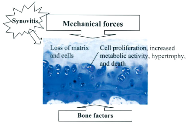
Factors acting on articular cartilage during the development of osteoarthritis (OA). The image of OA tissue is from a toluidine blue–stained section and demonstrates classic OA features including loss of matrix staining and loss of cells in the upper zone of the cartilage with clusters of chondrocytes in the deeper zone. These clusters can contain cells in various stages of cell division, hypertrophic differentiation, and death. Adapted from ref. .
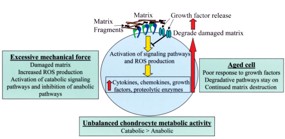
Theoretical model for pathways involved in cartilage destruction during the development of osteoarthritis. Excessive mechanical forces stimulate the chondrocyte directly or indirectly through signals generated by matrix damage, including generation of matrix fragments. The resultant activation of signaling pathways, including reactive oxygen species (ROS) generation, results in increased production of cytokines, chemokines, and proteolytic enzymes. This catabolic response to injury serves to degrade the damaged matrix. Matrix degradation results in release of growth factors stored in the matrix that would normally feed back on the cell and shut down the catabolic pathways. However, aged chondrocytes have an insufficient response to growth factor stimulation, and this results in continued matrix destruction from unbalanced catabolic and anabolic activity.
Comment on
-
Cellular events leading to chondrocyte death after cartilage impact injury.Arthritis Rheum. 2006 May;54(5):1509-17. doi: 10.1002/art.21812. Arthritis Rheum. 2006. PMID: 16649187
References
-
- Felson DT, Neogi T. Osteoarthritis: is it a disease of cartilage or of bone? [editorial] Arthritis Rheum. 2004;50:341–4. - PubMed
-
- Haywood L, McWilliams DF, Pearson CI, Gill SE, Ganesan A, Wilson D, et al. Inflammation and angiogenesis in osteoarthritis. Arthritis Rheum. 2003;48:2173–7. - PubMed
-
- Ayral X, Pickering EH, Woodworth TG, Mackillop N, Dougados M. Synovitis: a potential predictive factor of structural progression of medial tibiofemoral knee osteoarthritis—results of a 1 year longitudinal arthroscopic study in 422 patients. Osteoarthritis Cartilage. 2005;13:361–7. - PubMed
-
- Felson DT, Lawrence RC, Dieppe PA, Hirsch R, Helmick CG, Jordan JM, et al. Osteoarthritis: new insights. Part 1: the disease and its risk factors. Ann Intern Med. 2000;133:635–46. - PubMed
-
- Hunter DJ, Zhang Y, Niu J, Tu X, Amin S, Goggins J, et al. Structural factors associated with malalignment in knee osteoarthritis: the Boston osteoarthritis knee study. J Rheumatol. 2005;32:2192–9. - PubMed
Publication types
MeSH terms
Grants and funding
LinkOut - more resources
Full Text Sources
Other Literature Sources
Medical
