

Ionizing radiation enhances adenoviral vector expressing mda-7/IL-24-mediated apoptosis in human ovarian cancer
- PMID: 16646087
- PMCID: PMC2203216
- DOI: 10.1002/jcp.20663
Ionizing radiation enhances adenoviral vector expressing mda-7/IL-24-mediated apoptosis in human ovarian cancer
Abstract
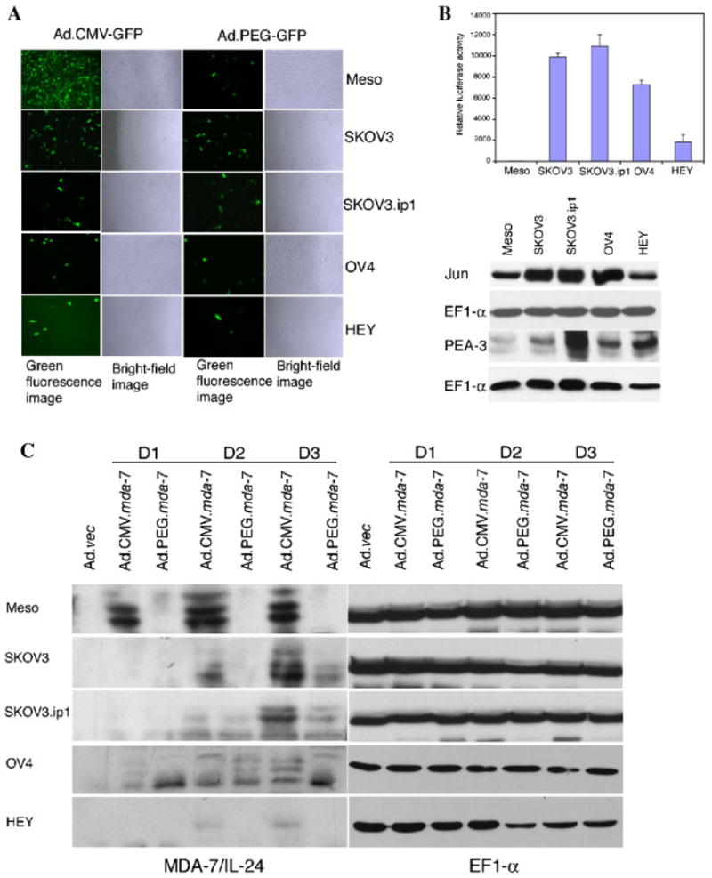
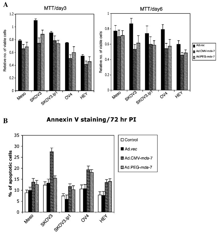
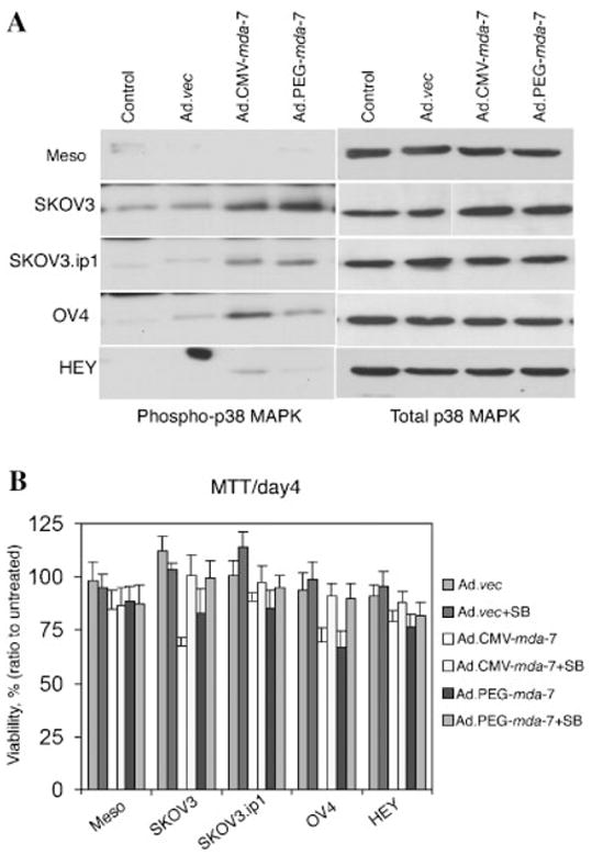
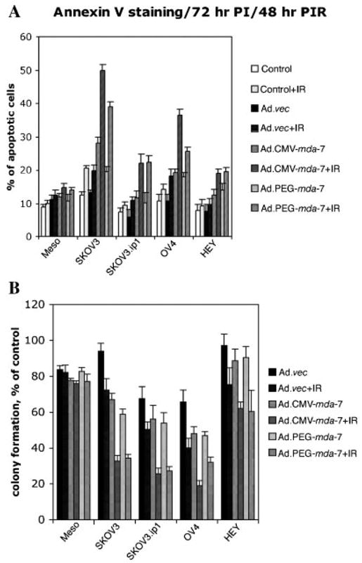
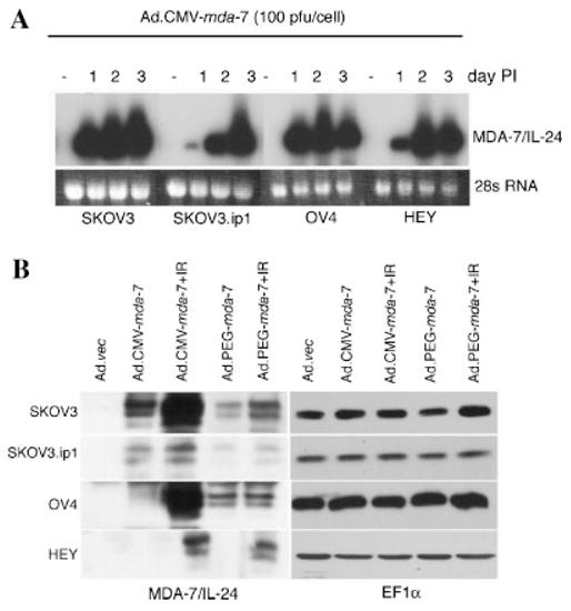
Ovarian cancer is the fifth most common cause of cancer-related death in women. Current interventional approaches, including debulking surgery, chemotherapy, and/or radiation have proven minimally effective in preventing the recurrence and/or mortality associated with this malignancy. Subtraction hybridization applied to terminally differentiating human melanoma cells identified melanoma differentiation associated gene-7/interleukin-24 (mda-7/IL-24), whose unique properties include the ability to selectively induce growth suppression, apoptosis, and radiosensitization in diverse cancer cells, without causing any harmful effects in normal cells. Previously, it has been shown that adenovirus-mediated mda-7/IL-24 therapy (Ad.mda-7) induces apoptosis in ovarian cancer cells, however, the apoptosis induction was relatively low. We now document that apoptosis can be enhanced by treating ovarian cancer cells with ionizing radiation (IR) in combination with Ad.mda-7. Additionally, we demonstrate that mda-7/IL-24 gene delivery, under the control of a minimal promoter region of progression elevated gene-3 (PEG-3), which functions selectively in diverse cancer cells with minimal activity in normal cells, displays a selective radiosensitization effect in ovarian cancer cells. The present studies support the use of IR in combination with mda-7/IL-24 as a means of augmenting the therapeutic benefit of this gene in ovarian cancer, particularly in the context of tumors displaying resistance to radiation therapy.
Figures
Similar articles
-
Infectivity enhanced adenoviral-mediated mda-7/IL-24 gene therapy for ovarian carcinoma.Gynecol Oncol. 2004 Aug;94(2):352-62. doi: 10.1016/j.ygyno.2004.04.028. Gynecol Oncol. 2004. PMID: 15297172
-
Ionizing radiation enhances therapeutic activity of mda-7/IL-24: overcoming radiation- and mda-7/IL-24-resistance in prostate cancer cells overexpressing the antiapoptotic proteins bcl-xL or bcl-2.Oncogene. 2006 Apr 13;25(16):2339-48. doi: 10.1038/sj.onc.1209271. Oncogene. 2006. PMID: 16331261
-
Unique aspects of mda-7/IL-24 antitumor bystander activity: establishing a role for secretion of MDA-7/IL-24 protein by normal cells.Oncogene. 2005 Nov 17;24(51):7552-66. doi: 10.1038/sj.onc.1208911. Oncogene. 2005. PMID: 16044151
-
mda-7/IL-24, novel anticancer cytokine: focus on bystander antitumor, radiosensitization and antiangiogenic properties and overview of the phase I clinical experience (Review).Int J Oncol. 2007 Nov;31(5):985-1007. Int J Oncol. 2007. PMID: 17912425 Review.
-
Melanoma differentiation associated gene-7/interleukin-24 (mda-7/IL-24): novel gene therapeutic for metastatic melanoma.Toxicol Appl Pharmacol. 2007 Nov 1;224(3):300-7. doi: 10.1016/j.taap.2006.11.021. Epub 2006 Nov 23. Toxicol Appl Pharmacol. 2007. PMID: 17208263 Free PMC article. Review.
Cited by
-
En Guard! The Interactions between Adenoviruses and the DNA Damage Response.Viruses. 2020 Sep 7;12(9):996. doi: 10.3390/v12090996. Viruses. 2020. PMID: 32906746 Free PMC article. Review.
-
Investigation of therapeutic potential of the Il24-p20 fusion protein against breast cancer: an in-silico approach.In Silico Pharmacol. 2024 Sep 18;12(2):84. doi: 10.1007/s40203-024-00252-x. eCollection 2024. In Silico Pharmacol. 2024. PMID: 39301086
-
Chapter seven--Cancer treatment with gene therapy and radiation therapy.Adv Cancer Res. 2012;115:221-63. doi: 10.1016/B978-0-12-398342-8.00007-0. Adv Cancer Res. 2012. PMID: 23021246 Free PMC article. Review.
-
MDA-7/IL-24 suppresses human ovarian carcinoma growth in vitro and in vivo.Mol Cancer. 2007 Feb 2;6:11. doi: 10.1186/1476-4598-6-11. Mol Cancer. 2007. PMID: 17274815 Free PMC article.
-
Combinatorial strategies based on CRAd-IL24 and CRAd-ING4 virotherapy with anti-angiogenesis treatment for ovarian cancer.J Ovarian Res. 2016 Jun 27;9(1):38. doi: 10.1186/s13048-016-0248-5. J Ovarian Res. 2016. PMID: 27349517 Free PMC article.
References
-
- Alvarez RD, Curiel DT. A phase I study of recombinant adenovirus vector-mediated delivery of an anti-erbB-2 single-chain (sFv) antibody gene for previously treated ovarian and extraovarian cancer patients. Hum Gene Ther. 1997;8:229–242. - PubMed
-
- Alvarez RD, Gomez-Navarro J, Wang M, Barnes MN, Strong TV, Arani RB, Arafat W, Hughes JV, Siegal GP, Curiel DT. Adenoviral-mediated suicide gene therapy for ovarian cancer. Mol Ther. 2000;2:524–530. - PubMed
-
- Barnes MN, Deshane JS, Rosenfeld M, Siegal GP, Curiel DT, Alvarez RD. Gene therapy and ovarian cancer: A review. Obstet Gynecol. 1997;89:145–155. - PubMed
-
- Casado E, Gomez-Navarro J, Yamamoto M, Adachi Y, Coolidge CJ, Arafat WO, Barker SD, Wang MH, Mahasreshti PJ, Hemminki A, Gonzalez-Baron M, Barnes MN, Pustilnik TB, Siegal GP, Alvarez RD, Curiel DT. Strategies to accomplish targeted expression of transgenes in ovarian cancer for molecular therapeutic applications. Clin Cancer Res. 2001;7:2496–2504. - PubMed
-
- Christian J, Thomas H. Ovarian cancer chemotherapy. Cancer Treat Rev. 2001;27:99–109. - PubMed
Publication types
MeSH terms
Substances
Grants and funding
LinkOut - more resources
Full Text Sources
Other Literature Sources
Medical
