. 2006;8(3):R77.
doi: 10.1186/ar1944. Epub 2006 Apr 28.

Folate-targeted immunotherapy effectively treats established adjuvant and collagen-induced arthritis

Affiliations

Folate-targeted immunotherapy effectively treats established adjuvant and collagen-induced arthritis

Chrystal M Paulos et al. Arthritis Res Ther. 2006.

Abstract

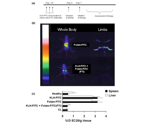
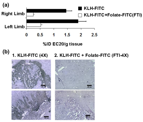
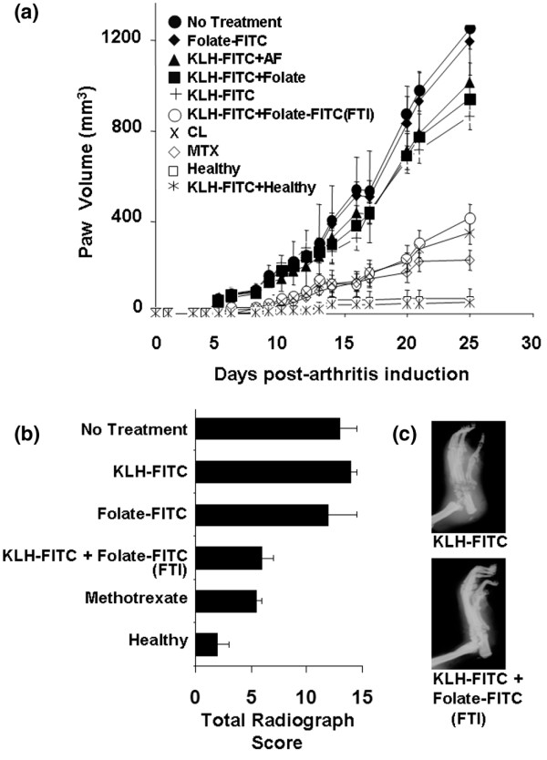
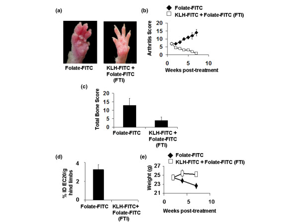
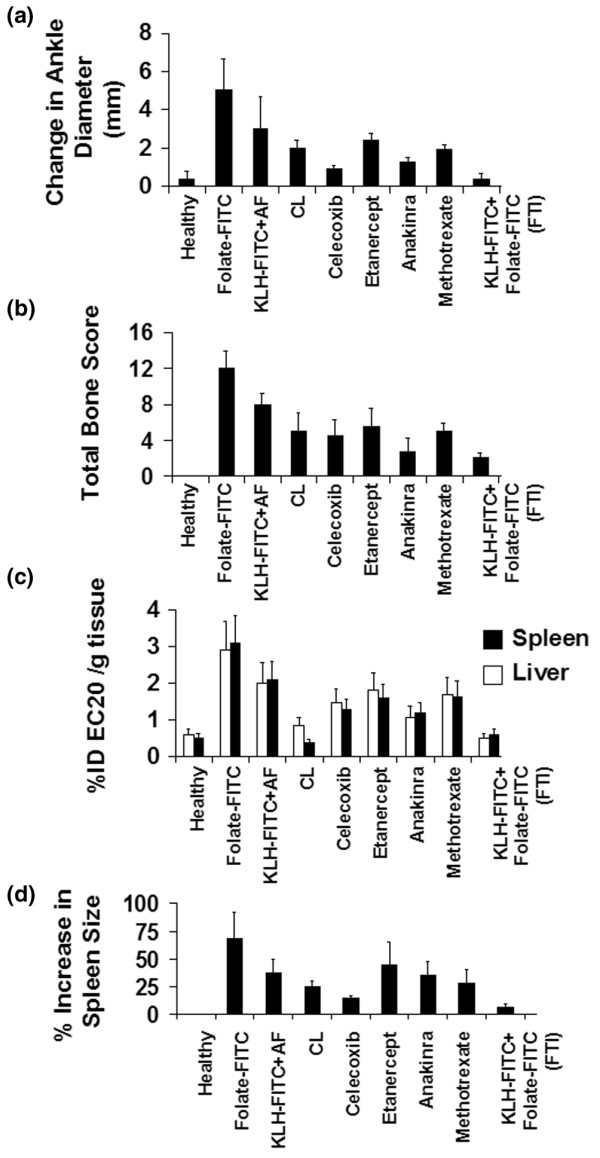
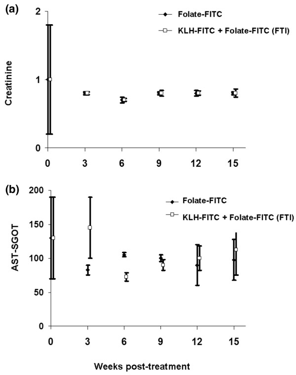
Activated macrophages express a cell surface receptor for the vitamin folic acid. Because this receptor is inaccessible or not measurably expressed on other normal cells, folic acid has been recently exploited to selectively deliver attached radio-emitters to sites of activated macrophage accumulation, allowing scintigraphic imaging of inflamed joints and organs of arthritic rats. We demonstrate here that folate-linked haptens can also be targeted to activated macrophages, decorating their cell surfaces with highly immunogenic molecules. Under conditions in which the rodent has already been immunized against keyhole limpet hemocyanine-(fluorescein isothiocyanate) FITC, activated macrophages are eliminated. Administration of folate-FITC conjugates to rodents with experimental arthritis attenuates (a) systemic and peri-articular inflammation, (b) bone and cartilage degradation, and (c) arthritis-related body weight loss. Treatment with folate-hapten conjugates is comparable to methotrexate, etanercept, anakinra, and celecoxib at alleviating the symptoms of arthritis. We conclude that reduction of activated macrophages by folate-targeted immunotherapy can ameliorate the symptoms of arthritis in two rodent models of the disease.


Figures

Figure 1
Figure 1
Folate-targeted immunotherapy reduces activated macrophages systemically in adjuvant-induced arthritic rats. (a) Treatment schedule for FTI. (b) The level of activated macrophages in internal organs (left) and limbs (right) of non-immunized arthritic rats (upper panel) and keyhole limpet hemocyanine-fluorescein isothiocyanate (KLH-FITC)-immunized arthritic rats (lower panel) 25 days after initiation of treatment with folate-FITC (375 nmole/kg twice a week until day 21) was imaged 4 hours after i.p. injection of 0.5 mg EC20. (c) Activated macrophage accumulation in the liver and spleen was evaluated by measuring EC20 uptake in healthy non-arthritic rats (Healthy), AIA rats immunized with KLH-FITC and left untreated (KLH-FITC), non-immunized rats injected i.p. with folate-FITC (375 nmole/kg twice a week), KLH-FITC-immunized AIA rats treated with folate-FITC (KLH-FITC + Folate-FITC [FTI], 375 nmole/kg twice a week), and AIA rats injected with clodronate liposomes ([CL]; 3.6 mg/kg on days 8, 16, and 21, i.p.). EC20 biodistribution was measured by removal of the indicated tissues, weighing and counting them for radioactivity; represented as percent injected dose of EC20 per gram tissue, for instance, %ID EC20/g tissue (mean ± standard deviation, n = 5 rats/group). Data are representative of three independent experiments. (c) folate-FITC versus KLH-FITC + folate-FITC (FTI), p < 0.001.
Figure 2
Figure 2
Folate-targeted immunotherapy reduces folate receptor expressing macrophages in inflamed tissue of AIA rats. (a) The level of folate receptor (FR)+ activated macrophages in the limbs of non-immunized (Folate-fluorescein isothiocyanate [FITC]) and keyhole limpet hemocyanine (KLH)-FITC-immunized AIA rats (KLH-FITC + Folate-FITC [FTI]) 25 days after initiation of treatment with folate-FITC was determined 4 hours after i.p. injection of 0.5 mg EC20. Folate-FITC was administered at 375 nmole/kg twice a week until day 23, and estimates of FR+ macrophage numbers were made by weighing and counting the limbs for EC20 radioactivity (± standard deviation for n = 5 rats/group; [folate-FITC] vs. [KLH-FITC + folate-FITC (FTI)], p < 0.001). (b) Histological analyses were conducted to obtain a visual assessment of the abundance of ED1+ macrophages remaining in the inflamed soft joint tissue of the ankle joints (inflamed soft tissue adjacent to bone) of AIA rats immunized with KLH-FITC and then treated with either phosphate-buffered saline (PBS) alone (KLH-FITC) or folate-FITC (KLH-FITC + Folate-FITC [FTI]). Representative images from the joint tissue of two PBS-treated and two folate-FITC (FTI)-treated rats are shown (n = 5 rats/group).
Figure 3
Figure 3
Effect of folate-targeted immunotherapy on joint inflammation and bone/cartilage destruction in AIA rats. Where indicated, AIA rats were immunized with a 1:1 suspension of keyhole limpet hemocyanine-fluorescein isothiocyanate (KLH-FITC) and TiterMax Gold adjuvant to generate a high anti-FITC antibody titer. On day 0, rats were induced to develop AIA via the footpad method. (a) Treatments began on the day of arthritis induction, and rats were given i.p. doses twice a week at 375 nmole folate-FITC/kg. Treatment groups included: AIA rats not immunized and not treated (No Treatment), non-immunized rats treated with folate-FITC (Folate-FITC), AIA rats immunized against fluorescein and treated with non-targeted aminofluorescein (KLH-FITC + AF), AIA rats immunized against fluorescein and treated with folic acid alone (KLH-FITC + Folate), AIA rats immunized against fluorescein but not further treated (KLH-FITC), AIA rats immunized against fluorescein and treated with folate-FITC (KLH-FITC + folate-FITC [FTI]), AIA rats treated on days 8, 16, and 23 with 3.6 mg/kg clodronate liposomes (CL), AIA rats treated with methotrexate (MTX; 0.75 mg/kg per week, i.p.), immunized healthy rats (KLH-FITC + Healthy), and untreated healthy rats (Healthy), respectively. Data are representative of two independent experiments (n = 5 rats/group; [folate-FITC] vs. [KLH-FITC + folate-FITC (FTI)], p < 0.01). The impact on bone and cartilage degradation was assessed both by analysis of radiological (RAD) scores (b) and visual inspection (c) on day 25 after arthritis induction (mean ± standard deviation, n = 4 or 5 rats/group). Data are representative of three independent experiments. (RAD scores: folate-FITC vs. KLH-FITC + folate-FITC (FTI), p < 0.05).
Figure 4
Figure 4
Effect of folate-targeted immunotherapy (FTI) on mice with established collagen-induced arthritis. Mice were immunized with keyhole limpet hemocyanine-fluorescein isothiocyanate (KLH-FITC) in an equal volume of TiterMax Gold adjuvant to generate a high anti-FITC antibody titer. Seven days after arthritis induction, when the average arthritis score had reached 7, mice were treated daily with 600 nmole/kg folate-FITC until day 38. (a) Differences between folate-FITC-treated and untreated mice were easily visualized. (b) Each limb was evaluated weekly by an individual blinded to the treatment groups and was assigned an arthritis score (see Methods and methods). (c) Bone erosion was analyzed on day 49 from radiographs by a radiologist blinded to the treatment groups. (d) Uptake of EC20 in both hind limbs was determined as a measure of activated macrophage accumulation on day 49, represented as percent injected dose of EC20 per gram hind limbs, for instance, %ID EC20/g hind limbs. (e) The change in weight of mice after arthritis induction was measured regularly.
Figure 5
Figure 5
Comparison of the effects of various arthritis therapies on disease severity in rats with established adjuvant-induced arthritic (AIA). Rats were left either unimmunized or immunized with keyhole limpet hemocyanine-fluorescein isothiocyanate (KLH-FITC) in an equal volume of alum adjuvant to generate a high anti-FITC antibody titer. AIA was induced by the base-of-tail method, and 7 days after induction each therapy was initiated. The therapies were folate-FITC in non-immunized rats (folate-FITC; 30 nmole/kg per day, i.p.), non-targeted aminofluorescein (AF) in immunized rats (KLH-FITC + AF; 30 nmole/kg per day, i.p.), clodronate liposomes (CL; 3.6 mg/kg on days 8, 16, and 23, i.p.), celecoxib (20 mg/kg every other day, oral gavage), etanercept (4 mg/kg per day, i.p), anakinra (120 mg/kg per day, continuous infusion, Alzet osmotic pump), methotrexate (MTX; 0.75 mg/kg per week, i.p.), or folate-targeted immunotherapy (FTI) in immunized rats (KLH-FITC + folate-FITC [FTI]; 30 nmole/kg per day, i.p.). All groups were measured for arthritis symptoms by monitoring changes in ankle diameter (a), bone and cartilage degradation (b), uptake of EC20 in the spleen and liver (c), and splenomegaly (d). Data are representative of two independent experiments (mean ± standard deviation, n = 5 rats/group).
Figure 6
Figure 6
No indication of toxicity was detected in rats treated with folate-targeted immunotherapy for 15 weeks. Toxicity analysis associated with continuous treatment of healthy keyhole limpet hemocyanine-fluorescein isothiocyanate (KLH-FITC)-immunized rats with folate-targeted immunotherapy (FTI) for 15 weeks. Non-immunized or KLH-FITC-immunized rats were treated with folate-FITC (300 nmole/kg, three times per week) and sacrificed at the indicated times. Serum was analyzed for indicators of kidney (creatinine) (a) and liver (aspartate aminotransferase [AST-SGOT])(b) function. Tissue samples, including liver, kidney, brain, lungs, and bone marrow, were simultaneously submitted for histopathological analyses. No indication of toxicity was detected by either type of analysis (tissue section data not shown) (mean ± standard deviation, n = 8 rats/group).

Similar articles

Cited by

References

    1. Kinne RW, Brauer R, Stuhlmuller B, Palombo-Kinne E, Burmester GR. Macrophages in rheumatoid arthritis. Arthritis Res. 2000;2:189–202. doi: 10.1186/ar86. - DOI - PMC - PubMed
    1. Tak PP, Smeets TJ, Daha MR, Kluin PM, Meijers KA, Brand R, Meinders AE, Breedveld FC. Analysis of the synovial cell infiltrate in early rheumatoid synovial tissue in relation to local disease activity. Arthritis Rheum. 1997;40:217–225. - PubMed
    1. Yanni G, Whelan A, Feighery C, Bresnihan B. Synovial tissue macrophages and joint erosion in rheumatoid arthritis. Ann Rheum Dis. 1994;53:39–44. - PMC - PubMed
    1. Mulherin D, Fitzgerald O, Bresnihan B. Synovial tissue macrophage populations and articular damage in rheumatoid arthritis. Arthritis Rheum. 1996;39:115–124. - PubMed
    1. Liu H, Pope RM. Phagocytes: mechanisms of inflammation and tissue destruction. Rheum Dis Clin North Am. 2004;30:19–39. doi: 10.1016/S0889-857X(03)00107-8. - DOI - PubMed

Publication types