

The net orientation of nicotinic receptor transmembrane alpha-helices in the resting and desensitized states
- PMID: 16648164
- PMCID: PMC1483077
- DOI: 10.1529/biophysj.106.082693
The net orientation of nicotinic receptor transmembrane alpha-helices in the resting and desensitized states
Abstract
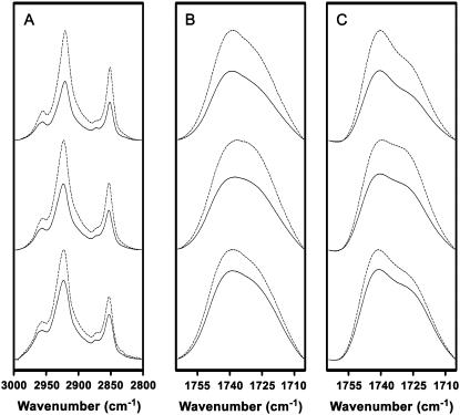
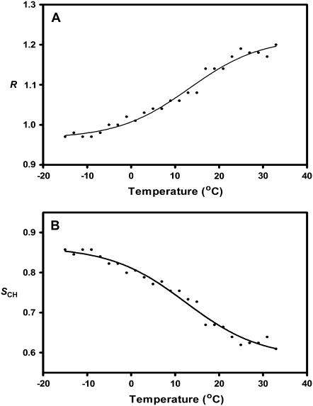
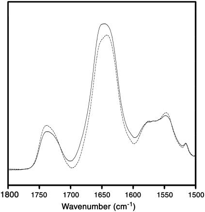
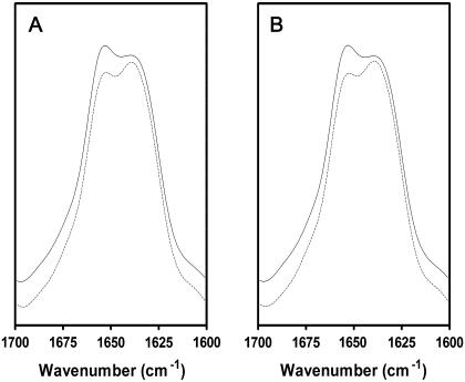
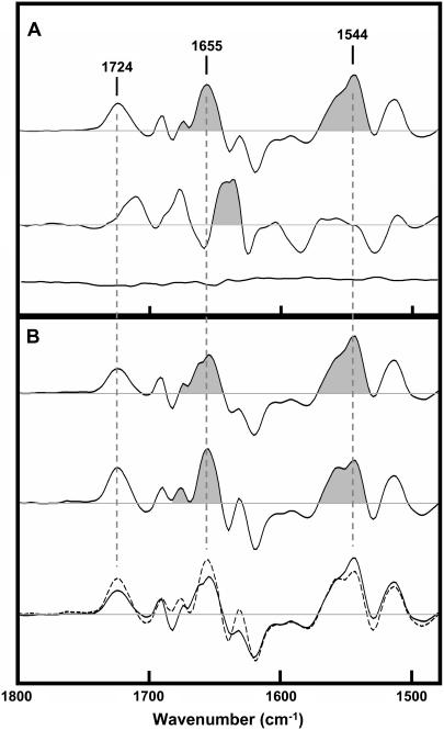
The net orientation of nicotinic acetylcholine receptor transmembrane alpha-helices has been probed in both the activatable resting and nonactivatable desensitized states using linear dichroism Fourier-transform infrared spectroscopy. Infrared spectra recorded from reconstituted nicotinic acetylcholine receptor membranes after 72 h exposure to (2)H2O exhibit an intense amide I component band near 1655 cm(-1) that is due predominantly to hydrogen-exchange-resistant transmembrane peptides in an alpha-helical conformation. The measured dichroism of this band is 2.37, suggesting a net tilt of the transmembrane alpha-helices of roughly 40 degrees from the bilayer normal, although this value overestimates the tilt angle because the measured dichroism at 1655 cm(-1) also reflects the dichroism of overlapping amide I component bands. Significantly, no change in the net orientation of the transmembrane alpha-helices is observed upon agonist binding. In fact, the main changes in structure and orientation detected upon desensitization involve highly solvent accessible regions of the polypeptide backbone. Our data are consistent with a capping of the ligand binding site by the solvent accessible C-loop with little change in the structure of the transmembrane domain in the desensitized state. Changes in structure at the interface between the ligand-binding and transmembrane domains may uncouple binding from gating.
Figures
Similar articles
-
Desensitization of the nicotinic acetylcholine receptor mainly involves a structural change in solvent-accessible regions of the polypeptide backbone.Biochemistry. 1997 Mar 25;36(12):3617-24. doi: 10.1021/bi962845m. Biochemistry. 1997. PMID: 9132013
-
Structure of the pore-forming transmembrane domain of a ligand-gated ion channel.J Biol Chem. 2001 Jun 29;276(26):23726-32. doi: 10.1074/jbc.M102101200. Epub 2001 Apr 27. J Biol Chem. 2001. PMID: 11328815
-
Structure of both the ligand- and lipid-dependent channel-inactive states of the nicotinic acetylcholine receptor probed by FTIR spectroscopy and hydrogen exchange.Biochemistry. 1995 Nov 21;34(46):15142-9. doi: 10.1021/bi00046a021. Biochemistry. 1995. PMID: 7578128
-
Nicotinic acetylcholine receptor-lipid interactions: Mechanistic insight and biological function.Biochim Biophys Acta. 2015 Sep;1848(9):1806-17. doi: 10.1016/j.bbamem.2015.03.010. Epub 2015 Mar 16. Biochim Biophys Acta. 2015. PMID: 25791350 Review.
-
Molecular sites of general anaesthetic action on acetylcholine receptors.Eur J Anaesthesiol. 1995 Jan;12(1):21-30. Eur J Anaesthesiol. 1995. PMID: 7535690 Review.
Cited by
-
Mammalian nicotinic acetylcholine receptors: from structure to function.Physiol Rev. 2009 Jan;89(1):73-120. doi: 10.1152/physrev.00015.2008. Physiol Rev. 2009. PMID: 19126755 Free PMC article. Review.
-
Desensitization of alpha7 nicotinic receptor is governed by coupling strength relative to gate tightness.J Biol Chem. 2011 Jul 15;286(28):25331-40. doi: 10.1074/jbc.M111.221754. Epub 2011 May 24. J Biol Chem. 2011. PMID: 21610071 Free PMC article.
-
Structural answers and persistent questions about how nicotinic receptors work.Front Biosci. 2008 May 1;13:5479-510. doi: 10.2741/3094. Front Biosci. 2008. PMID: 18508600 Free PMC article. Review.
References
-
- Karlin, A. 2002. Emerging structure of the nicotinic acetylcholine receptors. Nat. Rev. Neurosci. 3:102–114. - PubMed
-
- Unwin, N. 2003. Structure and action of the nicotinic acetylcholine receptor explored by electron microscopy. FEBS Lett. 555:91–95. - PubMed
-
- Lester, H. A., M. I. Dibas, D. S. Dahan, J. F. Leite, and D. A. Dougherty. 2004. Cys-loop receptors: new twists and turns. Trends Neurosci. 27:329–336. - PubMed
-
- Brejc, K., W. J. van Dijk, R. V. Klaassen, M. Schuurmans, J. van Der Oost, A. B. Smit, and T. K. Sixma. 2001. Crystal structure of an ACh-binding protein reveals the ligand-binding domain of nicotinic receptors. Nature. 411:269–276. - PubMed
-
- Celie, P. H., S. E. van Rossum-Fikkert, W. J. van Dijk, K. Brejc, A. B. Smit, and T. K. Sixma. 2004. Nicotine and carbamylcholine binding to nicotinic acetylcholine receptors as studied in AChBP crystal structures. Neuron. 41:907–914. - PubMed
Publication types
MeSH terms
Substances
LinkOut - more resources
Full Text Sources
