. 2006 May 9;103(19):7292-7.
doi: 10.1073/pnas.0602350103. Epub 2006 Apr 28.

Voltage sensor conformations in the open and closed states in ROSETTA structural models of K(+) channels

Affiliations

Voltage sensor conformations in the open and closed states in ROSETTA structural models of K(+) channels

Vladimir Yarov-Yarovoy et al. Proc Natl Acad Sci U S A. .

Abstract

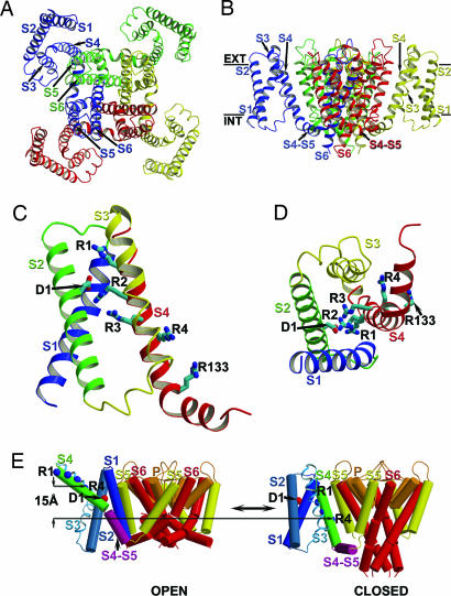
Voltage-gated ion channels control generation and propagation of action potentials in excitable cells. Significant progress has been made in understanding structure and function of the voltage-gated ion channels, highlighted by the high-resolution open-state structure of the voltage-gated potassium channel, K(v)1.2. However, because the structure of the closed state is unknown, the gating mechanism remains controversial. We adapted the rosetta membrane method to model the structures of the K(v)1.2 and KvAP channels using homology, de novo, and domain assembly methods and selected the most plausible models using a limited number of experimental constraints. Our model of K(v)1.2 in the open state is very similar in overall topology to the x-ray structure of this channel. Modeling of KvAP in the open state suggests that orientation of the voltage-sensing domain relative to the pore-forming domain is considerably different from the orientation in the K(v)1.2 open state and that the magnitude of the vertical movement of S4 is significantly greater. Structural modeling of closed state of K(v)1.2 suggests gating movement that can be viewed as a sum of two previously suggested mechanisms: translation (2-4 A) plus rotation ( approximately 180 degrees ) of the S4 segment as proposed in the original "sliding helix" or "helical screw" models coupled with a rolling motion of the S1-S3 segments around S4, similar to recent "transporter" models of gating. We propose a unified mechanism of voltage-dependent gating for K(v)1.2 and KvAP in which this major conformational change moves the gating charge across the electric field in an analogous way for both channels.


Conflict of interest statement

Conflict of interest statement: No conflicts declared.

Figures

Fig. 1.
Fig. 1.
Homology model of the transmembrane region of Kv1.2 in the open state. (A) Side view of the ribbon representation of the rosetta membrane model of a single subunit of Kv1.2. Regions of the Kv1.2 structure modeled using the backbone coordinates of the unidentified residues in the x-ray structure of Kv1.2 as a template by the rosetta membrane homology method are shown in red (see Methods). Regions of the Kv1.2 structure modeled using the identified residues in the x-ray structure of Kv1.2 (9) as a template by the rosetta membrane homology method are shown in blue. Regions of the Kv1.2 structure modeled using the rosetta membrane de novo method are shown in green. Transmembrane segments are labeled from S1 to S6, the selectivity filter helix is labeled P, and the position of the S4–S5 linker is indicated by an arrow. N and C termini residues of the transmembrane region are labeled N and C, respectively. (B) Side view of the VSD segments S1–S4 only (colored individually) of the model shown in A. Side chains of gating-charge-carrying arginines in S4 (labeled R1–R4) and E226 in S2 (labeled E1) are shown in stick representation. Blue, red, and cyan atoms in the side chains shown represent nitrogen, oxygen, and carbon atoms, respectively. (C) View of the model shown in B from the extracellular side of the membrane. All structural figures presented in this work were generated using molscript (61) and raster3d (62).
Fig. 2.
Fig. 2.
The rosetta membrane domain assembly model of the open state of Kv1.2. (A) View of the Kv1.2 model from the extracellular side of the membrane. All four subunits are colored individually. Segments S1–S6 for the blue-colored subunit and S5–S6 for the green-colored subunit are labeled accordingly. (B) View of the model in A from the side of the membrane. Segments S1–S4, S6, and S4–S5 linker for blue- and yellow-colored subunits are labeled accordingly. Approximate position of the first residue in the S4–S5 linker is indicated by an arrow for blue- and yellow-colored subunits. Extracellular and intracellular edges of the hydrocarbon core of the membrane are marked by solid bars and labeled “EXT” and “INT,” respectively. (C) View from the extracellular side of the membrane of the Kv1.2 structure (shown in blue) and the best rosetta membrane model (shown in orange) of the open state of Kv1.2 superimposed over the PD residues. Only a single VSD is shown attached to the tetramer of the PD for clarity. (D) View of the models shown in C from the side of the membrane.
Fig. 3.
Fig. 3.
The rosetta membrane domain assembly model of the closed state of Kv1.2. (A) View of the Kv1.2 model from the extracellular side of the membrane. The model is colored and labeled as in Fig. 2A. (B) View of the model in A from the side of the membrane. The model is colored and labeled as in Fig. 2B. The S4–S5 linker for blue and red subunits is labeled accordingly. (C) Side view of the VSD only of the model in A. The model is colored and labeled as in Fig. 1B. (D) View of the model in C from the intracellular side of the membrane. (E) Kv1.2 models of the closed and open states shown in cylinder representation. Only a single VSD is shown attached to the tetramer of the PD for clarity. Transmembrane segments S1–S6 and P-loop are colored by a rainbow scheme from blue to red. The S4–S5 linker is in purple. Approximate positions of the Cα atoms of the first and fourth gating charge-carrying arginines in S4 (labeled as R1 and R4 and colored in blue) and E226 in S2 (labeled E1 and colored in red) are shown in sphere representation. All intracellular and extracellular loops, except for the S4–S5 linker, are represented by curved lines for simplicity. Vertical translation of R4 between the closed and open states along the membrane normal vector and relative to the plane of the membrane is indicated by arrows. S3 is represented by ribbon to show clearly the positions of the gating-charge-carrying arginines in S4.
Fig. 4.
Fig. 4.
The rosetta membrane domain assembly model of an open/inactivated state of KvAP. (A) View of the KvAP model from the extracellular side of the membrane. The model is colored and labeled as in Fig. 2A. (B) View of the model in A from the side of the membrane. The model is colored and labeled as in Fig. 2B. Approximate position of the first residue in the S4–S5 linker is indicated by arrow for blue and yellow subunits. (C) Side view of the VSD only (colored individually) of the model in A. Side chains of gating-charge-carrying arginines in S4 (labeled R1–R4), R133 in S4, and D62 in S2 (labeled D1) are shown in stick representation. Atoms in the side chains shown are colored as in Fig. 1B. (D) View of the model in C from the extracellular side of the membrane.
Fig. 5.
Fig. 5.
The rosetta membrane homology model of KvAP in closed state. (A) View of the KvAP model from the extracellular side of the membrane. The model is colored and labeled as in Fig. 2A. (B) View of the model in A from the side of the membrane. The model is colored and labeled as in Fig. 2B. The S4–S5 linker for blue and red subunits is labeled accordingly. (C) Side view of the VSD only of the model in A. The model is colored and labeled as in Fig. 4C. (D) View of the model in C from the intracellular side of the membrane. (E) KvAP models of the closed and open/inactivated (labeled as “open”) states shown in cylinder representation and as described in Fig. 3E. Approximate positions of the Cα atoms of the first and fourth gating-charge-carrying arginines in S4 (labeled R1 and R4), R133 in S4, and D62 in S2 (labeled D1) are shown in sphere representation.

References

    1. Hille B. Ion Channels of Excitable Membranes. Sunderland, MA: Sinauer Associates; 2001.
    1. Yu F. H., Catterall W. A. Sci. STKE. 2004;2004:re15. - PubMed
    1. Armstrong C. M., Bezanilla F. J. Gen. Physiol. 1974;63:533–552. - PMC - PubMed
    1. Bezanilla F. Physiol. Rev. 2000;80:555–592. - PubMed
    1. Sigworth F. J. Q. Rev. Biophys. 1994;27:1–40. - PubMed

Publication types

MeSH terms

Substances