. 2006 May 9;103(19):7500-5.
doi: 10.1073/pnas.0503465103. Epub 2006 Apr 28.

Localization of cardiac L-type Ca(2+) channels to a caveolar macromolecular signaling complex is required for beta(2)-adrenergic regulation

Affiliations

Localization of cardiac L-type Ca(2+) channels to a caveolar macromolecular signaling complex is required for beta(2)-adrenergic regulation

Ravi C Balijepalli et al. Proc Natl Acad Sci U S A. .

Abstract

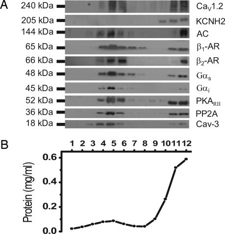
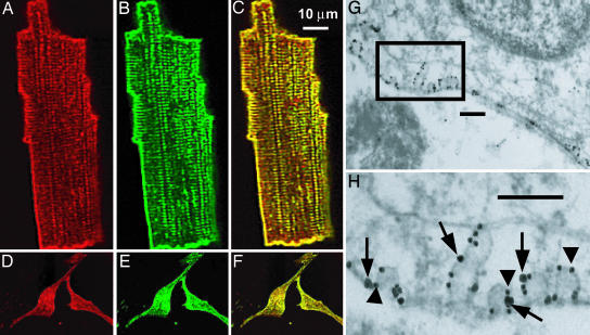
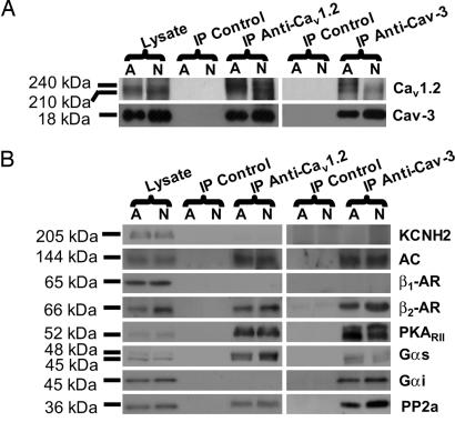
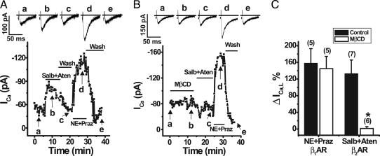
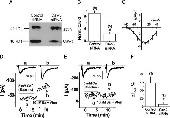
L-type Ca(2+) channels play a critical role in regulating Ca(2+)-dependent signaling in cardiac myocytes, including excitation-contraction coupling; however, the subcellular localization of cardiac L-type Ca(2+) channels and their regulation are incompletely understood. Caveolae are specialized microdomains of the plasmalemma rich in signaling molecules and supported by the structural protein caveolin-3 in muscle. Here we demonstrate that a subpopulation of L-type Ca(2+) channels is localized to caveolae in ventricular myocytes as part of a macromolecular signaling complex necessary for beta(2)-adrenergic receptor (AR) regulation of I(Ca,L). Immunofluorescence studies of isolated ventricular myocytes using confocal microscopy detected extensive colocalization of caveolin-3 and the major pore-forming subunit of the L-type Ca channel (Ca(v)1.2). Immunogold electron microscopy revealed that these proteins colocalize in caveolae. Immunoprecipitation from ventricular myocytes using anti-Ca(v)1.2 or anti-caveolin-3 followed by Western blot analysis showed that caveolin-3, Ca(v)1.2, beta(2)-AR (not beta(1)-AR), G protein alpha(s), adenylyl cyclase, protein kinase A, and protein phosphatase 2a are closely associated. To determine the functional impact of the caveolar-localized beta(2)-AR/Ca(v)1.2 signaling complex, beta(2)-AR stimulation (salbutamol plus atenolol) of I(Ca,L) was examined in pertussis toxin-treated neonatal mouse ventricular myocytes. The stimulation of I(Ca,L) in response to beta(2)-AR activation was eliminated by disruption of caveolae with 10 mM methyl beta-cyclodextrin or by small interfering RNA directed against caveolin-3, whereas beta(1)-AR stimulation (norepinephrine plus prazosin) of I(Ca,L) was not altered. These findings demonstrate that subcellular localization of L-type Ca(2+) channels to caveolar macromolecular signaling complexes is essential for regulation of the channels by specific signaling pathways.


Conflict of interest statement

Conflict of interest statement: No conflicts declared.

Figures

Fig. 1.
Fig. 1.
Cav1.2 is enriched in caveolar membranes together with β2-AR, AC, Gαs, PKA, PP2A, and Cav-3. Caveolar membranes were fractionated from neonatal cardiomyocytes by homogenizing in sodium carbonate buffer and centrifugation to equilibrium in sucrose gradients. (A) One-milliliter fractions were collected from the top of the gradient and analyzed by SDS/PAGE and immunoblot analysis with antibodies to Cav1.2, KCNH2, AC, β1-AR, β2-AR, Gαs, Gαi, PKARII, PP2A, and Cav-3. (B) Protein recovery in each of the gradient fractions. Results are representative of data from six separate experiments.
Fig. 2.
Fig. 2.
Cav1.2 and Cav-3 are colocalized in adult canine and neonatal mouse ventricular myocytes. Isolated adult canine ventricular myocytes (A-C) and neonatal mouse myocytes (D-F) were immunolabeled with anti-Cav1.2 (A and D) and anti-Cav-3 (B and E) antibodies. Both proteins are detected on the surface membrane and in adult myocytes also in punctuate areas consistent with T-tubule localization. (C and F) Merged images with yellow regions indicating colocalization of Cav1.2 and Cav-3. (G and H) Immunogold colocalization of the Cav1.2 subunit of L-type Ca2+ channel (large particle, arrows) and Cav-3 (small particle, arrowheads) in the caveolae in isolated mouse cardiomyocytes. (Scale bars: 200 nm.).
Fig. 3.
Fig. 3.
Cav1.2 channels are associated with Cav-3 and components of β2-AR/AC/PKA signaling cascade in mouse hearts. Adult (A) and neonatal (N) mouse myocyte homogenates were subjected to immunoprecipitation with either anti-Cav1.2 or anti-Cav-3 antibodies, and the immunoprecipitates were analyzed by immunoblotting. Both Cav1.2 and Cav-3 are detected in the immunoprecipitates with either of the two antibodies, whereas control IgG does not immunoprecipitate the proteins (A), indicating an association between the two proteins. In B, immunoprecipitation of Cav1.2 or Cav-3 led to coprecipitation of AC, β2-AR, Gαs, PKARII, and PP2A but not β1-AR and KCNH2. A protein band for Gαi was detected only in the anti-Cav-3 immunoprecipitate. Results are representative of six different experiments.
Fig. 4.
Fig. 4.
Caveolar disruption with MβCD eliminated β2-AR but not β1-AR stimulation of ICa,L in neonatal mouse ventricular myocytes. Perforated patch whole-cell voltage clamp recordings of ICa,L were performed by using a holding potential of −40 mV with 50-ms test pulses to +20 mV every 15 s in myocytes pretreated with PTX. (A) Peak ICa,L was reversibly increased by β2-AR activation with 10 μM Salb plus 10 μM Aten and by β1-AR activation with 1 μM norepinephrine (NE) plus 1 μM prazosin (praz) in a representative cell (whole-cell capacitance = 13.3 pF). (B) Application of 10 mM MβCD eliminated the β2-AR but not β1-AR stimulation of ICa,L in a representative myocyte (whole-cell capacitance = 22.0 pF). (C) Average effect of β1-AR and β2-AR stimulation on ICa,L in cells with and without MβCD treatment. The number of cells tested is shown in parentheses. ∗, P < 0.005, MβCD-treated relative to control.
Fig. 5.
Fig. 5.
siRNA-mediated Cav-3 inhibition eliminated β2-AR stimulation of ICa,L in neonatal mouse ventricular myocytes. (A) Representative Western blot of Cav-3 and sarcomeric actin expression in myocytes with transfected with control and Cav-3 siRNA. (B) Densitometric analysis of Cav-3 expression normalized to sarcomeric actin (n = 5). Perforated patch whole-cell voltage clamp recordings of ICa,L were performed by using a holding potential of −40 mV with 50-ms test pulses to +20 mV every 15 s in myocytes treated with PTX. (C) Average current-voltage relationship of control siRNA (n = 6, ■) and Cav-3 siRNA (n = 6, ●). (D) Peak ICa,L is increased by β2-AR activation with 10 μM Salb plus 10 μM Aten in a representative control siRNA-treated myocyte (whole-cell capacitance = 7.7 pF). (E) siRNA-mediated Cav-3 inhibition eliminated β2-AR stimulation of ICa,L in a representative myocyte (whole-cell capacitance = 11.9 pF). (F) Average effect of β2-AR stimulation on ICa,L in myocytes with and without Cav-3 siRNA inhibition. ∗, P < 0.001 relative to control.

Comment in

References

    1. Balijepalli R. C., Lokuta A. J., Maertz N. A., Buck J. M., Haworth R. A., Valdivia H. H., Kamp T. J. Cardiovasc. Res. 2003;59:67–77. - PubMed
    1. Carl S. L., Felix K., Caswell A. H., Brandt N. R., Ball W. J., Jr., Vaghy P. L., Meissner G., Ferguson D. G. J. Cell Biol. 1995;129:673–682. - PMC - PubMed
    1. Sun X. H., Protasi F., Takahashi M., Takeshima H., Ferguson D. G., Franzini-Armstrong C. J. Cell Biol. 1995;129:659–671. - PMC - PubMed
    1. Franzini-Armstrong C., Protasi F., Ramesh V. Ann. N.Y. Acad. Sci. 1998;853:20–30. - PubMed
    1. Tang Z., Scherer P. E., Okamoto T., Song K., Chu C., Kohtz D. S., Nishimoto I., Lodish H. F., Lisanti M. P. J. Biol. Chem. 1996;271:2255–2261. - PubMed

Publication types

MeSH terms