

Gender differences in the response of CD-1 mouse bone to parathyroid hormone: potential role of IGF-I
- PMID: 16648295
- PMCID: PMC10745196
- DOI: 10.1677/joe.1.06351
Gender differences in the response of CD-1 mouse bone to parathyroid hormone: potential role of IGF-I
Abstract
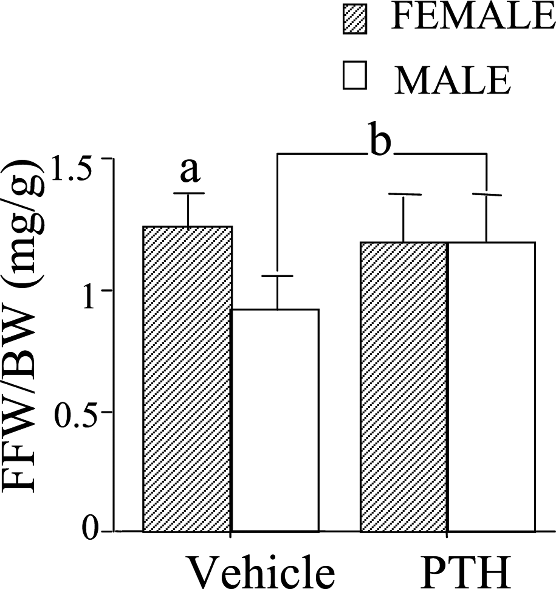
Parathyroid hormone (PTH) exerts both catabolic and anabolic actions on bone. Studies on the skeletal effects of PTH have seldom considered the effects of gender. Our study was designed to determine whether the response of mouse bone to PTH differed according to sex. As a first step, we analyzed gender differences with respect to bone mass and structural properties of 4 month old PTH treated (80 microg/kg per day for 2 weeks) male and female CD-1 mice. PTH significantly increased fat free weight/body weight, periosteal bone formation rate, mineral apposition rate, and endosteal single labeling surface, while significantly decreasing medullary area in male mice compared with vehicle treated controls, but induced no significant changes in female mice. We then analyzed the gender differences in bone marrow stromal cells (BMSC) isolated from 4 month old male and female CD-1 mice following treatment with PTH (80 microg/kg per day for 2 weeks). PTH significantly increased the osteogenic colony number and the alkaline phosphatase (ALP) activity (ALP/cell) by day 14 in cultures of BMSCs from male and female mice. PTH also increased the mRNA level of receptor activator of nuclear factor kappaB ligand in the bone tissue (marrow removed) of both females and males. However, PTH increased the mRNA levels of IGF-I and IGF-IR only in the bones of male mice. Our results indicate that on balance a 2-weeks course of PTH is anabolic on cortical bone in this mouse strain. These effects are more evident in the male mouse. These differences between male and female mice may reflect the greater response to PTH of IGF-I and IGF-IR gene expression in males enhancing the anabolic effect on cortical bone.
Figures


Similar articles
-
Insulin-like growth factor I is required for the anabolic actions of parathyroid hormone on mouse bone.J Bone Miner Res. 2002 Sep;17(9):1570-8. doi: 10.1359/jbmr.2002.17.9.1570. J Bone Miner Res. 2002. PMID: 12211426
-
Anabolic action of parathyroid hormone is skeletal site specific at the tissue and cellular levels in mice.J Bone Miner Res. 2002 May;17(5):808-16. doi: 10.1359/jbmr.2002.17.5.808. J Bone Miner Res. 2002. PMID: 12009011
-
Skeletal unloading alleviates the anabolic action of intermittent PTH(1-34) in mouse tibia in association with inhibition of PTH-induced increase in c-fos mRNA in bone marrow cells.J Bone Miner Res. 2004 Nov;19(11):1813-20. doi: 10.1359/JBMR.040808. Epub 2004 Aug 16. J Bone Miner Res. 2004. PMID: 15476581
-
Insulin like growth factor-I: a critical mediator of the skeletal response to parathyroid hormone.Curr Mol Pharmacol. 2012 Jun;5(2):135-42. Curr Mol Pharmacol. 2012. PMID: 21787292 Free PMC article. Review.
-
Role of Gut Microbiota in the Skeletal Response to PTH.J Clin Endocrinol Metab. 2021 Mar 8;106(3):636-645. doi: 10.1210/clinem/dgaa895. J Clin Endocrinol Metab. 2021. PMID: 33254225 Free PMC article. Review.
Cited by
-
Gender-Specific Differences in the Skeletal Response to Continuous PTH in Mice Lacking the IGF1 Receptor in Mature Osteoblasts.J Bone Miner Res. 2015 Jun;30(6):1064-76. doi: 10.1002/jbmr.2433. J Bone Miner Res. 2015. PMID: 25502173 Free PMC article.
-
Regulation of Ligand and Shear Stress-induced Insulin-like Growth Factor 1 (IGF1) Signaling by the Integrin Pathway.J Biol Chem. 2016 Apr 8;291(15):8140-9. doi: 10.1074/jbc.M115.693598. Epub 2016 Feb 10. J Biol Chem. 2016. PMID: 26865633 Free PMC article.
-
The osteocytic actions of glucocorticoids on bone mass, mechanical properties, or perilacunar remodeling outcomes are not rescued by PTH(1-34).Front Endocrinol (Lausanne). 2024 Jul 18;15:1342938. doi: 10.3389/fendo.2024.1342938. eCollection 2024. Front Endocrinol (Lausanne). 2024. PMID: 39092287 Free PMC article.
-
Overexpression of Bone Morphogenetic Protein-1 Promotes Osteogenesis of Bone Marrow Mesenchymal Stem Cells In Vitro.Med Sci Monit. 2020 Feb 21;26:e920122. doi: 10.12659/MSM.920122. Med Sci Monit. 2020. PMID: 32084123 Free PMC article.
-
Repeated irradiation from micro-computed tomography scanning at 2, 4 and 6 months of age does not induce damage to tibial bone microstructure in male and female CD-1 mice.Bonekey Rep. 2017 Jan 13;6:855. doi: 10.1038/bonekey.2016.87. eCollection 2017. Bonekey Rep. 2017. PMID: 28277563 Free PMC article.
References
-
- Alexander JM, Bab I, Fish S, Muller R, Uchiyama T, Gronowicz G, Nahounou M, Zhao Q, White DW, Chorev M et al. 2001. Human parathyroid hormone 1–34 reverses bone loss in ovariectomized mice. Journal of Bone and Mineral Research 16 1665–1673. - PubMed
-
- Bikle D, Majumdar S, Laib A, Powell-Braxton L, Rosen C, Beamer W, Nauman E, Leary C & Halloran B 2001. The skeletal structure of insulin-like growth factor I-deficient mice. Journal of Bone and Mineral Research 16 2320–2329. - PubMed
-
- Bikle DD, Sakata T, Leary C, Elalieh H, Ginzinger D, Rosen CJ, Beamer W, Majumdar S & Halloran BP 2002. Insulin-like growth factor I is required for the anabolic actions of parathyroid hormone on mouse bone. Journal of Bone and Mineral Research 17 1570–1578. - PubMed
-
- Dempster DW, Cosman F, Kurland ES, Zhou H, Nieves J, Woelfert L, Shane E, Plavetic K, Muller R, Bilezikian J et al. 2001. Effects of daily treatment with parathyroid hormone on bone micro-architecture and turnover in patients with osteoporosis: a paire d biopsy study. Journal of Bone and Mineral Research 16 1846–1853. - PubMed
-
- Eisman JA 1999. Genetics of osteoporosis. Endocrine Reviews 20 788–804. - PubMed
Publication types
MeSH terms
Substances
Grants and funding
LinkOut - more resources
Full Text Sources
Other Literature Sources
Research Materials
