. 2006 May;26(10):3718-27.
doi: 10.1128/MCB.26.10.3718-3727.2006.

Nuclear recycling of the pre-60S ribosomal subunit-associated factor Arx1 depends on Rei1 in Saccharomyces cerevisiae

Affiliations

Nuclear recycling of the pre-60S ribosomal subunit-associated factor Arx1 depends on Rei1 in Saccharomyces cerevisiae

Nai-Jung Hung et al. Mol Cell Biol. 2006 May.

Abstract

Arx1 and Rei1 are found on late pre-60S ribosomal particles containing the export adaptor Nmd3. Arx1 is related to methionine aminopeptidases (MetAPs), and Rei1 is a C2H2 zinc finger protein whose function in ribosome biogenesis has not been previously characterized. Arx1 and Rei1 localized predominately to the nucleus and cytoplasm, respectively, but could be coimmunoprecipitated, suggesting that they are transiently in the same 60S complex. arx1delta mutants showed a modest accumulation of 60S subunits in the nucleus, suggesting that Arx1 enhances 60S export. Deletion of REI1 led to cold sensitivity and redistribution of Arx1 to the cytoplasm, where it remained bound to free 60S subunits. However, deletion of ARX1 or the fusion of enhanced GFP (eGFP) to Rpl25 suppressed the cold sensitivity of an rei1delta mutant. The presence of eGFP on Rpl25 or its neighboring protein Rpl35 reduced the binding of Arx1 to 60S subunits, suggesting that Arx1 binds to 60S subunits in the vicinity of the exit tunnel. Mutations in Arx1 that disrupted its binding to 60S also suppressed an rei1delta mutant and restored the normal nuclear localization of Arx1. These results indicate that the cold sensitivity of rei1delta cells is due to the persistence of Arx1 on 60S subunits in the cytoplasm. Furthermore, these results suggest that Rei1 is needed for release of Arx1 from nascent 60S subunits after export to the cytoplasm but not for the subsequent nuclear import of Arx1.


Figures

FIG. 1.
FIG. 1.
Sequence alignment of Arx1 and MetAP2. Sequences of Arx1, yeast MetAP2 (yMetAP2), and human MetAP2 (hMetAP2) were aligned using ClustalW1.8 (http://serachlauncher.bcm.tmc.edu/). Identical residues in at least two sequences are shaded in black, and similar residues are shaded in gray. Residues responsible for MetAP2 enzymatic activity are indicated by asterisks. The insertions within the Arx1 sequence are indicative of the Arx1 protein family. Based on threading the Arx1 sequence onto the structure of MetAP, the insertions are predicted to lie predominately on one face of the protein.
FIG. 2.
FIG. 2.
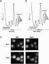
Sucrose gradient sedimentation and cellular localization of Arx1 and Rei1. (A and B) Lysates were prepared in the presence of cycloheximide (50 μg/ml) from room-temperature cultures of strains AJY1905 (ARX1-3HA) (A) and AJY1910 (REI1-13myc) (B) and fractioned on 7% to 47% sucrose gradients by ultracentrifugation. Buffer conditions were as described in Materials and Methods, except that extracts for AJY1910 (REI1-13myc) were prepared in a low-ionic-strength buffer (20 mM HEPES, 10 mM KCl, 2.5 mM MgCl2, 1 mM DTT, 50 μg/ml cycloheximide), because Rei1-60S association is salt sensitive. Fractions were collected, and the absorbance at 254 nm was monitored continuously. Proteins were precipitated with trichloroacetic acid, separated by SDS-polyacrylamide gel electrophoresis, transferred to nitrocellulose membrane, and immunoblotted for HA (Covance) or myc (Covance) for visualization of Arx1 or Rei1, respectively. (C) Cultures of AJY1909 (ARX1-GFP) and AJY1902 (rei1Δ) with pAJ1017 (REI1-GFP) were grown to mid-log phase at room temperature, and the in vivo localizations of Arx1-GFP and Rei1-GFP were monitored by fluorescence microscopy. DAPI staining was used to identify the position of nuclei.
FIG. 3.
FIG. 3.
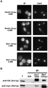
Rei1 does not shuttle in a Crm1-dependent fashion but can be found in the Arx1 particle. (A) The LMB-sensitive strain AJY1539 (CRM1T539C) containing plasmid pAJ538 (Nmd3-13myc) or pAJ1018 (REI1-13myc) was cultured in selective media at room temperature. Cells were concentrated 20-fold in fresh media, LMB was added to a final concentration of 0.1 μg/ml, and cultures were incubated for 30 min. Cells were then fixed with 3.7% formaldehyde (final concentration) and subjected to indirect immunofluorescence microscopy (IF). (B) Extract from strain AJY1907 (rei1Δ Arx1-HA) with pAJ1018 (REI1-13myc) was incubated without addition of antibody (N/A) or with anti-HA or anti-myc antibody and protein A beads. Precipitated proteins were eluted from protein A beads in sample buffer and separated by SDS-polyacrylamide gel electrophoresis. Total protein extract (T) was included as a loading control for the immunoprecipitations (IP). Western blotting was performed against the HA or c-myc epitopes.
FIG. 4.
FIG. 4.
Deletion of ARX1 or REI1 affects 60S subunit levels and export. Extracts, prepared from mid-log-phase cultures of strain AJY1911 (wild type [WT]) grown at 25°C (A), AJY1901 (arx1Δ) grown at room temperature (B), and strain AJY1902 (rei1Δ) grown at the temperatures indicated (C) were fractionated by sucrose gradient sedimentation as described in the legend to Fig. 2. Polysome profiles of the wild type grown at 30°C or 37°C were virtually indistinguishable from that shown in panel A (data not shown). Culture of AJY1911 (WT), AJY1901 (arx1Δ), or AJY1902 (rei1Δ) carrying plasmid pAJ908 (RPL25-eGFP) was incubated at the indicated temperature, and the in vivo localization of Rpl25-eGFP was monitored by fluorescence microscopy.
FIG. 5.
FIG. 5.
Deletion of ARX1 or the fusion of eGFP to RPL25 suppresses the cold sensitivity and 60S subunit deficiency of rei1Δ mutant cells. (A) Tenfold serial dilutions of saturated cultures were spotted onto yeast extract-peptone-glucose plates and incubated for 3 days at the indicated temperatures. The strains used were the following: AJY1905 (wild-type [WT] ARX1-HA), AJY1901 (arx1Δ), AJY1907 (rei1Δ ARX1-HA), AJY1903 (arx1Δ rei1Δ), AJY1907 (rei1Δ ARX1-HA) carrying plasmid pAJ908 (RPL25-eGFP), AJY1906 (rpl25Δ ARX1-HA pAJ908), and AJY1908 (rei1Δ rpl25Δ ARX1-HA pAJ908). (B) Extracts were prepared from mid-log-phase cultures of strains AJY1911 (WT), AJY1907 (rei1Δ), AJY1903 (arx1Δ rei1Δ), and AJY1908 (rei1Δ rpl25Δ Rpl25-eGFP) at room temperature (25°C) and fractionated by sucrose gradient sedimentation as described in the legend to Fig. 2.
FIG. 6.
FIG. 6.
Rpl25-eGFP and Rpl35A-GFP and Rpl35B-GFP alter Arx1-60S subunit association. (A) Lysates were prepared in the presence of cycloheximide from room-temperature cultures of strain AJY1905 (ARX1-HA) and AJY1906 (rpl25Δ ARX1-HA Rpl25-eGFP) and fractioned on sucrose gradients as described in the legend to Fig. 2. Fractions were collected, and proteins were precipitated with trichloroacetic acid, separated on SDS-polyacrylamide gel electrophoresis gels, transferred to nitrocellulose membrane, and immunoblotted for HA. (B) Cartoon of ribosomal proteins surrounding the exit tunnel (adapted from reference 45). (C) Cell extracts from strains AJY2128 (Rpl19A-GFP Rpl19B-GFP), AJY2125 (Rpl35A-GFP Rpl35B-GFP), and an isogenic wild type carrying Arx1-13myc (pAJ1016) plasmids were prepared and incubated with anti-myc antibodies and protein A beads. Precipitated proteins were eluted from protein A beads in sample buffer and separated by SDS-polyacrylamide gel electrophoresis. Western blotting was performed against myc or Rpl8. (D) Cell extracts were prepared from Rpl35A-GFP Rpl35B-GFP strain AJY2125 expressing c-myc tagged Lsg1 (pAJ901), Nmd3 (pAJ1001), Tif6 (pAJ1010), Nog2 (pAJ1014), and Arx1 (pAJ1026). The tagged proteins were immunoprecipitated, and their association with 60S subunits was monitored by blotting for Rpl8. IP, immunoprecipitation.
FIG. 7.
FIG. 7.
Cellular localization and sucrose gradient sedimentation of Arx1 in an rei1Δ strain. (A) Cultures of strains AJY1905 (ARX1-HA) and AJY1907 (rei1Δ ARX1-HA) were grown to mid-log phase at room temperature, and the localization of Arx1-HA was monitored by indirect immunofluorescence microscopy as described in Materials and Methods. Cells were treated with 1 μg/ml DAPI to visualize nuclear DNA. (B) Lysates were prepared in the presence of cycloheximide from room-temperature cultures of strain AJY1907 (rei1Δ ARX1-HA) and fractioned on sucrose gradients as described in the legend to Fig. 2. Fractions were collected, and proteins were precipitated with trichloroacetic acid, separated on SDS-polyacrylamide gel electrophoresis gels, transferred to nitrocellulose membrane, and immunoblotted for HA.
FIG. 8.
FIG. 8.
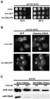
An arx1 mutant that suppresses rei1Δ exhibited reduced binding to 60S subunits. (A) Tenfold serial dilutions of saturated cultures were spotted onto Leu dropout plates and incubated for 3 days at the indicated temperatures. The strains used were AJY1903 (arx1Δ rei1Δ) carrying empty vector (pRS416), Arx1-GFP (pAJ1015), and Arx1(B6)-GFP (pAJ1463). (B) Cultures from strain AJY1903 (arx1Δ rei1Δ) carrying Arx1-GFP (pAJ1015) or Arx1(B6)-GFP (pAJ1463) were grown to mid-log phase. Cultures were incubated for a further 30 min in the presence of 4 μM Hoechst 33342 dye to stain nuclei. Cells were then immediately taken to monitor in vivo localizations of Arx1-GFP by fluorescence microscopy. (C) Cell extracts from strain AJY1901 (arx1Δ) carrying Arx1-13myc (pAJ1916) or Arx1(B6)-13myc (pAJ1464) plasmids were prepared and incubated with anti-myc antibodies and protein A beads. Precipitated proteins were eluted from protein A beads in sample buffer and separated by SDS-polyacrylamide gel electrophoresis. Western blotting was performed against myc (Arx1) or Rpl8 as a marker for 60S subunits.

Similar articles

Cited by

References

    1. Bassler, J., P. Grandi, O. Gadal, T. Lessmann, E. Petfalski, D. Tollervey, J. Lechner, and E. Hurt. 2001. Identification of a 60S preribosomal particle that is closely linked to nuclear export. Mol. Cell 8:517-529. - PubMed
    1. Basu, U., K. Si, J. R. Warner, and U. Maitra. 2001. The Saccharomyces cerevisiae TIF6 gene encoding translation initiation factor 6 is required for 60S ribosomal subunit biogenesis. Mol. Cell. Biol. 21:1453-1462. - PMC - PubMed
    1. Beckmann, R., C. M. Spahn, N. Eswar, J. Helmers, P. A. Penczek, A. Sali, J. Frank, and G. Blobel. 2001. Architecture of the protein-conducting channel associated with the translating 80S ribosome. Cell 107:361-372. - PubMed
    1. Ceci, M., C. Gaviraghi, C. Gorrini, L. A. Sala, N. Offenhauser, P. C. Marchisio, and S. Biffo. 2003. Release of eIF6 (p27BBP) from the 60S subunit allows 80S ribosome assembly. Nature 426:579-584. - PubMed
    1. Ciufo, L. F., and J. D. Brown. 2000. Nuclear export of yeast signal recognition particle lacking Srp54p by the Xpo1p/Crm1p NES-dependent pathway. Curr. Biol. 10:1256-1264. - PubMed

Publication types

MeSH terms

Substances