Comparative Study
. 2006 May;26(10):3824-34.
doi: 10.1128/MCB.26.10.3824-3834.2006.

Determinants that control the specific interactions between TAB1 and p38alpha

Affiliations
Comparative Study

Determinants that control the specific interactions between TAB1 and p38alpha

Huamin Zhou et al. Mol Cell Biol. 2006 May.

Abstract

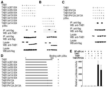
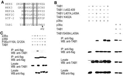
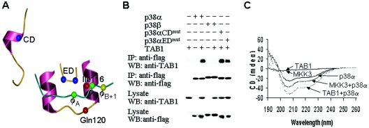
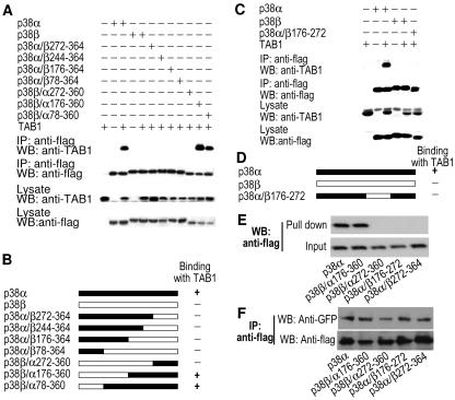
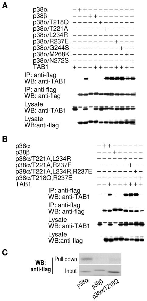
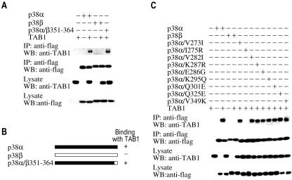
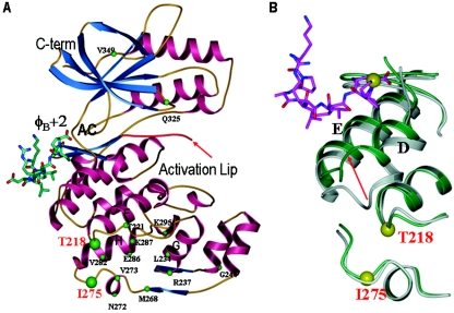
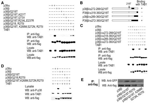
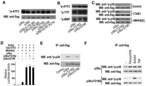
Previous studies have revealed that transforming growth factor-beta-activated protein kinase 1 (TAB1) interacts with p38alpha and induces p38alpha autophosphorylation. Here, we examine the sequence requirements in TAB1 and p38alpha that drive their interaction. Deletion and point mutations in TAB1 reveal that a proline residue in the C terminus of TAB1 (Pro412) is necessary for its interaction with p38alpha. Furthermore, a cryptic D-domain-like docking site was identified adjacent to the N terminus of Pro412, putting Pro412 in the phi(B)+3 position of the docking site. Through mutational analysis, we found that the previously identified hydrophobic docking groove in p38alpha is involved in this interaction, whereas the CD domain and ED domain are not. Furthermore, chimeric analysis with p38beta (which does not bind to TAB1) revealed a previously unidentified locus of p38alpha comprising Thr218 and Ile275 that is essential for specific binding of p38alpha to TAB1. Converting either of these residues to the corresponding amino acid of p38beta abolishes p38alpha interaction with TAB1. These p38alpha mutants still can be fully activated by p38alpha upstream activating kinase mitogen-activated protein kinase kinase 6, but their basal activity and activation in response to some extracellular stimuli are reduced. Adjacent to Thr218 and Ile275 is a site where large conformational changes occur in the presence of docking-site peptides derived from p38alpha substrates and activators. This suggests that TAB1-induced autophosphorylation of p38alpha results from conformational changes that are similar but unique to those seen in p38alpha interactions with its substrates and activating kinases.


Figures

FIG. 1.
FIG. 1.
Pro412 of TAB1 is required for TAB1-p38α interaction. (A) TAB1 C-terminal truncated mutants TAB1/Δ387-504, TAB1/Δ395-504, TAB1/Δ408-504, and TAB1/Δ419-504 were coexpressed with Flag-p38α in 293 cells. The cells were lysed 24 h after transfection. One-third of the cell lysates was analyzed by Western blotting (WB) with anti-TAB1 and anti-Flag antibodies. The rest of the cell lysates were subjected to immunoprecipitation (IP) with anti-Flag antibodies, and the immunoprecipitates were then analyzed by Western blotting with anti-TAB1 and anti-Flag. (B) The same experiments as shown in panel A except TAB1 mutants with deletions after amino acids 409 (TAB1/Δ410-504), 411 (TAB1/Δ412-504), and 413 (TAB1/Δ414-504) were used in the coexpression. (C) The interaction between TAB1 or TAB1 mutants and p38α was analyzed as described in the legend to panel A. The TAB1 mutants with P412 changed to alanine (TAB1/P412A), S413 changed to alanine (TAB1/S413A), and P412 and S413 changed to alanine (TAB1/P412A/S413A) are used. (D) The TAB1 mutants used in panels A to C are summarized with bar graphs and their abilities to bind with p38α are indicated by + and −. (E) 293 cells were transfected with the NF-κB reporter plasmid together with expression vectors of TAK1, TAB1, or TAB1/P412A in different combinations as indicated. Luciferase activity was measured 24 h after transfection.
FIG. 2.
FIG. 2.
K-X4A-X-φB motif in TAB1 is involved in TAB1-p38α interaction. (A) Sequence alignments of the K-X4A-X-φB motif near P412 in TAB1 compared to the docking sites of some known p38α activators and substrates. (B) TAB1 or TAB1 mutants with a deletion of amino acids 402 to 409 (TAB1/Δ402-409), L407 and L409 changed to alanine (TAB1/L407A,L409A), K402 changed to alanine (TAB1/K402A), or S408 and L409 changed to alanine (TAB1/S409A,L410A) were coexpressed with p38α in 293 cells. The interaction between p38α and TAB1 or the TAB1 mutant was analyzed as in Fig. 1A. (C) p38α, p38α mutated in the docking groove (p38α/I116A/Q120A), or p38β was coexpressed with TAB1. The interaction between TAB1 and p38α, the p38α mutant, or p38β was analyzed as in described in the legend to Fig. 1A.
FIG. 3.
FIG. 3.
p38α's interaction with TAB1 is different from its interaction with upstream kinases and substrates. (A) Part of the p38α structure shows the CD domain, the ED domain, and the hydrophobic docking groove bound to the MEF2A peptide (p38α/pepMEF2A; Protein Data Bank [PDB] file 1LEW). (B) p38α, p38α mutated in the CD domain (p38αCDmut) or ED site (p38αEDmut), or p38β was coexpressed with TAB1. The interaction between TAB1 and p38α, p38α mutants, or p38β was analyzed as in Fig. 1A. (C) MKK3 peptide SKGKSKRKKDLRISCNSK or TAB1 peptide SSAQSTSKTSVTLSLVMPSQ and p38α were incubated alone or together, and the CD spectra were analyzed.
FIG. 4.
FIG. 4.
The two regions in p38α that distinguish p38α from p38β in TAB1 binding. (A) p38α, the p38α-p38β chimeras, or p38β was coexpressed with TAB1. The interaction between TAB1 and p38α, the chimeras, or p38β was analyzed as in Fig. 1A. (B) Diagrams of the chimeras of p38α and p38β and a summary of their binding with TAB1. (C) p38α, p38α/β176-272, or p38β was coexpressed with TAB1. The interaction between TAB1 and p38α, p38α/β176-272, or p38β was analyzed as described in the legend to Fig. 1A. (D) Diagram of the chimera p38α/β176-272. (E) A total of 40 ml GST-TAB1β bound to glutathione-agarose was added to cell lysates from the 293 cells transfected with the expression vector of Flag-p38α, Flag-p38β/α176-360, Flag-p38β/α272-360, Flag-p38α/β176-272, or Flag-p38α/β272-364. The agarose beads were washed and subjected to Western blot analysis with anti-Flag antibodies. The levels of these Flag-tagged proteins in the cell lysates were also determined by Western blot analysis with anti-Flag-antibodies. (F) The GFP-MK2 expression vector was cotransfected with Flag-p38α, Flag-p38β/α176-360, Flag-p38β/α272-360, Flag-p38α/β176-272, or Flag-p38α/β272-364 expression plasmids. Immunoprecipitations were performed with anti-Flag antibodies 24 h after transfection. Immunoprecipitates were analyzed by Western blot with anti-GFP and anti-Flag antibodies.
FIG. 5.
FIG. 5.
Thr218 in p38α is required for its interaction with TAB1. (A) p38α, p38β, or p38α with point mutations at T218, T221, L234, R237, G244, M268, or N272 (to the corresponding amino acids from p38β at the same positions) was coexpressed with TAB1. Their interaction with TAB1 was analyzed as described in the legend to Fig. 1A. (B) p38α, p38β, or p38α with two or three point mutations at T218, T221, L234, or R237 (to the corresponding amino acids from p38β at the same positions) was coexpressed with TAB1. Their interaction with TAB1 was analyzed as described in the legend to Fig. 1A. (C) In vitro pull-down of Flag-p38α, Flag-p38β, or Flag-p38α/T218Q by TAB1β was performed as described in the legend to Fig. 4E.
FIG. 6.
FIG. 6.
Ile275 in p38α is required for its interaction with TAB1. (A) p38α, p38β, or p38α/β351-364 was coexpressed with TAB1. Their interaction with TAB1 was analyzed as described in the legend to Fig. 1A. (B) Diagram of p38α/β351-364. (C) p38α, p38β, or p38α with single point mutations at V273, I275, V282, K287, E286, K295, E301, Q325, or V349 (to the corresponding amino acids from p38β at the same positions) was coexpressed with TAB1. Their interaction with TAB1 was analyzed as described in the legend to Fig. 1A.
FIG. 7.
FIG. 7.
(A) A summary of point mutations on the three-dimensional structure of p38α complexed with pepMEF2A (PDB file 1LEW). Mutations affecting TAB1 binding are shown as large green spheres. Mutations that do not affect TAB1 binding are shown as small spheres. Placement of the φB+2 residue in pepMEF2A is labeled. φB+3 could bind in the active site. Note that the activation loop is missing in 1LEW. (B) The location of residues Thr218 and Ile275 in relation to the p38α docking groove and the MEF2A docking-site peptide (PDB file 1LEW; green) overlaid with uncomplexed p38α (1P38; gray). Note that the peptide-induced conformational changes in p38α helices D and E and the linker between them.
FIG. 8.
FIG. 8.
The interaction between p38β mutants and TAB1. (A) p38α, p38β, or p38β with point mutations at Q218; Q218 and A221; Q218 and I273; Q218, R243 and E237; Q218 and R275; or Q218, K268, S272, and R275 (to the corresponding amino acids from p38α at the same positions) was coexpressed with TAB1. The interaction with TAB1 was analyzed as described in the legend to Fig. 1A. (B) Diagrams of p38β mutants with Q218 mutated to threonine and C-terminal fragments replaced by sequences from p38α. (C) p38α, p38β, or the p38β mutants described in the legend to panel B were coexpressed with TAB1. Their interaction with TAB1 was analyzed as described in the legend to Fig. 1A. (D) p38α, p38β, or p38β with point mutations at Q218, Q218, and R275 or at Q218, K268, S272, and R275 (to the corresponding amino acids from p38α at the same positions) was coexpressed with TAB1. The phosphorylation of p38α, p38β, or the p38β mutants was determined by Western blotting analysis with anti-phospho-p38 antibodies. (E) GFP-MK2 was coexpressed with Flag-p38β, Flag-p38β/Q218T, Flag-p38β/Q218T,R275I, or Flag-p38β/Q218T,K268M,S272N,R275I. Immunoprecipitations were performed with anti-Flag antibodies 24 h after transfection. Immunoprecipitates were analyzed by Western blotting with anti-GFP and anti-Flag antibodies.
FIG. 9.
FIG. 9.
Activity and activation of p38α mutants that cannot interact with TAB1. (A) Expression vector of Flag-p38α, Flag-p38β, Flag-p38β/α176-360, Flag-p38α/β176-264, Flag-p38α/T218Q, Flag-p38α/I275R, or Flag-p38β/Q218T,R275I was transfected into in 293 cells. At 24 h after transfection, the cells were treated with 0.4 M sorbitol for 30 min. Flag-tagged proteins were immunoprecipitated with anti-Flag antibodies. An in vitro kinase assay was performed using the immunoprecipitates as kinases and GST-ATF2 as a substrate. The levels of immunoprecipitated Flag-proteins were determined by Western blot analysis with anti-Flag antibodies. (B) Flag-p38α, Flag-p38α/T218Q, and Flag-p38α/I275R were prepared by immunoprecipitations as described in the legend to panel A and used as kinases. GST-ATF2, tristetraprolin, or myelin basic protein was used as a substrate in the kinase assays. (C) Flag-p38α, Flag-p38α/T218Q, or Flag-p38α/I275R was coexpressed with control (empty vector), TAB1, or MKK6(E) in 293 cells as indicated. Total cell lysates were analyzed 24 h after transfection by Western blotting with anti-phospho-p38 and anti-Flag antibodies. (D) Expression vectors of a luciferase reporter gene under the control of 5× GAL4-binding site (5xGal), GAL4-binding domain fused with ATF2 activation domain (Gal-ATF2), MKK6(E), Flag-p38α, Flag-p38α/T218Q, or Flag-p38α/I275R were transfected into 293 cells in different combinations as indicated. Luciferase activity was measured 24 h later. (E) 293 cells were transfected with empty (control), Flag-p38α, Flag-p38α/T218Q, or Flag-p38α/I275R expression vectors. Flag-tagged proteins were immunoprecipitated with anti-Flag antibodies 48 h after transfection. The immunoprecipitates were analyzed by Western blotting using anti-phospho-p38 and anti-Flag antibodies. (F) Flag-p38α or Flag-p38α/T218Q was expressed in 293 cells. At 48 h after transfection, the cells were treated with peroxynitrite (500 μM), tumor necrosis factor (100 ng/ml), or sorbitol (0.4 M) for 5, 30, and 30 min, respectively. Flag-p38α or Flag-p38α/T218Q was immunoprecipitated with anti-Flag antibodies. The phosphorylation and the levels of Flag-p38α or Flag-p38α/T218Q were analyzed by Western blotting with anti-phospho-p38 and anti-Flag antibodies.

Similar articles

Cited by

References

    1. Anderson, N. G., J. L. Maller, N. K. Tonks, and T. W. Sturgill. 1990. Requirement for integration of signals from two distinct phosphorylation pathways for activation of MAP kinase. Nature 343:651-653. - PubMed
    1. Barsyte-Lovejoy, D., A. Galanis, and A. D. Sharrocks. 2002. Specificity determinants in MAPK signaling to transcription factors. J. Biol. Chem. 277:9896-9903. - PubMed
    1. Bell, M., R. Capone, I. Pashtan, A. Levitzki, and D. Engelberg. 2001. Isolation of hyperactive mutants of the MAPK p38/Hog1 that are independent of MAPK kinase activation. J. Biol. Chem. 276:25351-25358. - PubMed
    1. Brunner, D., N. Oellers, J. Szabad, W. H. Biggs III, S. L. Zipursky, and E. Hafen. 1994. A gain-of-function mutation in Drosophila MAP kinase activates multiple receptor tyrosine kinase signaling pathways. Cell 76:875-888. - PubMed
    1. Chang, C. I., B. E. Xu, R. Akella, M. H. Cobb, and E. J. Goldsmith. 2002. Crystal structures of MAP kinase p38 complexed to the docking sites on its nuclear substrate MEF2A and activator MKK3b. Mol. Cell 9:1241-1249. - PubMed

Publication types

MeSH terms

LinkOut - more resources