Comparative Study
. 2006 May;26(10):3917-34.
doi: 10.1128/MCB.26.10.3917-3934.2006.

Furin-, ADAM 10-, and gamma-secretase-mediated cleavage of a receptor tyrosine phosphatase and regulation of beta-catenin's transcriptional activity

Affiliations
Comparative Study

Furin-, ADAM 10-, and gamma-secretase-mediated cleavage of a receptor tyrosine phosphatase and regulation of beta-catenin's transcriptional activity

Lars Anders et al. Mol Cell Biol. 2006 May.

Abstract

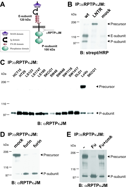
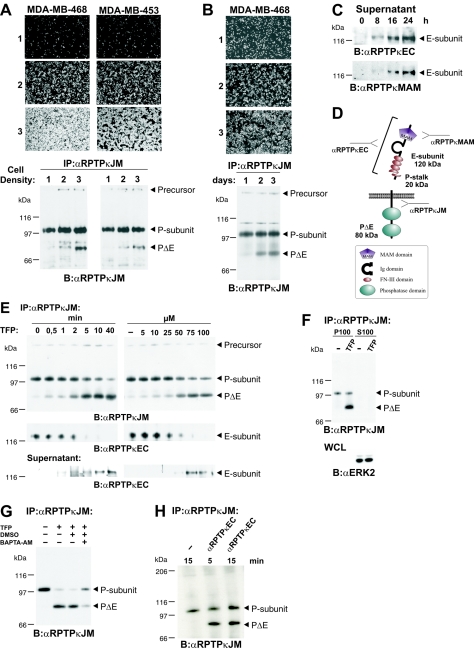
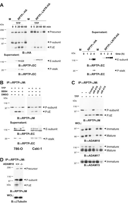
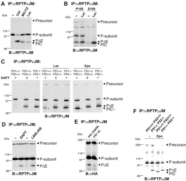
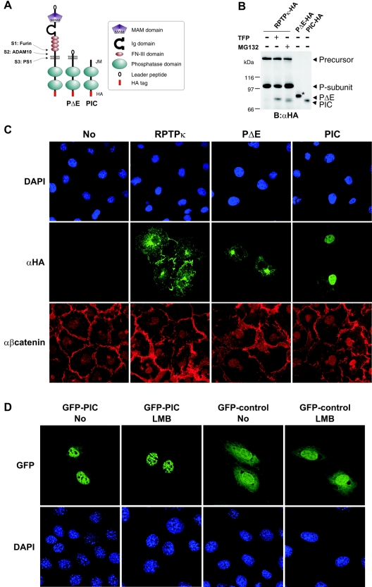
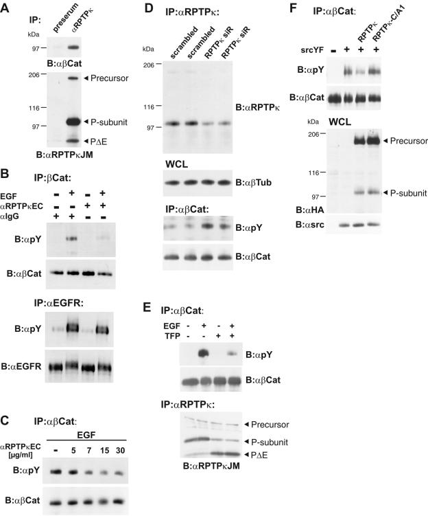
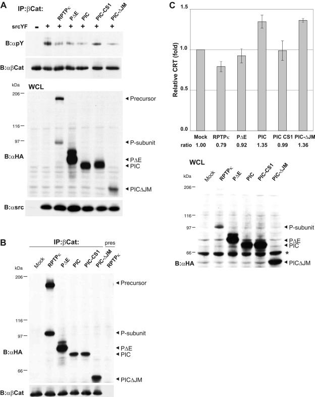
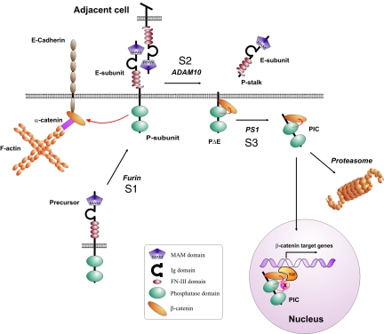
Several receptor protein tyrosine phosphatases (RPTPs) are cell adhesion molecules involved in homophilic interactions, suggesting that RPTP outside-in signaling is coupled to cell contact formation. However, little is known about the mechanisms by which cell density regulates RPTP function. We show that the MAM family prototype RPTPkappa is cleaved by three proteases: furin, ADAM 10, and gamma-secretase. Cell density promotes ADAM 10-mediated cleavage and shedding of RPTPkappa. This is followed by gamma-secretase-dependent intramembrane proteolysis of the remaining transmembrane part to release the phosphatase intracellular portion (PIC) from the membrane, thereby allowing its translocation to the nucleus. When cells were treated with leptomycin B, a nuclear export inhibitor, PIC accumulated in nuclear bodies. PIC is an active protein tyrosine phosphatase that binds to and dephosphorylates beta-catenin, an RPTPkappa substrate. The expression of RPTPkappa suppresses beta-catenin's transcriptional activity, whereas the expression of PIC increases it. Notably, this increase required the phosphatase activity of PIC. Thus, both isoforms have acquired opposing roles in the regulation of beta-catenin signaling. We also found that RPTPmu, another MAM family member, undergoes gamma-secretase-dependent processing. Our results identify intramembrane proteolysis as a regulatory switch in RPTPkappa signaling and implicate PIC in the activation of beta-catenin-mediated transcription.


Figures

FIG. 1.
FIG. 1.
RPTPκ is a two-subunit receptor at the cell surface after furin-mediated processing. (A) Scheme depicting the two-subunit structure of RPTPκ. The fragment sizes are indicated. Ig, immunoglobulin. (B) Cell surface-presented RPTPκ is a two-subunit enzyme, composed of the E subunit and the PTP domain-containing P subunit. 293 cells were transfected with wild-type RPTPκ and the convertase cleavage site mutant RPTPκ-LNTR, in which the dibasic sequence motif RTKR located in the membrane-proximal fibronectin type III domain was replaced by LNTR. Cells were surface biotinylated prior to lysis under standard conditions as described in Materials and Methods. RPTPκ was immunoprecipitated by an antibody to the intracellular juxtamembrane part (anti-RPTPκJM), followed by blotting with horseradish peroxidase (HRP)-conjugated streptavidin. (C) Accumulation of the precursor in LoVo cells that are devoid of functional furin. RPTPκ protein was analyzed in a panel of colon carcinoma cell lines by immunoprecipitation and blotting with anti-RPTPκJM antibody. (D) Stable expression of furin in LoVo cells restores processing of RPTPκ. The phosphatase was immunoprecipitated from two LoVo cell clones stably expressing human furin and from a vector-transfected clone (mock) for comparison. (E) Purified furin cleaves RPTPκ within the membrane-proximal fibronectin type III domain at the sequence RTKR in vitro. Furin-null, LoVo cell-derived RPTPκ was immunoprecipitated and incubated for 1 h at 37°C with phosphate-buffered saline (PBS) (−), purified recombinant mouse furin (Fu) or purified recombinant furin previously treated with the inhibitor decRVKR-cmk (Fu+cmk). Abbreviations: α, anti; B, blotting; IP, immunoprecipitation.
FIG. 2.
FIG. 2.
Proteolytic processing at a second site (S2 cleavage) results in generation of PΔE and shedding of RPTPκ. (A) Mammary carcinoma cell lines MDA-MB-468 and MDA-MB-453 were seeded at increasing cell densities (10, 50, and 100% [panels and lanes 1, 2, and 3, respectively]) and incubated for 24 h in serum-free medium containing 2% (vol/vol) Ultroser G. RPTPκ was immunoprecipitated with anti-RPTPκJM antibody and analyzed as indicated. (B) MDA-MB-468 cells were seeded at equal cell densities in medium containing 10% fetal calf serum and incubated for 1, 2, or 3 days (panels 1, 2, and 3, respectively). (C) Time-dependent accumulation of the E subunit in cell supernatants. Conditioned media were collected from confluent MDA-MB-468 cells at indicated time points and concentrated by TCA precipitation. The E subunit was detected with antibodies to the extracellular part (anti-RPTPκEC) and the MAM domain of RPTPκ (anti-RPTPκMAM). (D) Scheme depicting the expected fragments generated by cleavage at S1 and S2. The fragment sizes are indicated. (E) Time and concentration dependence of TFP-induced S2 cleavage and shedding. MDA-MB-468 cells were seeded at confluence, washed, and incubated in serum-free medium. The cells were treated with 100 μM TFP for different incubation times (left panel) or for 20 min with various concentrations of TFP as indicated (right panel). RPTPκ was immunoprecipitated from cell lysate with anti-RPTPκJM antibody (upper panel). The blot was reprobed with an antibody to the extracellular part of RPTPκ (anti-RPTPκEC) (middle panel). Conditioned media were collected, proteins concentrated by TCA precipitation and probed with anti-RPTPκEC antibody (lower panel). (F) PΔE is bound to the plasma membrane. 786-O cells were incubated with 100 μM TFP for 30 min and resuspended in hypotonic buffer. Soluble (S100) and membrane-bound (P100) proteins were separated by membrane fractionation. RPTPκ was immunoprecipitated from both fractions (upper panel). As a control, fractions were analyzed for the soluble protein ERK2 (lower panel). WCL, whole-cell lysate. (G) Inhibitory effect of the membrane-permeable calcium chelator BAPTA-AM on TFP-induced S2 processing. Cells were incubated for 2 h with (+) or without (−) 10 μM BAPTA-AM. DMSO, dimethyl sulfoxide. (H) Anti-RPTPκEC antibody treatment of MDA-MB-468 cells leads to rapid cleavage of endogenously expressed RPTPκ. Cells were starved and incubated with or without (−) anti-RPTPκEC antibody at 10 μg/ml as indicated. Abbreviations: α, anti; B, blotting; IP, immunoprecipitation.
FIG. 3.
FIG. 3.
ADAM 10 is involved in S2 processing. (A) RPTPκ subunit dissociation does not contribute to TFP-induced or basal shedding. RPTPκ constructs used here are HA tagged at the C terminus. COS-7 cells were transfected with cDNAs of RPTPκ, RPTPκ-LNTR, or pRK5 vector (M). Left panel, cells were washed, incubated in serum-free medium, and stimulated with 100 μM TFP for the times indicated. Upper panel, cell lysates were blotted and probed with anti-HA antibody. Lower panels, detection of shed RPTPκ fragments. Conditioned media were collected, and proteins were concentrated by TCA precipitation and probed with anti-RPTPκEC antibody. Right panel, time course of RPTPκ basal shedding at a high cell density. Transfected COS-7 cells were incubated in serum-free medium for the times indicated in the text. Conditioned media were processed as described above. (B) Metalloprotease inhibitor BB-94 diminishes S2 cleavage and shedding of RPTPκ in 786-O and Caki-1 cells. Prior to stimulation, cells were washed and serum-free medium was added. Cells were pretreated either with (+) or without (−) the metalloprotease inhibitor BB-94 (5 μM) or dimethyl sulfoxide (DMSO) and were then stimulated with 100 μM TFP for 15 min. Upper panel, RPTPκ was immunoprecipitated with anti-RPTPκJM antibody. Lower panel, conditioned media were collected, and proteins were concentrated by TCA precipitation, blotted, and probed with anti-RPTPκEC antibody. (C) ADAM 10 is involved in TFP-induced S2 cleavage. Caki-1 cells were transfected with siRNA (siR) directed against ADAM 10 (A10), ADAM 15 (A15), and ADAM 17 (A17). After 48 h, cells were treated or not treated (−) with 100 μM TFP for 15 min. Upper panel, RPTPκ was immunoprecipitated with anti-RPTPκJM antibody. Lower panels, specific silencing of ADAM expression was confirmed by immunoblot analyses of whole-cell lysate with antibodies to ADAM 10, ADAM 15, and ADAM 17. While immature and mature isoforms were detected for ADAM 10 and ADAM 17, no mature isoform was observed for ADAM 15. (D) ADAM 10-mediated cleavage at a high cell density. RPTPκ was immunoprecipitated from confluent ADAM 10+/+ and ADAM 10−/− fibroblasts. Abbreviations: α, anti; B, blotting; IP, immunoprecipitation; WCL, whole-cell lysate.
FIG. 4.
FIG. 4.
The S2 cleavage product PΔE is subject to PS1-dependent processing, leading to generation of PIC. (A) Specific inhibition of the proteasome results in accumulation of an additional RPTPκ-specific isoform (PIC) that is slightly smaller than PΔE. RPTPκ was immunoprecipitated from HEK293 cells treated with the peptide aldehyde MG132 (5 μM) and the specific proteasomal inhibitor lactacystin (Lac, 5 μM) for 16 h. Note the accumulation of PIC upon lactacystin treatment and of both PΔE and PIC upon incubation with MG132. For comparison, PΔE generation was induced by TFP. −, no treatment. (B) PIC is a soluble RPTPκ isoform. HEK293 cells were treated with or without (−) lactacystin (Lac) for 16 h, and cellular proteins were separated into membrane (P100) and cytosolic (S100) fractions. (C) Accumulation of the ADAM product PΔE and lack of PIC in the absence of PS (PIC is marked by asterisks). RPTPκ was immunoprecipitated from untreated (−) PS1+/+/PS2+/+ and PS1−/−/PS2−/− fibroblasts or PS1+/+/PS2+/+ fibroblasts treated (+) with the γ-secretase inhibitor DAPT (2 μM) for 16 h. Additionally, cells were incubated with 5 μM lactacystin (Lac, middle panel) or 5 μM epoxomycin (Epo, right panel) for 16 h. Note the accumulation of PΔE upon inhibition of PS activity (left) concomitantly with the lack of stabilized PIC (middle and right). (D) PS-mediated processing of PΔE in renal carcinoma cells. 768-O cells grown to confluence were treated with dimethyl sulfoxide or the γ-secretase inhibitors DAPT (2 μM) and L-685458 (5 μM) for 8 h. (E) HEK293 cells expressing wild-type PS1 or PS1 D385N were transiently transfected with HA-tagged RPTPκ. (F) PS-mediated cleavage of RPTPμ. PS1+/+PS2+/+ and PS1−/−PS2−/− fibroblasts were incubated with or without (−) epoxomycin (5 μM) for 12 h and endogenously expressed RPTPμ was immunoprecipitated. RPTPμ shows a characteristic precursor band at 200 kDa and its P subunit at 100 kDa (5). Note that the accumulation of PIC strictly depends on PS activity upon inhibition of the proteasome. Abbreviations: α, anti; B, blotting; IP, immunoprecipitation.
FIG. 5.
FIG. 5.
Exogenously expressed PIC localizes to the nucleus. (A) Schematic representation of ADAM-derived, transmembrane PΔE- and PS1-derived soluble PIC. All constructs used in this study were HA tagged at the C terminus. Ig, immunoglobulin. (B) Comparison of PS1-derived and recombinant PIC starting at residue 773 at the membrane-cytoplasm interface. COS-7 cells were transfected with HA-tagged RPTPκ and treated with (+) or without (−) TFP or MG132 to induce PΔE and PIC generation. Additionally, recombinant PΔE and PIC constructs were expressed in these cells. For comparison, only 1/5 of PΔE and 1/10 of PIC were transfected. The increase in size of recombinant PΔE (marked by an asterisk) is due to glycosylation (data not shown). B, blotting; α, anti. (C) COS-7 cells were transfected with the indicated HA-tagged RPTPκ isoforms, fixed and immunostained by using anti-HA antibody and AlexaFluor 488-labeled secondary antibody (green). Endogenous β-catenin was detected with a polyclonal antibody to β-catenin and AlexaFluor 546-labeled secondary antibody (red). Nuclear staining with DAPI (4′,6′-diamidino-2-phenylindole) is shown in the upper panels. Note the detection of PIC in the nucleus by confocal microscopy. Due to overexpression, transmembrane PΔE localizes predominantly in the endoplasmic reticulum and Golgi complex. α, anti. (D) Accumulation of PIC in nuclear bodies after the inhibition of CRM1-dependent nuclear export. NIH 3T3 cells were transfected with either GFP-PIC (left panels) or GFP control vector (right panels) and treated or not treated with leptomycin B (LMB) at 25 ng/ml for 3 h. After fixation, cells were stained with DAPI (4′,6′-diamidino-2-phenylindole) and observed with a Leica confocal microscope. Note the specific accumulation of GFP-PIC in nuclear bodies upon leptomycin B treatment.
FIG. 6.
FIG. 6.
β-Catenin is a cellular substrate of RPTPκ. (A) Coprecipitation of β-catenin with RPTPκ in 786-O renal carcinoma cells. Upper panel, detection of β-catenin in anti-RPTPκJM antibody immunoprecipitates. Preserum was used as a negative control. Lower panel, the blot was reprobed with anti-RPTPκJM antibody. (B) Stimulation of cells with anti-RPTPκEC antibody leads to decreased tyrosine phosphorylation of β-catenin but not of EGFR. MDA-MB-468 cells were treated with anti-RPTPκEC antibody (10 μg/ml) for 30 min prior to stimulation with EGF (200 ng/ml) for 5 min as indicated. −, no treatment. Immunoglobulin G-Fc antibody was used as a control. Upper panel, β-catenin was immunoprecipitated and probed for tyrosine phosphorylation with antibody 4G10 (anti-pY). Lower panel, EGFR was immunoprecipitated and probed for tyrosine phosphorylation. (C) Reduction in β-catenin tyrosine phosphorylation is antibody concentration dependent. Cell stimulation was performed with 200 ng/ml EGF for 5 min and various concentrations of anti-RPTPκEC antibody for 30 min as indicated. (D) Short interfering RNA (siR)-mediated knockdown of RPTPκ increases β-catenin tyrosine phosphorylation. Stably transfected ACHN cells were analyzed for RPTPκ expression andtyrosine phosphorylation of β-catenin. All cells were stimulated with EGF (200 ng/ml) for 5 min to induce the tyrosine phosphorylation of β-catenin. RPTPκ (top panel) and β-catenin (bottom panel) were immunoprecipitated and probed with indicated antibodies. Tubulin was used as loading control (middle panel). WCL, whole-cell lysates. (E) Effect of TFP treatment on β-Catenin tyrosine phosphorylation. Cells were incubated with (+) or without (−) TFP (100 μM) for 10 min prior to stimulation with EGF (200 ng/ml) for 5 min as indicated. β-Catenin (upper panel) and RPTPκ (lower panel) were immunoprecipitated and analyzed. (F) β-Catenin dephosphorylation depends on catalytic activity of the membrane-proximal PTP domain of RPTPκ. HEK293 cells were cotransfected with constitutive active Src (SrcYF) (+) or empty vector (−) and either RPTPκ-HA or catalytically inactive RPTPκ-C/A1-HA. To obtain maximal tyrosine phosphorylation of β-catenin, cells were starved for 12 h and the protein was immunoprecipitated and probed with the indicated antibody (upper panels). Whole-cell lysates (WCL) were analyzed for the expression of RPTPκ and SrcYF. Abbreviations: α, anti; B, blotting; IP, immunoprecipitation.
FIG. 7.
FIG. 7.
PIC is an active protein tyrosine phosphatase that promotes β-catenin-mediated transcription. (A) Dephosphorylation of β-catenin by PIC. HEK293 cells were transfected with empty vector (−) or constitutive active Src (SrcYF) (+) to induce tyrosine phosphorylation of β-catenin. All RPTPκ constructs used here are HA tagged at the C terminus. RPTPκ, PΔE, PIC, PIC-CS1, and PICΔJM were cotransfected, and tyrosine phosphorylation of β-catenin was analyzed (upper panel). Transfection controls are shown below. PIC-CS1 is a catalytically inactive version; PICΔJM is devoid of the juxtamembrane sequence. (B) Coprecipitation of PIC and PICΔJM with β-catenin. HEK293 cells were transfected with either empty vector (mock) or the indicated HA-tagged constructs, and β-catenin was immunoprecipitated. Preserum was used as a control (right lane). RPTPκ-derived constructs were detected with anti-HA antibody. (C) PIC enhances transcriptional activation of β-catenin, whereas RPTPκ suppresses it. HCT116 cells were cotransfected with β-catenin/TCF reporter constructs and either respective RPTPκ plasmids or empty vector (Mock). PIC-CS1 is a catalytically inactive PIC version that harbors a cysteine-to-alanine transition in the proximal PTP domain. Luciferase activity was determined using the dual luciferase kit (Promega), and the data were normalized to the cotransfected cytomegalovirus Renilla plasmid. Error bars represent the standard deviations of triplicate assays. The expression of the RPTPκ constructs is shown in the lower panel (an unspecific bovine serum albumin signal is marked by an asterisk). Abbreviations: α, anti; B, blotting; IP, immunoprecipitation.
FIG. 8.
FIG. 8.
Hypothetical model of the RPTP S1/S2/S3 cleavage mechanism and activation of β-catenin-mediated transcription. Furin-mediated cleavage constitutively yields two subunit RPTPκ proteins at the cell surface. Homophilic binding between RPTPκ proteins expressed in trans at high cell densities may induce S2 cleavage by ADAM 10, resulting in the release of the homophilic binding site into the cell supernatant. The remaining part, PΔE, is subject to γ-secretase/PS1-dependent intramembrane proteolysis that allows translocation of catalytically active PIC to the nucleus. Proteasomal destruction decreases the level of PIC in cells and may prohibit its nuclear import. PIC dephosphorylates the coactivator β-catenin and, unlike RPTPκ, increases TCF-mediated transcription. Note that PIC may additionally dephosphorylate transcriptional regulatory proteins associated with the β-catenin/TCF complex. Ig, immunoglobulin.

References

    1. Aicher, B., M. M. Lerch, T. Muller, J. Schilling, and A. Ullrich. 1997. Cellular redistribution of protein tyrosine phosphatases LAR and PTPσ by inducible proteolytic processing. J. Cell Biol. 138:681-696. - PMC - PubMed
    1. Borden, K. L. B. 2002. Pondering the promyelocytic leukemia protein (PML) puzzle: possible functions for PML nuclear bodies. Mol. Cell. Biol. 22:5259-5269. - PMC - PubMed
    1. Bradykalnay, S. M., A. J. Flint, and N. K. Tonks. 1993. Homophilic binding of PTP mu, a receptor-type protein tyrosine phosphatase, can mediate cell-cell aggregation. J. Cell Biol. 122:961-972. - PMC - PubMed
    1. Bradykalnay, S. M., and N. K. Tonks. 1995. Protein tyrosine phosphatases as adhesion receptors. Curr. Opin. Cell Biol. 7:650-657. - PubMed
    1. Campan, M., M. Yoshizumi, N. G. Seidah, M. E. Lee, C. Bianchi, and E. Haber. 1996. Increased proteolytic processing of protein tyrosine phosphatase μ in confluent vascular endothelial cells—the role of pc5, a member of the subtilisin family. Biochemistry 35:3797-3802. - PubMed

Publication types

MeSH terms

Substances

LinkOut - more resources