Comparative Study
. 2006 May;26(10):3935-41.
doi: 10.1128/MCB.26.10.3935-3941.2006.

Early embryonic lethality due to targeted inactivation of DNA ligase III

Affiliations
Comparative Study

Early embryonic lethality due to targeted inactivation of DNA ligase III

Nahum Puebla-Osorio et al. Mol Cell Biol. 2006 May.

Abstract

DNA ligases catalyze the joining of strand breaks in the phosphodiester backbone of duplex DNA and play essential roles in DNA replication, recombination, repair, and maintenance of genomic integrity. Three mammalian DNA ligase genes have been identified, and their corresponding ligases play distinct roles in DNA metabolism. DNA ligase III is proposed to be involved in the repairing of DNA single-strand breaks, but its precise role has not yet been demonstrated directly. To determine its role in DNA repair, cellular growth, and embryonic development, we introduced targeted interruption of the DNA ligase III (LIG3) gene into the mouse. Mice homozygous for LIG3 disruption showed early embryonic lethality. We found that the mutant embryonic developmental process stops at 8.5 days postcoitum (dpc), and excessive cell death occurs at 9.5 dpc. LIG3 mutant cells have relatively normal XRCC1 levels but elevated sister chromatid exchange. These findings indicate that DNA ligase III is involved in essential DNA repair activities required for early embryonic development and therefore cannot be replaced by other DNA ligases.


Figures

FIG. 1.
FIG. 1.
Targeted disruption of murine DNA ligase III. (A) Partial genomic structure of the mouse LIG3 gene. There is a total of 20 exons in this gene; exons 7 to 14 are shown. A 5.3-kb fragment containing exons 8 to 13 is replaced by a neo-resistant gene in a reversed orientation. Probes (both 3′ and 5′) used for genotyping by Southern blotting are shown, and the primers for PCR genotyping are marked with arrows. Restriction sites on the map: X, XbaI; P, PstI; H, HindIII; R1, EcoRI; R5, EcoRV. (B) Results of genotyping of ES cells by Southern blotting. WT, germ line band of 19.3 kb. MUT, targeted band of 7.4 kb identified by the 3′ probe. (C) Results of genotyping of embryos by PCR. Primers specific for germ line sequences and targeted mutant sequences are shown.
FIG. 2.
FIG. 2.
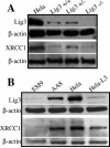
Western blotting analysis. (A) Extracts from 8.5-dpc embryos with different genotypes were detected for their DNA ligase III and XRCC1 expression, with HeLa cell extract as a control. (B) Detection of DNA ligase III and XRCC1 in cell extracts from XRCC1-deficient EM9 cells and its wild-type control, AA8, as well as LIG3 knockdown cells and its control, HeLa cells.
FIG. 3.
FIG. 3.
LIG3 deficiency and the resultant disruption of embryonic development at 8.5 dpc and excessive apoptosis at 9.5 dpc. A morphological comparison of wild-type (left) and mutant (right) embryos at 9.5 dpc (A) and 10.5 dpc (B). Yellow bars, 1 mm. (C) Section of wild-type (left) and mutant (right) embryos at 9.5 dpc stained with hematoxylin and eosin. TUNEL assays were performed on sections of wild-type (D) and mutant (E) embryos. Apoptotic cells are visible as green fluorescence, and nuclei are labeled with phosphatidylinositol staining. As a positive control, a section of wild-type embryos was treated with DNase I prior to the TUNEL assay (F).
FIG. 4.
FIG. 4.
SCE assays. Depicted are HeLa cells (A) and their counterpart LIG3 knockdown cells (B) showing elevated levels of SCE in LIG3 knockdown cells.

References

    1. Barnes, D. E., G. Stamp, I. Rosewell, A. Denzel, and T. Lindahl. 1998. Targeted disruption of the gene encoding DNA ligase IV leads to lethality in embryonic mice. Curr. Biol. 8:1395-1398. - PubMed
    1. Caldecott, K. W. 2001. Mammalian DNA single-strand break repair: an X-ra(y)ted affair. Bioessays 23:447-455. - PubMed
    1. Caldecott, K. W., S. Aoufouchi, P. Johnson, and S. Shall. 1996. XRCC1 polypeptide interacts with DNA polymerase beta and possibly poly (ADP-ribose) polymerase, and DNA ligase III is a novel molecular ‘nick-sensor’ in vitro. Nucleic Acids Res. 24:4387-4394. - PMC - PubMed
    1. Caldecott, K. W., C. K. McKeown, J. D. Tucker, S. Ljungquist, and L. H. Thompson. 1994. An interaction between the mammalian DNA repair protein XRCC1 and DNA ligase III. Mol. Cell. Biol. 14:68-76. - PMC - PubMed
    1. Caldecott, K. W., J. D. Tucker, L. H. Stanker, and L. H. Thompson. 1995. Characterization of the XRCC1-DNA ligase III complex in vitro and its absence from mutant hamster cells. Nucleic Acids Res. 23:4836-4843. - PMC - PubMed

Publication types

MeSH terms