. 2006 May;26(10):3942-54.
doi: 10.1128/MCB.26.10.3942-3954.2006.

Functional interaction of CP2 with GATA-1 in the regulation of erythroid promoters

Affiliations

Functional interaction of CP2 with GATA-1 in the regulation of erythroid promoters

Francesca Bosè et al. Mol Cell Biol. 2006 May.

Abstract

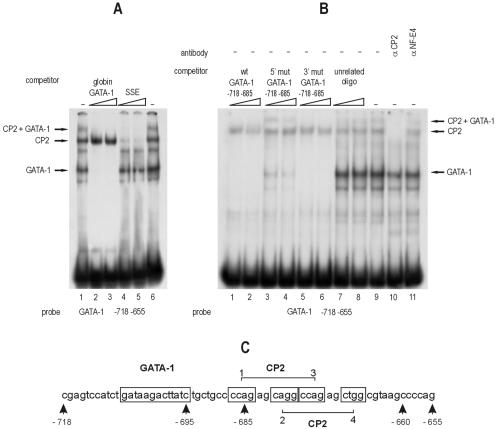
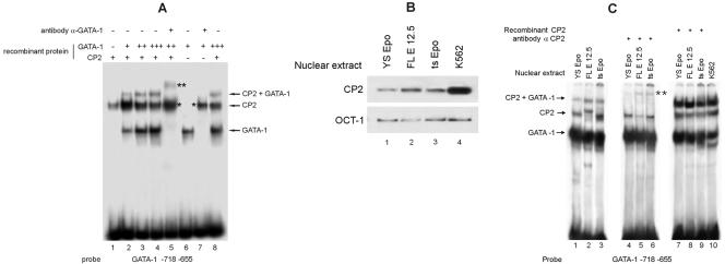
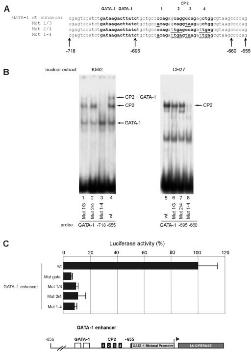
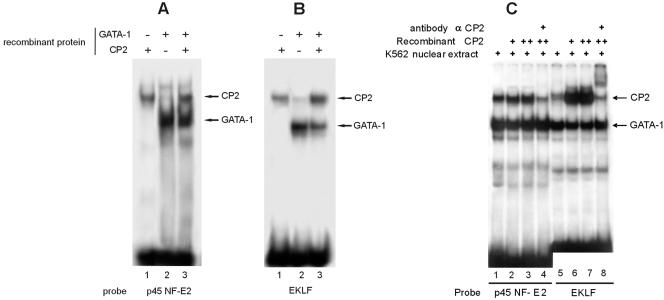
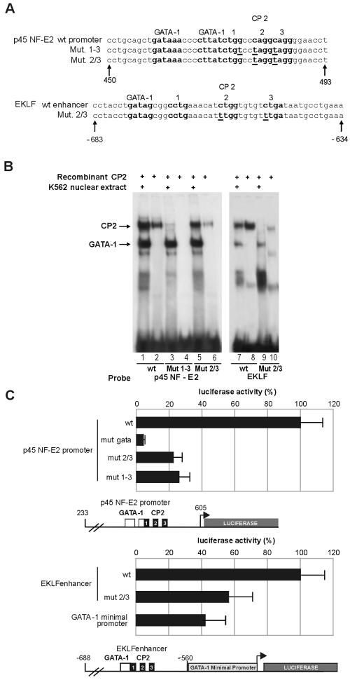
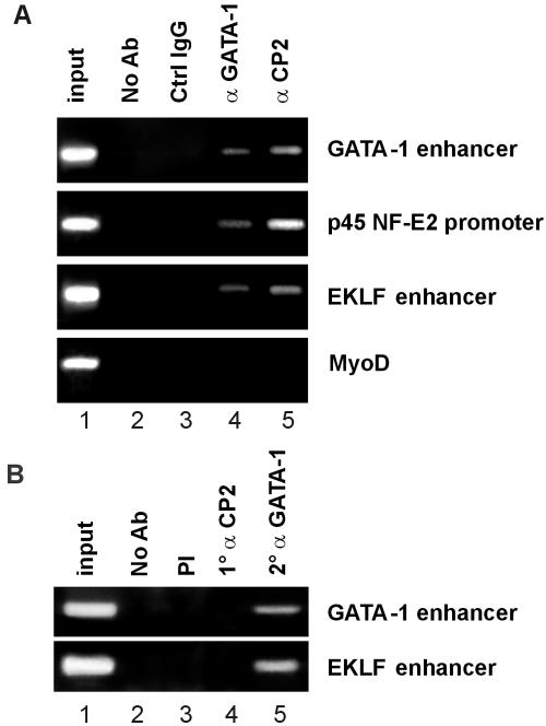
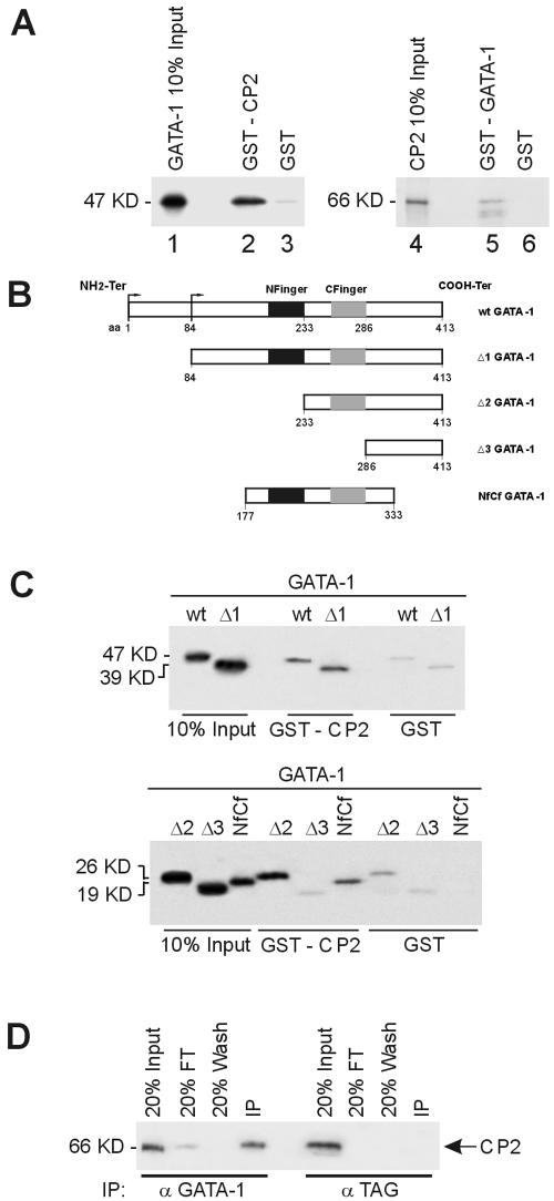
We observed that binding sites for the ubiquitously expressed transcription factor CP2 were present in regulatory regions of multiple erythroid genes. In these regions, the CP2 binding site was adjacent to a site for the erythroid factor GATA-1. Using three such regulatory regions (from genes encoding the transcription factors GATA-1, EKLF, and p45 NF-E2), we demonstrated the functional importance of the adjacent CP2/GATA-1 sites. In particular, CP2 binds to the GATA-1 HS2 enhancer, generating a ternary complex with GATA-1 and DNA. Mutations in the CP2 consensus greatly impaired HS2 activity in transient transfection assays with K562 cells. Similar results were obtained by transfection of EKLF and p45 NF-E2 mutant constructs. Chromatin immunoprecipitation with K562 cells showed that CP2 binds in vivo to all three regulatory elements and that both GATA-1 and CP2 were present on the same GATA-1 and EKLF regulatory elements. Adjacent CP2/GATA-1 sites may represent a novel module for erythroid expression of a number of genes. Additionally, coimmunoprecipitation and glutathione S-transferase pull-down experiments demonstrated a physical interaction between GATA-1 and CP2. This may contribute to the functional cooperation between these factors and provide an explanation for the important role of ubiquitous CP2 in the regulation of erythroid genes.


Figures

FIG. 1.
FIG. 1.
Binding of CP2 to the GATA-1 HS2 enhancer element. (A) EMSA using an oligonucleotide (positions −718 to −655) encompassing the GATA-1 CP2 binding motifs present in the HS2 enhancer with K562 cell nuclear extract. The effects of excess unlabeled GATA-1 binding (globin GATA-1) and CP2 binding (SSE) competitors are shown. The positions of the GATA-1, CP2, and GATA-1-plus-CP2 complexes are indicated. Note that both competitors abolish the upper band, demonstrating that it contains both CP2 and GATA-1. (B) Unlabeled oligonucleotides comprising the wild-type (wt) or mutated GATA-1 palindrome do not affect the binding of CP2 to the GATA-1 HS2 enhancer oligonucleotide in competition experiments. Note that the weak GATA-1 binding mutant (5′ mut GATA-1, lanes 3 to 4) competes almost completely for the GATA-1 band, while it leaves the upper CP2-plus-GATA-1 band essentially intact. A specific anti-CP2 antibody, but not an anti-NF-E4 antibody, abolishes the CP2 band (lanes 10 to 11). The positions of the GATA-1, CP2, and GATA-1-plus-CP2 complexes are indicated. (C) Schematic representation of the GATA-1 and CP2 binding sites on the mouse GATA-1 promoter.
FIG. 2.
FIG. 2.
Binding of GATA-1 and CP2 to the GATA-1 HS2 enhancer element. (A) Studies of recombinant GATA-1 and CP2 confirm that both proteins bind to the HS2 enhancer element. Note that antibody against GATA-1 supershifts both the GATA-1 and the GATA-1-plus-CP2 bands (lanes 5 and 7, asterisks). The positions of the GATA-1, CP2, and GATA-1-plus-CP2 complexes are indicated. (B) Western analysis of extracts from primary fetal liver cells from E12.5 day embryos (FL E 12.5) and immortalized mouse yolk sac (YS Epo) and bone marrow (ts Epo) growth factor-dependent cell lines with anti-CP2 antibody. Anti-OCT-1 antibody served as the loading control. (C) The GATA-1-plus-CP2 band was obtained using extracts from various hematopoietic cell types (lanes 1 to 3); compare lanes 1 to 3 with lanes 4 to 6, where the anti-CP2 antibody was added. Recombinant CP2 added to nuclear extracts from the same hematopoietic cell extracts generates a strong GATA-1-plus-CP2 band (lanes 7 to 9); compare lanes 7 to 9 with lane 10, K562 nuclear extracts.
FIG. 3.
FIG. 3.
CP2 sites on the GATA-1 HS2 enhancer substantially contribute to the transcriptional activity of the GATA-1 gene. (A) Schematic of the wild-type (wt) and mutated GATA-1 HS2 enhancer constructs. The GATA-1 and CP2 consensus sites are boldface, and the mutated bases in the CP2 consensus sites are underlined. (B) The effect of mutations on protein binding. EMSAs with the wild-type and mutated oligonucleotides were performed with extracts from K562 (erythroid) or CH27 (nonerythroid) cells. The positions of the GATA-1, CP2, and GATA-1-plus-CP2 complexes are indicated. The most extensive mutation (Mut 1-4) totally abolished CP2 binding (lanes 3 and 8), but mutations in the single CP2 boxes (Mut 1/3 and Mut 2/4) still allowed significant CP2 binding on the intact CP2 site (lanes 1, 2, 6, and 7). The CH27 nuclear extracts contain CP2 but not GATA-1. (C) Functional luciferase reporter assays with K562 cells of mutants shown in panel A and an additional construct carrying a mutation of the GATA-1 binding site. All three CP2 mutations greatly reduced the ability of the HS2 enhancer linked to the GATA-1 minimal promoter to drive the luciferase reporter, shown schematically in the lower panel.
FIG. 4.
FIG. 4.
Sequences from the fetal 1b proximal p45-NF-E2 promoter and EKLF erythroid hypersensitive site 1 bind CP2. Binding of recombinant CP2 and GATA-1 to the p45 NF-E2 promoter (A) and the EKLF promoter (B) was assessed by EMSA. The positions of the GATA-1 and CP2 complexes are indicated. (C) The effects of the addition of recombinant CP2 and anti-CP2 antibody on GATA-1 and CP2 complex formation on the p45 NF-E2 and EKLF promoters were assessed by EMSA. The positions of the GATA-1 and CP2 complexes are indicated.
FIG. 5.
FIG. 5.
Mutations in the CP2 binding sites in the p45-NF-E2 and EKLF promoters inhibit their transcriptional activity in K562 cells. (A) Schematic of the wild-type (wt) and mutated p45 NF-E2 and EKLF promoters. The GATA-1 and CP2 binding sites are boldface and labeled, and the mutated bases are underlined. (B) EMSA with K562 nuclear extract or recombinant CP2 and the wild-type and mutant oligonucleotides shown in panel A. The mutations abolished CP2 binding but did not affect GATA-1 binding, as demonstrated by both direct binding (lanes 1 to 10) and competition experiments (not shown). (C) Functional luciferase reporter assays with K562 cells of mutants shown in panel A. In experiments with the p45 NF-E2 promoter, a construct carrying a mutation of the GATA-1 binding site was also tested. In the experiments with the EKLF promoter, the GATA-1 minimal promoter was tested as a control. Mutations in the CP2 binding sites significantly reduced the transcriptional activity of the transfected constructs, which are shown schematically below the respective bar graphs.
FIG. 6.
FIG. 6.
Binding of CP2 and GATA-1 to erythroid gene regulatory elements. (A) ChIP with anti-GATA-1 or anti-CP2 antibody. Chromatin from K562 cells was immunoprecipitated using antiserum to CP2 or GATA-1. Normal rabbit serum (Ctrl IgG) samples and samples without antibody (No Ab) served as the controls. Quantitative PCR was performed with primer pairs to amplify the erythroid gene regulatory elements (GATA-1 enhancer, p45 NF-E2 promoter, and EKLF enhancer) or the MyoD gene as a control. The input chromatin is shown. (B) ChIP/Re-ChIP with anti-GATA-1 and anti-CP2 antibodies. Chromatin from K562 cells was sequentially immunoprecipitated with anti-CP2 and then anti-GATA-1 antibodies prior to quantitative PCR with the erythroid gene regulatory elements shown. Controls were as described for panel A.
FIG. 7.
FIG. 7.
Direct physical interaction between GATA-1 and CP2. (A) Purified GST or GST fusion proteins containing full-length CP2 (GST-CP2) preadsorbed to glutathione-Sepharose beads were incubated with 35S-labeled in vitro-transcribed/translated GATA-1 (lanes 1 to 3). Specifically bound protein was eluted from washed beads and visualized by autoradiography after SDS-PAGE. Input represents 10% of the in vitro-translated GATA-1 used in the assay. Lanes 4 to 6 utilized glutathione-Sepharose-bound GST-GATA-1 and 35S-labeled CP2. (B) Schematic of truncation mutants used to map the GATA-1 regions responsible for the interaction with CP2. (C) GST or GST-CP2 protein coupled to glutathione-Sepharose was incubated with 35S-labeled wild type or truncated GATA-1 mutants shown in panel A. Input represents 10% of the in vitro-translated GATA-1 or mutant protein used in the assay. The molecular masses of the proteins are shown. (D) K562 cell extract was immunoprecipitated with either anti-GATA-1 or an unrelated antibody (anti-TAG) as the control. Immunoprecipitates were fractionated by SDS-PAGE and immunoblotted with anti-CP2 antibody. CP2 protein was detected in precipitates from anti-GATA-1 antibody but not from the control.

References

    1. Anderson, K. P., S. C. Crable, and J. B. Lingrel. 1998. Multiple proteins binding to a GATA-E box-GATA motif regulate the erythroid Kruppel-like factor (EKLF) gene. J. Biol. Chem. 273:14347-14354. - PubMed
    1. Anderson, K. P., S. C. Crable, and J. B. Lingrel. 2000. The GATA-E box-GATA motif in the EKLF promoter is required for in vivo expression. Blood 95:1652-1655. - PubMed
    1. Bekri, S., A. May, P. D. Cotter, A. I. Al Sabah, X. Guo, G. S. Masters, and D. F. Bishop. 2003. A promoter mutation in the erythroid-specific 5-aminolevulinate synthase (ALAS2) gene causes X-linked sideroblastic anemia. Blood 102:698-704. - PubMed
    1. Cairns, L., M. Ciro, M. Minuzzo, F. Morle, J. Starck, S. Ottolenghi, and A. Ronchi. 2003. Induction of globin mRNA expression by interleukin-3 in a stem cell factor-dependent SV-40 T-antigen-immortalized multipotent hematopoietic cell line. J. Cell. Physiol. 195:38-49. - PubMed
    1. Cantor, A. B., and S. H. Orkin. 2001. Hematopoietic development: a balancing act. Curr. Opin. Genet. Dev. 11:513-519. - PubMed

Publication types

MeSH terms

LinkOut - more resources