. 2006 Jul;173(3):1357-64.
doi: 10.1534/genetics.106.057463. Epub 2006 Apr 28.

Metabolic disruption in Drosophila bang-sensitive seizure mutants

Affiliations

Metabolic disruption in Drosophila bang-sensitive seizure mutants

Tim Fergestad et al. Genetics. 2006 Jul.

Abstract

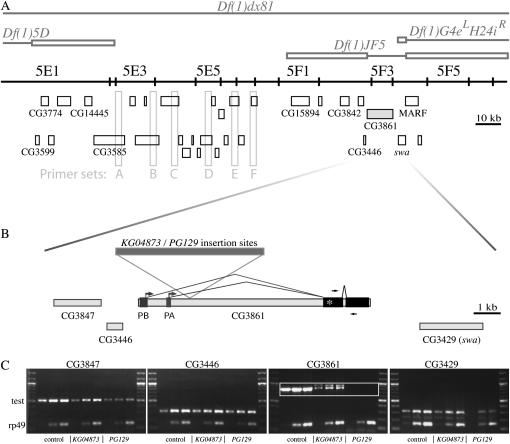
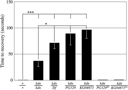
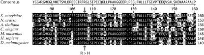
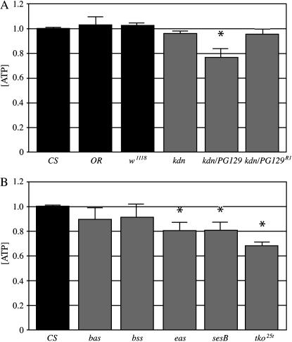
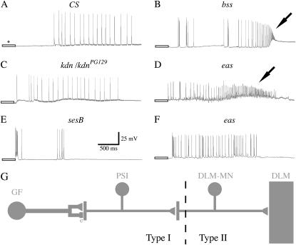
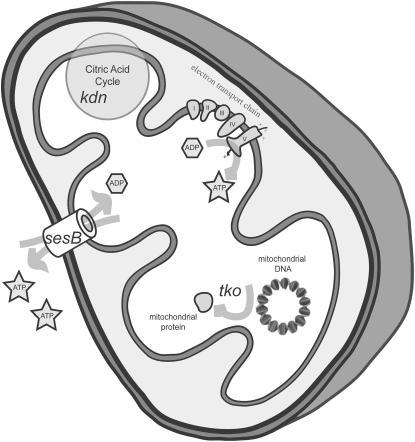
We examined a number of Drosophila mutants with increased susceptibility to seizures following mechanical or electrical stimulation to better understand the underlying factors that predispose neurons to aberrant activity. Several mutations in this class have been molecularly identified and suggest metabolic disruption as a possible source for increased seizure susceptibility. We mapped the bang-sensitive seizure mutation knockdown (kdn) to cytological position 5F3 and identified citrate synthase as the affected gene. These results further support a role for mitochondrial metabolism in controlling neuronal activity and seizure susceptibility. Biochemical analysis in bang-sensitive mutants revealed reductions in ATP levels consistent with disruption of mitochondrial energy production in these mutants. Electrophysiological analysis of mutants affecting mitochondrial proteins revealed an increased likelihood for a specific pattern of seizure activity. Our data implicate cellular metabolism in regulating seizure susceptibility and suggest that differential sensitivity of neuronal subtypes to metabolic changes underlies distinct types of seizure activity.


Figures

F<sc>igure</sc> 1.—
Figure 1.—
Genetic mapping of the kdn locus. (A) Complementation testing of kdn bang sensitivity using X chromosome deficiencies in the 5A–6A region resulted in failure of complementation by two deficiencies, Df(1)dx81 and Df(1)JF5. Several of these deficiencies were described as having breakpoints in the 5E3–E5 region. However, PCR analysis of genomic DNA from homozygous deficiency animals revealed that this region was intact in all deficiencies except Df(1)dx81 (primer sets A–F). Empirically defined breakpoint limits based on our PCR analysis are depicted as boxes and deleted segments are represented as solid lines. The results of these experiments placed the kdn locus cytologically at 5F1–4 and between CG15894 and CG3861. (B) The failure of two independently generated P-element insertion lines, PG129 and KG04873, to complement the kdn bang-sensitive phenotype implicates gene CG3861 as the affected gene. Two unique transcripts of CG3861 have been identified and suggest two alternative initial exons. Both P-element insertion sites map to within 20 bp of each other and are located in between the alternative starting exons and the constitutive remaining exons. * denotes the location of the only observed sequence alteration in this region of the original kdn chromosome and is in the first constitutive exon of CG3861. (C) Semiquantitative RT–PCR gene expression studies revealed a reduction only in the CG3861 transcript. cDNA reverse transcribed from isolated homozygous P-element and control animal RNA was used as PCR template (see materials and methods). Bands shown are samples taken from the PCR reactions at cycles 30, 35, and 40. The bottom band is an internal control for RNA and the top band is a product generated from experimental intron-spanning primers specific to the gene under study. The specific reduction of CG3861 transcript in KG04873 and PG129 homozygous lines supports the specific disruption of CG3861 expression due to the element insertions. The larger band in KG04873 corresponds to the genomic size PCR product amplified from remaining genomic DNA and the absence of cDNA template. The DNA ladder in all gels is 100 bp.
F<sc>igure</sc> 2.—
Figure 2.—
Behavioral analysis of various kdn genotypes. Animals of the genotype indicated were observed for their time to recover following 15 sec of mechanical stimulation (n > 10 for each sample; error bars represent SEM). Wild-type and control animals right themselves instantly, while bang-sensitive mutants display different durations of paralysis or immobility prior to delayed discharge spasms and recovery. Flies containing the kdn mutation over a more severe allele, such as a deletion or P-element insertion, require significantly longer periods to recover from paralysis. Animals heterozygous for kdn and a revertant that precisely excises the P element (PG129R1 and KG04873R1) exhibit no sensitivity to mechanical stress. Nonparametric Mann–Whitney U analyses were performed, with * and *** representing statistical significance of P < 0.05 and P < 0.001, respectively.
F<sc>igure</sc> 3.—
Figure 3.—
Sequence alignment of citrate synthase in the region of the kdn mutation. This region is highly conserved in all known eukaryotic citrate synthase sequences. The arginine 95 residue is mutated to a histidine in kdn as the result of a G-to-A base pair change. This residue is completely conserved in all sequences examined, from yeast to human.
F<sc>igure</sc> 4.—
Figure 4.—
ATP levels are reduced in bang-sensitive mutants. (A) Wild-type strains Canton-S and Oregon-R, as well as w1118 controls, exhibit no significant change in ATP levels, while kdn mutants exhibit a reduction in ATP. kdn/PG129 heterozygotes with a strong bang-sensitive phenotype exhibited a significant reduction in ATP levels, whereas ATP levels in kdn homozygotes with mild and variable bang sensitivity were not significantly reduced. Similarly, kdn/PG129R1 animals, which are not bang sensitive, displayed normal ATP levels. (B) Other bang-sensitive mutants had significant reductions in ATP levels. Animals homozygous for eas, sesB, and tko all exhibited significantly reduced levels of ATP. Reductions in ATP levels were observed in bas and bss mutants, although these reductions were not quite significant. N values ≥6 were used for each genotype. U-test analyses revealing significant changes in ATP levels are denoted with * (P < 0.01).
F<sc>igure</sc> 5.—
Figure 5.—
Representative traces of distinct types of initial seizure activity in the GF-DLM neuronal circuit from bang-sensitive mutants. (A) Application of high-frequency electrical stimulation to the brain in Canton-S elicits a behavioral seizure with corresponding bursts of activity in DLMs. Inducing the seizure phenotypes in wild-type animals requires that the voltage of the pulses be increased (denoted with an *) during the stimulation train (depicted as open boxes). (B) Following light brain stimulation, bss mutants exhibit bursts of spike activity in DLMs that ultimately increase in frequency and decrease in amplitude (arrows) indicative of type II seizures. (C and E) kdn and sesB exhibit initial DLM spiking with constant amplitude and variable firing rates typical of type I seizures. (D and F) eas mutants predominately exhibit type II seizure activity, although type I activity was also commonly observed. (G) A diagram of the giant fiber (GF) to dorsal longitudinal muscle (DLM) escape reflex circuit (King and Wyman 1980). Some of the en passant chemical and electrical (e) synaptic outputs of the giant fiber neuron directly stimulate the PSI interneuron. The PSI neuron in turn innervates multiple DLM motor neurons that synapse onto distinct DLMs. The increasing frequency and decreasing amplitude of DLM spikes seen in type II seizure activity reflect electrical activity of the motor neuron because attenuation in synaptic output from the PSI should evoke “all-or-nothing” action potentials in the motor neuron with corresponding DLM activity.
F<sc>igure</sc> 6.—
Figure 6.—
Sites of mitochondrial disruption in bang-sensitive mutants. Mutations in technical knockout (tko) are thought to disrupt translation of mitochondrial proteins. Mutations in stress-sensitive B (sesB), which encodes the ADP/ATP translocase, may disrupt nucleotide transport and signaling in mitochondria. Mutations in the knockdown (kdn) gene disrupt an enzyme implicated in metabolism. All of these mutations are associated with reduction in ATP levels suggesting that metabolic disruption may be a general mechanism underlying bang-sensitive seizures in Drosophila.

Similar articles

Cited by

References

    1. Attwell, D., and S. B. Laughlin, 2001. An energy budget for signaling in the grey matter of the brain. J. Cereb. Blood Flow Metab. 21: 1133–1145. - PubMed
    1. Beal, M. F., B. T. Hyman and W. Koroshetz, 1993. Do defects in mitochondrial energy metabolism underlie the pathology of neurodegenerative diseases? Trends Neurosci. 16: 125–131. - PubMed
    1. Benzer, S., 1971. From the gene to behavior. JAMA 218: 1015–1022. - PubMed
    1. Bourbon, H. M., G. Gonzy-Treboul, F. Peronnet, M. F. Alin, C. Ardourel et al., 2002. A P-insertion screen identifying novel X-linked essential genes in Drosophila. Mech. Dev. 110: 71–83. - PubMed
    1. De Meirleir, L., 2002. Defects of pyruvate metabolism and the Krebs cycle. J. Child Neurol. 17(Suppl. 3): 3S26–33. - PubMed

Publication types

Substances