. 2006 Jul;173(3):1275-86.
doi: 10.1534/genetics.106.056721. Epub 2006 Apr 30.

The molecular identities of the Caenorhabditis elegans intraflagellar transport genes dyf-6, daf-10 and osm-1

Affiliations

The molecular identities of the Caenorhabditis elegans intraflagellar transport genes dyf-6, daf-10 and osm-1

Leslie R Bell et al. Genetics. 2006 Jul.

Abstract

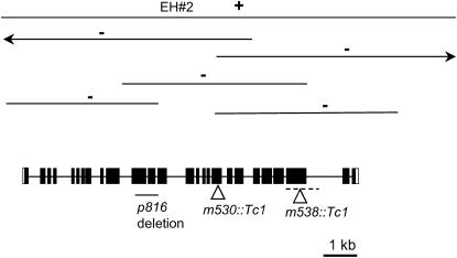
The Caenorhabditis elegans genes dyf-6, daf-10, and osm-1 are among the set of genes that affect chemotaxis and the ability of certain sensory neurons to take up fluorescent dyes from the environment. Some genes in this category are known to be required for intraflagellar transport (IFT), which is the bidirectional movement of raft-like particles along the axonemes of cilia and flagella. The cloning of dyf-6, daf-10, and osm-1 are described here. The daf-10 and osm-1 gene products resemble each other and contain WD and WAA repeats. DYF-6, the product of a complex locus, lacks known motifs, but orthologs are present in flies and mammals. Phenotypic analysis of dyf-6 mutants expressing an OSM-6::GFP reporter indicates that the cilia of the amphid and phasmid dendritic endings are foreshortened. Consistent with genetic mosaic analysis, which indicates that dyf-6 functions in neurons of the amphid sensilla, DYF-6::GFP is expressed in amphid and phasmid neurons. Movement of DYF-6::GFP within the ciliated endings of the neurons indicates that DYF-6 is involved in IFT. In addition, IFT can be observed in dauer larvae.


Figures

F<sc>igure</sc> 1.—
Figure 1.—
Aberrant pattern of OSM-6∷GFP in dyf-6 mutants. (A) The wild-type pattern of an amphid sensillum in dyf-6(+). Note the tapering of the fluorescence into a single unit toward the anterior tip of the animal (anterior is up in all micrographs). The single unit corresponds to the channel of the ciliary endings through the amphid sheath and body cuticle. The transition zone between the dendritic bodies and the dendritic endings is indicated with an arrowhead. (B) An abnormal pattern typically seen with the dyf-6(m175) mutation. Note the failure of the fluorescence to extend to the tip of the animal. (C) The other mutation, dyf-6(mn346), confers a similar defect at the anterior end of the amphid neurons. (D) Expression of OSM-6∷GFP in a phasmid sensillum in the tail of a dyf-6(+) animal. The transition zone at the junction between the dendritic bodies and the ciliated endings that penetrate the cuticle is indicated with an arrowhead. (E) A much shorter region of fluorescence below the arrowhead indicates aberrant, apparently truncated, dendritic endings of the phasmid neurons in a dyf-6(mm175) animal. (F) Aberrant endings are also apparent in dyf-6(mn346) mutants. Bar, 5 μm.
F<sc>igure</sc> 2.—
Figure 2.—
The dyf-6 gene. Diagrammed are the overlapping transcripts of the dyf-6 complex locus. The location of the dyf-6(m175) mutation is shown. At the bottom are shown the rescuing PCR fragment 1040 and the DYF-6∷GFP plasmid.
F<sc>igure</sc> 3.—
Figure 3.—
Comparison of DYF-6B with orthologs from human (GenBank NP 06538) and fly (Drosophila melanogaster, GenBank NP 609890). The N-terminal 50 amino acids of the Drosophila protein are not shown, as they contain no sequence similarity. We note that the Drosophila gene is a prediction, unsupported by EST data. The position of the dyf-6(m175) mutation is indicated.
F<sc>igure</sc> 4.—
Figure 4.—
Expression of DYF-6∷GFP. (A) DYF-6∷GFP is in the dendritic endings of amphid neurons. As for OSM-6∷GFP, note the anterior tapering of the endings. Both the left and the right amphid sensilla are visible in this dorsal–ventral view. (B) The corresponding Nomarski image of the animal in A. (C) Expression of DYF-6∷GFP in the dendritic endings, delineated by arrows, of phasmid neurons. The view is slightly dorsal–ventral, and the other phasmid sensillum can also be seen. (D) The corresponding Nomarski image of C. Bars, 10 μm.
F<sc>igure</sc> 5.—
Figure 5.—
Anterograde IFT of DYF-6∷GFP in the amphid neurons. Time-lapse images taken at 0.5-sec intervals show the anterograde movement of DYF-6∷GFP (arrow) relative to an immobile particle at the transition zone (arrowhead). Bar, 5 μm.
F<sc>igure</sc> 6.—
Figure 6.—
Expression of DYF-6∷GFP in amphid neurons of dauer larvae. (A) Accumulation of fluorescence in the dendritic endings of amphid neurons. An arrow marks the transition zone. Fainter fluorescence can be seen in the dendritic bodies (arrowhead). (B) The corresponding Nomarski image of A. (C) Dorsal–ventral view of bright fluorescence in the cell bodies of amphid neurons in another dauer larva. Fainter fluorescence can also be seen in dendritic bodies (arrowhead). An arrow delimits autofluorescent gut granules just posterior to the terminal bulb of the pharynx. Both specimens had been in the dauer state for >3 days. Bars, 10 μm.
F<sc>igure</sc> 7.—
Figure 7.—
The daf-10 gene. (A) A schematic of the mRNA structure of daf-10. Another gene, flp-1 (F23B2.5), is located within the first intron of daf-10 (shaded arrow). The positions of the two sequenced daf-10 mutations, p821 and e1387, are shown. (B) The position of daf-10(p821), which is a G-to-A transition at the 3′ splice site of exon 13. Intron sequence is in lowercase; exon is in uppercase.
F<sc>igure</sc> 8.—
Figure 8.—
DAF-10 and OSM-1 are structurally related. (A) Diagram of DAF-10 and OSM-1, showing regions of WD and WAA repeats. The most highly conserved region is indicated with the shaded box labeled “similarity,” which corresponds to the sequence alignment in C. (B) DAF-10 and OSM-1 contain WAA repeats. The WAA consensus sequence is from Pederson et al. 2005. Identical amino acids are shaded. The WAA sequences present in OSM-1 are derived from the published alignment with Chlamydomonas IFT172 (Pedersen et al. 2005) with the exception of 1a, which we propose to be an additional WAA repeat. (C) Comparison of DAF-10 and OSM-1 in the region of highest similarity. This alignment corresponds to the shaded region labeled “similarity” in A. The three WAA repeats in this region are underlined.
F<sc>igure</sc> 9.—
Figure 9.—
Identification of the osm-1 gene. Schematic of the λ-clone, EH#2, that rescues osm-1 (indicated by +), and additional λ- and plasmid clones that do not rescue (indicated by −). The transcript structure is shown below. The positions of the three analyzed mutations are indicated; the dashed line shows the region within which m538∷Tc1 falls.

References

    1. Albert, P. S., and D. L. Riddle, 1983. Developmental alterations in sensory neuroanatomy of the Caenorhabditis elegans dauer larva. J. Comp. Neurol. 219: 461–481. - PubMed
    1. Albert, P. S., S. J. Brown and D. L. Riddle, 1981. Sensory control of dauer larva formation in Caenorhabditis elegans. J. Comp. Neurol. 198: 435–451. - PubMed
    1. Altschul, S. F., T. L. Madden, A. A. Schaffer, J. Zhang, Z. Zhang et al., 1997. Gapped BLAST and PSI-BLAST: a new generation of protein database search programs. Nucleic Acids Res. 25: 3389–3402. - PMC - PubMed
    1. Avidor-Reiss, T., A. M. Maer, E. Koundakjian, A. Polyanovsky, T. Keil et al., 2004. Decoding cilia function: defining specialized genes required for compartmentalized cilia biogenesis. Cell 117: 527–539. - PubMed
    1. Bargmann, C. I., and H. R. Horvitz, 1991. Control of larval development by chemosensory neurons in Caenorhabditis elegans. Science 251: 1243–1246. - PubMed

Publication types

MeSH terms

Substances