. 2006 May;12(5):557-67.
doi: 10.1038/nm1400. Epub 2006 Apr 30.

Cytokine-mediated deployment of SDF-1 induces revascularization through recruitment of CXCR4+ hemangiocytes

Affiliations

Cytokine-mediated deployment of SDF-1 induces revascularization through recruitment of CXCR4+ hemangiocytes

David K Jin et al. Nat Med. 2006 May.

Erratum in

  • Nat Med. 2006 Aug;12(8):978

Abstract

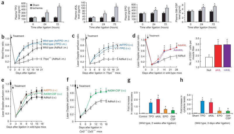
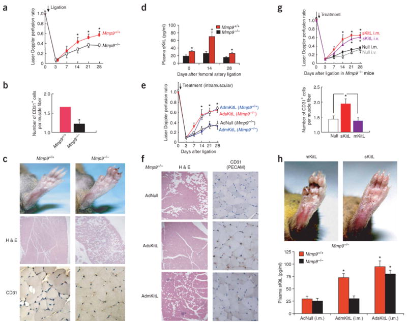
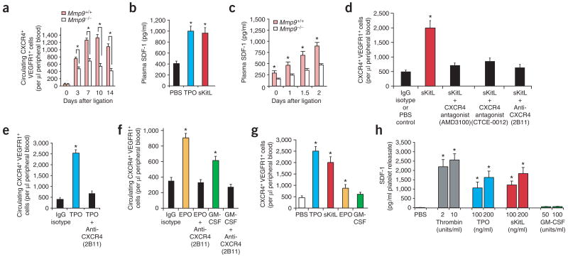
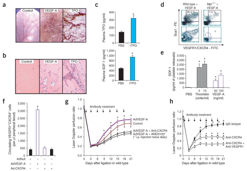
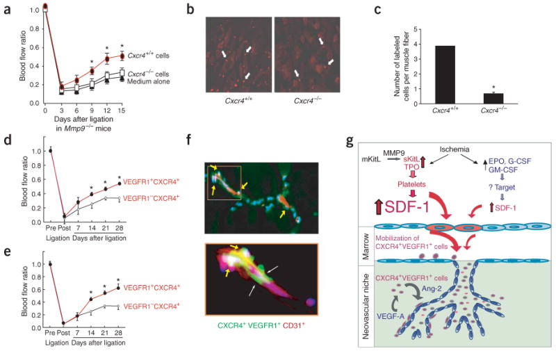
The mechanisms through which hematopoietic cytokines accelerate revascularization are unknown. Here, we show that the magnitude of cytokine-mediated release of SDF-1 from platelets and the recruitment of nonendothelial CXCR4+ VEGFR1+ hematopoietic progenitors, 'hemangiocytes,' constitute the major determinant of revascularization. Soluble Kit-ligand (sKitL), thrombopoietin (TPO, encoded by Thpo) and, to a lesser extent, erythropoietin (EPO) and granulocyte-macrophage colony-stimulating factor (GM-CSF) induced the release of SDF-1 from platelets, enhancing neovascularization through mobilization of CXCR4+ VEGFR1+ hemangiocytes. Although revascularization of ischemic hindlimbs was partially diminished in mice deficient in both GM-CSF and G-CSF (Csf2-/- Csf3-/-), profound impairment in neovascularization was detected in sKitL-deficient Mmp9-/- as well as thrombocytopenic Thpo-/- and TPO receptor-deficient (Mpl-/-) mice. SDF-1-mediated mobilization and incorporation of hemangiocytes into ischemic limbs were impaired in Thpo-/-, Mpl-/- and Mmp9-/- mice. Transplantation of CXCR4+ VEGFR1+ hemangiocytes into Mmp9-/- mice restored revascularization, whereas inhibition of CXCR4 abrogated cytokine- and VEGF-A-mediated mobilization of CXCR4+ VEGFR1+ cells and suppressed angiogenesis. In conclusion, hematopoietic cytokines, through graded deployment of SDF-1 from platelets, support mobilization and recruitment of CXCR4+ VEGFR1+ hemangiocytes, whereas VEGFR1 is essential for their angiogenic competency for augmenting revascularization. Delivery of SDF-1 may be effective in restoring angiogenesis in individuals with vasculopathies.


Conflict of interest statement

Competing Interests Statement: The authors declare competing financial interests (see the Nature Medicine website for details).

Figures

Figure 1
Figure 1
Hematopoietic cytokines promote ischemic revascularization. Hindlimb ischemia model was used to assess the role of hematopoietic cytokines in neoangiogenesis. (a) After unilateral femoral artery ligation, plasma TPO and sKitL were upregulated within 72 h. Plasma EPO and GM-CSF were increased to a lesser extent (n = 5 per group, *P < 0.05). (b) Intramuscular (i.m.) delivery of AdTPO (108 plaque-forming units (p.f.u.)) or rTPO (200 ng every 3 d) into the ischemic hindlimb accelerated blood perfusion compared to AdNull-treated controls. After ligation, hindlimb perfusion in untreated Thpo−/− mice was one-third that of wild-type mice (n = 5 per group, *P < 0.05). (c) Intravenous (i.v.) delivery of AdTPO or rTPO (200 ng every 3 d) to Thpo−/− mice induced a similar recovery of perfusion in ischemic hindlimb as did local delivery in wild-type mice (n = 5 per group, *P < 0.05). (d) Wild-type (Mmp9+/+) mice injected intravenously with AdNull, AdmKitL or AdsKitL (108 p.f.u. each) after ligation. Systemic delivery of sKitL or mKitL accelerated restoration of hindlimb blood flow (46% increase) compared to AdNull (left graph; n = 6 per group, *P < 0.05). Vessel density (CD31+ cells/muscle fiber) increased twofold in Mmp9+/+ mice injected with AdsKitL or AdmKitL compared to AdNull (right graph; n = 6 per group, *P < 0.05). (e) After ligation, wild-type mice were injected intravenously with 108 p.f.u. of AdNull, AdEPO or AdGM-CSF. AdEPO or AdGM-CSF accelerated blood flow by 30% 18 d after ligation compared to AdNull (n = 5, *P < 0.05). (f) Csf2−/−Csf3−/− mice were injected intravenously with 108 p.f.u. of AdNull or AdGM-CSF. Ischemic revascularization was slightly decreased in AdNull-treated Csf2−/−Csf3−/− mice, but accelerated in AdGM-CSF–treated Csf2−/−Csf3−/− mice (n = 5 per group, *P < 0.05). (g) At 2 weeks after femoral artery ligation, TPO and sKitL increased perfusion by 1.8- to 2-fold in ischemic hindlimb compared to controls. EPO and GM-CSF increased ischemic blood flow by 1.3-fold (n = 5 per group, *P < 0.05). (h) At 3 d after ligation, plasma TPO and sKitL increased 3.6-fold, whereas EPO and GM-CSF increased only 1.5-fold (n = 5 per group, *P < 0.05).
Figure 2
Figure 2
sKitL but not mKitL restores ischemic revascularization in angiogenesis-defective Mmp9−/− mice. (a) The Laser Doppler perfusion ratio was impaired in Mmp9−/− mice compared to Mmp9+/+ controls (n = 12 per group, *P < 0.05). (b) Vessel density (CD31+ cells/muscle fiber) decreased significantly in Mmp9−/− mice compared to Mmp9+/+ controls (n = 12 per group, *P < 0.005). (c) At 28 d after ligation, ischemic footpads showed profound swelling, ulceration and necrosis in Mmp9−/− compared to Mmp9+/+ mice (upper panels). H&E staining of Mmp9+/+ mice showed restoration of angiomyogenesis (middle panels; original magnification, ×100). Mmp9−/− mice showed loss of viable tissues followed by necrosis and adipose replacement. CD31 (PECAM-1) staining of hindlimb muscle sections showed decreased vessel density in Mmp9−/− mice (bottom panels; original magnification, ×400). (d) Hindlimb ischemia induced elevation of sKitL in plasma in Mmp9+/+ but not in Mmp9−/− (n = 6 per group, *P < 0.05). (e) Adenoviral delivery of mKitL (AdmKitL, single dose of 108 p.f.u.) augmented blood flow in Mmp9+/+ but not Mmp9−/− mice (n = 5 per group; P < 0.05). Adenoviral delivery of sKitL (AdsKitL, single dose of 108 p.f.u.) accelerated ischemic revascularization in Mmp9−/− mice. Control mice were injected with 108 p.f.u. of AdNull. (f) H&E (left panels; original magnification, ×100) and CD31 (right panels; original magnification, ×400) staining of lower limb tissue of Mmp9−/− mice injected with AdNull (upper panels), AdsKitL (middle panels) and AdmKitL (bottom panels) 28 d after ligation. Loss of viable tissues with adipose replacement and muscle necrosis was seen after treatment with AdNull or AdmKitL, but not in the AdsKitL-treated group, in which there was restoration of angiogenesis. (g) Intramuscular (i.m.) or intravenous (i.v.) delivery of AdsKitL (single dose of 108 p.f.u.) into Mmp9−/− mice accelerated revascularization comparably in ischemic hindlimbs (upper graph). Vessel density increased in AdsKitL-treated Mmp9−/− mice compared to AdNull- or AdmKitL-treated Mmp9−/− mice (lower graph) (n = 6 per group, *P < 0.05). (h) Reversal of ischemia-induced vasculopathy was observed in Mmp9−/− mice treated with sKitL, but not with mKitL (upper panels). Intramuscular AdmKitL (108 p.f.u.) injected into ischemic hindlimb increased sKitL plasma levels in Mmp9+/+ but not in Mmp9−/− mice 3 d after ligation (n = 4 per group, *P < 0.05). AdsKitL (108 p.f.u.) increased plasma sKitL threefold in Mmp9+/+ and Mmp9−/− mice compared to AdNull (n = 4 per group, *P < 0.05).
Figure 3
Figure 3
Soluble hematopoietic cytokines induce release of SDF-1 from platelets and recruitment of CXCR4+VEGFR1+ cells, accelerating ischemic revascularization. (a) Mobilized CXCR4+VEGFR1+ cells isolated from peripheral blood of Mmp9+/+ and Mmp9−/− mice after induction of hindlimb ischemia were analyzed by two-color flow cytometry. Mobilization of CXCR4+VEGFR1+ cells was impaired in Mmp9−/− compared to wild-type mice (n = 4 per group, *P < 0.05). (b) Plasma SDF-1 levels were induced 2.5-fold 3 d after intravenous injection of recombinant TPO or sKitL compared to PBS-treated controls (n = 4 per group, *P < 0.05). (c) Response of plasma SDF-1 levels to hindlimb ischemia over time. In Mmp9+/+ mice, the peak plasma SDF-1 after ligation was 1.7- to 1.9-fold higher than in Mmp9−/− mice (n = 4 per group, *P < 0.05). (d) Circulating CXCR4+VEGFR1+ cells were measured 3 d after intravenous injection of recombinant sKitL (100 ng) in wild-type mice with or without CXCR4 blockade (CXCR4-specific monoclonal antibody, nonpeptide antagonist AMD3100 or peptidomimetic antagonist CTCE-0012). sKitL induced a fourfold increase in the number of mobilized CXCR4+VEGFR1+ cells compared to PBS- or IgG isotype–treated controls. Administration of sKitL with CXCR4-specific monoclonal antibody, AMD3100 or CTCE-0012 suppressed mobilization of CXCR4+VEGFR1+ cells by threefold compared to sKitL alone (n = 4 per group, *P < 0.05). (e) Wild-type mice were injected intravenously with recombinant TPO (100 ng) or a combination of TPO and CXCR4-specific monoclonal antibody (20 μg/mouse, clone 2B11). After 3 d, CXCR4-specific monoclonal antibody decreased TPO-induced mobilization of CXCR4+VEGFR1+ cells by 83% (n = 4 per group, *P < 0.05). (f) Wild-type mice were injected intravenously with recombinant EPO (100 units) or GM-CSF (100 ng) with or without administration of CXCR4-specific monoclonal antibody (20 μg; clone 2B11) along with PBS- and IgG-treated mice as controls. Neutralizing CXCR4-specific monoclonal antibody completely blocked EPO- and GM-CSF–induced mobilization of CXCR4+VEGFR1+ cells as compared to cytokine-treated groups (n = 4 per group, *P < 0.05). (g) TPO or sKitL conferred a 2.5-fold higher mobilization activity of CXCR4+VEGFR1+ cells than either EPO or GM-CSF (n = 4 per group; P < 0.05). (h) Platelets from wild-type mice were isolated from peripheral blood, stimulated with thrombin or cytokines as indicated, and SDF-1 levels in platelet releasate were measured by ELISA. TPO and sKitL induced a similar dose-dependent release of SDF-1 from platelets, whereas GM-CSF had a minimal effect. Data represent SDF-1 detected in 1 ml platelet releasate obtained from 1 × 10⁷ platelets (n = 3 per group, *P < 0.05).
Figure 4
Figure 4
SDF-1 reverses the neoangiogenesis defects in Mmp9−/− and Thpo−/− mice in a dose-dependent manner. (a) Hindlimb ischemia after femoral artery ligation was performed in Mpl−/−, Thpo−/− or wild-type mice. Plasma levels of SDF-1 were measured at indicated time points by ELISA. Plasma SDF-1 levels were persistently higher (3.2-fold at 48 h) in wild-type mice as compared to Mpl−/− or Thpo−/− mice during the first 72 h after femoral artery ligation (n = 6 per group, *P < 0.05). (b) The number of circulating CXCR4+VEGFR1+ cells in wild-type mice was 5.5-fold and 5-fold higher than that of Mpl−/− mice 3 and 8 d after femoral artery ligation, respectively (n = 4 per group, *P < 0.05). (c) Administration of AdSDF-1 (low dose, 108 p.f.u. intravenously) to Thpo−/− mice after femoral artery ligation restored perfusion in the ischemic hindlimb by twofold as compared to the AdNull-treated controls. A higher dose (5 × 10⁸ p.f.u. intravenously) improved ischemic perfusion by 2.8-fold compared to AdNull-treated controls (n = 4 per group, *P < 0.05). (d) There was increased vessel density (CD31+ vessels/gastrocnemius or adductor muscle fiber, n = 4 per group) and less muscle necrosis in SDF-1–treated Thpo−/− mice compared to AdNull-treated controls (n = 4 per group, data represent average from five separate high-power fields (HPFs)). Original magnification, ×400. (e) AdSDF-1 injection (low dose, 108 p.f.u. intravenously) in Mmp9−/− mice restored perfusion in the ischemic hindlimb by 1.95-fold compared to AdNull-treated controls. A higher dose (5 × 10⁸ p.f.u. intravenously) improved ischemic perfusion further by 2.5-fold compared to AdNull-treated controls (n = 4 per group, *P < 0.05).
Figure 5
Figure 5
Inhibition of CXCR4 and, to a lesser degree, of VEGFR1 blocks VEGF-A–induced mobilization of hemangiocytes and ischemic revascularization. (a) In the mouse ear pinna angiogenesis model, subcutaneous recombinant TPO (200 ng every 3 d) induced substantial formation and sprouting of neovessels similar to subcutaneous treatment with VEGF-A but with less edema formation (original magnification, ×10). PBS-injected ear pinna was used as a control. (b) Matrigel plugs with recombinant TPO showed substantial formation and branching of vascular channels comparable to VEGF-A (original magnification, ×400). Matrigel plugs loaded with PBS were used as controls. (c) Subcutaneous injection of low-dose recombinant TPO into ear pinna led to a twofold increase in plasma TPO and SDF-1 levels compared to PBS-treated controls (n = 4 per group, *P < 0.05). (d) Three days after intravenous AdVEGF-A (single dose of 108 p.f.u.), a fivefold decrease in circulating CXCR4+VEGFR1+ cells also expressing Sca1 in Mpl−/− versus wild-type mice was detected by flow cytometry. Upper, cytometric distribution of Sca1+VEGFR1+CXCR4+ cells. Lower, corresponding contour mapping of fluorescent signaling intensity. (e) Thrombin and VEGF-A induced dose-dependent release of SDF-1 from wild-type platelets in vitro. Data represent amounts of SDF-1 in 1 ml of platelet releasate obtained from 1 × 10⁷ platelets (n = 3 per group, *P < 0.05). (f) Monoclonal antibody to CXCR4 (clone 2B11, 20 μg intravenously) blocked VEGF-A–induced mobilization of CXCR4+VEGFR1+ cells by 80% in wild-type compared to AdNull-treated mice (n = 4 per group, *P < 0.05). (g) Wild-type C57Bl/6 mice after femoral artery ligation were treated intravenously with AdVEGF-A (single dose of 108 p.f.u.) alone or in combination with either CXCR4-specific monoclonal antibody (clone 2B11, 20 μg intravenously every 3 d) or AMD3100 (1.25 mg/kg intraperitoneally twice daily). CXCR4 blockade with either CXCR4-specific monoclonal antibody (2B11) or AMD3100 aborted enhanced vascular recovery conferred by VEGF-A. Concurrent treatment of AdVEGF-A with either 2B11 or AMD3100 reduced perfusion restoration by 36% and 42.3%, respectively, compared to treatment with AdVEGF-A alone (n = 4 per group, *P < 0.05). Control mice were treated with IgG and AdNull. (h) CXCR4-specific monoclonal antibody (20 μg intravenously every 3 d) reduced the restoration of perfusion by 50% in wild-type mice (n = 4 per group, *P < 0.05), whereas coadministration of CXCR4-specific (20 μg intravenously every 3 d) and VEGFR1-specific (400 μg intraperitoneally every 3 d) antibodies further diminished recovery of perfusion by 63% compared to IgG-isotype controls.
Figure 6
Figure 6
Retention and incorporation of CXCR4+VEGFR1+ cells, but not CXCR4 or VEGFR1 cells, restore functional neoangiogenesis in Mmp9−/− mice. (a) Hematopoietic cells from liver of Cxcr4+/+ or Cxcr4−/− embryos (E13.5) were expanded ex vivo with KitL, fluorescently labeled with PKH26 and subsequently transplanted locally into the ischemic hindlimb of Mmp9−/− mice 24 h after femoral artery ligation. Implantation of Cxcr4+/+ cells (5 × 10⁵, filled circles), but not Cxcr4−/− cells (5 × 10⁵, open squares) or vehicle medium (filled triangles) in ischemic hindlimb of Mmp9−/− mice conferred a 33% increase in perfusion recovery (n = 3 per group, *P < 0.05). (b) PKH26 fluorescently labeled Cxcr4+/+ and Cxcr4−/− cells (white arrows) localized within ischemic limbs 15 d after femoral artery ligation. Original magnification, ×200. (c) The retention of hematopoietic Cxcr4+/+ cells within the ischemic hindlimb in Mmp9−/− mice was fourfold higher than that of Cxcr4−/− cells (n = 3 per group, *P < 0.05). (d,e) Delivery of CXCR4+VEGFR1+ cells (3 × 10⁵), but not CXCR4+VEGFR1 cells (3 × 10⁵), either intramuscularly (d) or intravenously (e) into ischemic hindlimb, significantly improved the rate and extent of revascularization in Mmp9−/− mice (n = 6 per group; P < 0.05). (f) Donor-derived CXCR4+VEGFR1+ cells (green, labeled with PKH2GL) were coimmunostained with CD31 (PECAM-1)-PE (red) and counterstained with DAPI (blue). CXCR4+VEGFR1+ cells were predominantly localized to the perivascular and interstitial region on the abluminal aspect of the collateral CD31+ neovessels 7 d after transplantation (f, arrows in upper panels; original magnification, ×100). The same pattern was observed in all tested ischemic hindlimbs (n = 6 per group). Higher magnification of the indicated area in the upper panel is shown in the lower panel (original magnification, ×400). (g) Schematic model: SDF1-CXCR4 as the molecular hub for hematopoietic cytokine-induced neovascularization.

References

    1. Rafii S, Lyden D. Therapeutic stem and progenitor cell transplantation for organ vascularization and regeneration. Nat Med. 2003;9:702–712. - PubMed
    1. Coussens LM, Werb Z. Inflammation and cancer. Nature. 2002;420:860–867. - PMC - PubMed
    1. Coussens LM, Tinkle CL, Hanahan D, Werb Z. MMP-9 supplied by bone marrow-derived cells contributes to skin carcinogenesis. Cell. 2000;103:481–490. - PMC - PubMed
    1. Takakura N, et al. A role for hematopoietic stem cells in promoting angiogenesis. Cell. 2000;102:199–209. - PubMed
    1. Aicher A, et al. Essential role of endothelial nitric oxide synthase for mobilization of stem and progenitor cells. Nat Med. 2003;9:1370–1376. - PubMed

Publication types

MeSH terms