Contrast sensitivity, first-order motion and Initial ocular following in demyelinating optic neuropathy
- PMID: 16649097
- PMCID: PMC2408647
- DOI: 10.1007/s00415-006-0200-5
Contrast sensitivity, first-order motion and Initial ocular following in demyelinating optic neuropathy
Abstract
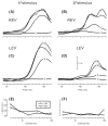
The ocular following response (OFR) is a measure of motion vision elicited at ultra-short latencies by sudden movement of a large visual stimulus. We compared the OFR to vertical sinusoidal gratings (spatial frequency 0.153 cycles/ degrees or 0.458 cycles/ degrees) of each eye in a subject with evidence of left optic nerve demyelination due to multiple sclerosis (MS). The subject showed substantial differences in vision measured with stationary low-contrast Sloan letters (20/63 OD and 20/200 OS at 2.5% contrast) and the Lanthony Desaturated 15-hue color test (Color Confusion Index 1.11 OD and 2.14 OS). Compared with controls, all of the subject's OFR to increasing contrast showed a higher threshold. The OFR of each of the subject's eyes were similar for the 0.153 cycles/ degrees stimulus, and psychophysical measurements of his ability to detect these moving gratings were also similar for each eye. However, with the 0.458 cycles/ degrees stimulus, the subject's OFR was asymmetric and the affected eye showed decreased responses (smaller slope constant as estimated by the Naka-Rushton equation). These results suggest that, in this case, optic neuritis caused a selective deficit that affected parvocellular pathways mediating higher spatial frequencies, lower-contrast, and color vision, but spared the field-holding mechanism underlying the OFR to lower spatial frequencies. The OFR may provide a useful method to study motion vision in individuals with disorders affecting anterior visual pathways.
Figures


References
-
- Abadi RV, Gowen E. Characteristics of saccadic intrusions. Vision Res. 2004;44:2675–2690. - PubMed
-
- Albrecht DG, Geisler WS, Frazor RA, Crane AM. Visual cortex neurons of monkeys and cats: temporal dynamics of the contrast response function. J Neurophysiol. 2002;88:888–913. - PubMed
-
- Albrecht DG, Hamilton DB. Striate cortex of monkey and cat: contrast response function. J Neurophysiol. 1982;48:217–237. - PubMed
-
- Asselman P, Chadwick DW, Marsden CD. Visual evoked responses in the diagnosis and management of patients suspected of multiple sclerosis. Brain. 1975;98:261–282. - PubMed
-
- Balcer LJ, Baier ML, Cohen JA, et al. Contrast letter acuity as a visual component for the Multiple Sclerosis Functional Composite. Neurology. 2003;61:1367–1373. - PubMed
Publication types
MeSH terms
Grants and funding
LinkOut - more resources
Full Text Sources
Medical

