

Methamphetamine-induced cell death: selective vulnerability in neuronal subpopulations of the striatum in mice
- PMID: 16650608
- PMCID: PMC2882192
- DOI: 10.1016/j.neuroscience.2006.02.055
Methamphetamine-induced cell death: selective vulnerability in neuronal subpopulations of the striatum in mice
Abstract
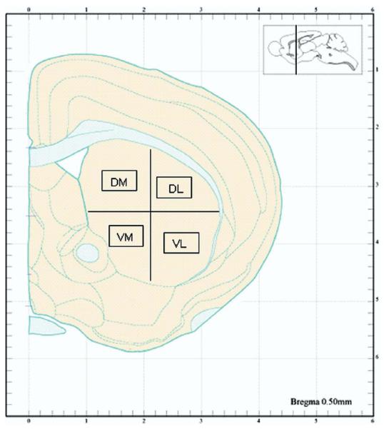
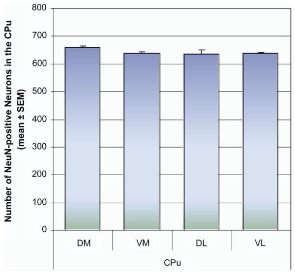
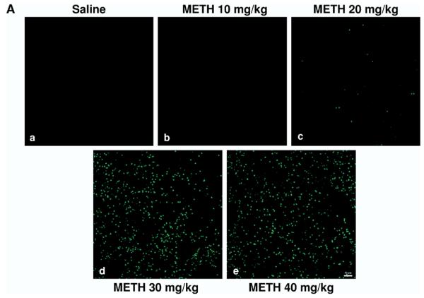
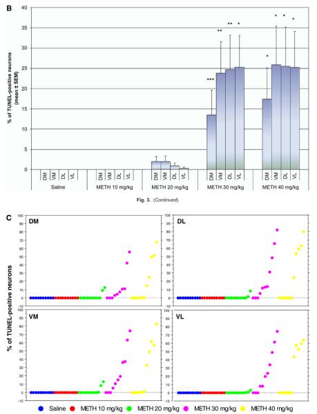
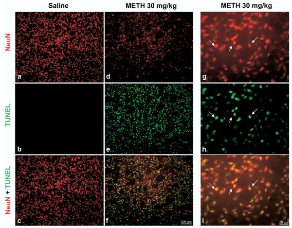
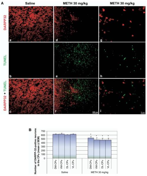
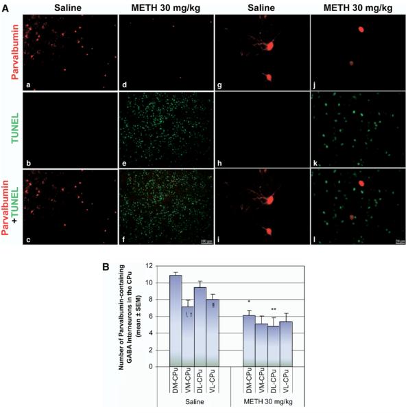
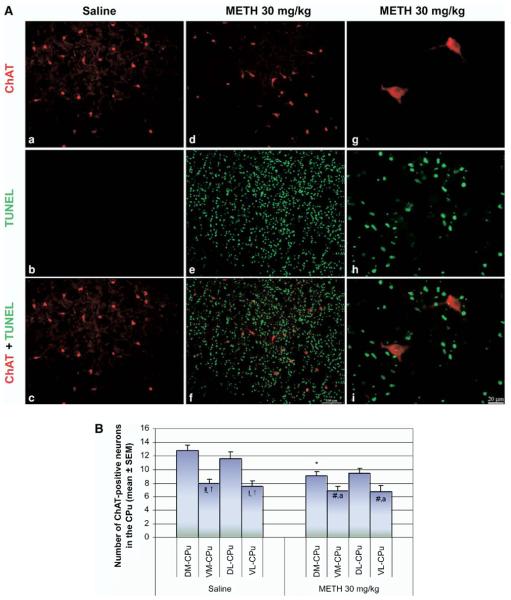
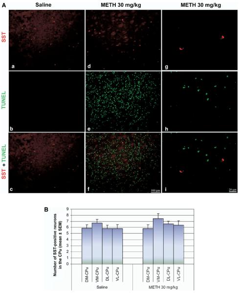
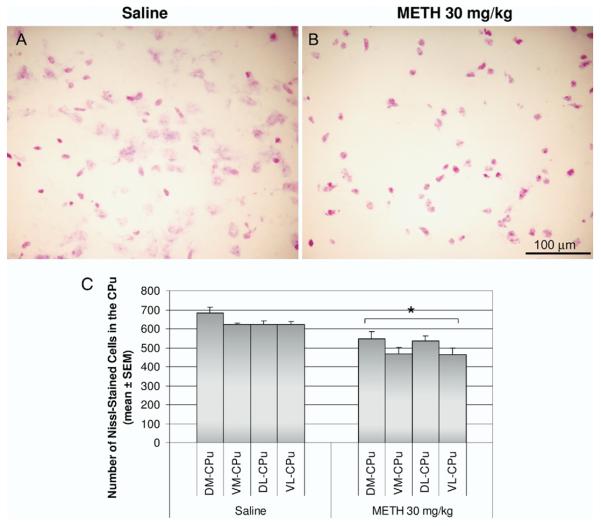
Methamphetamine (METH) is an illicit and potent psychostimulant, which acts as an indirect dopamine agonist. In the striatum, METH has been shown to cause long lasting neurotoxic damage to dopaminergic nerve terminals and recently, the degeneration and death of striatal cells. The present study was undertaken to identify the type of striatal neurons that undergo apoptosis after METH. Male mice received a single high dose of METH (30 mg/kg, i.p.) and were killed 24 h later. To demonstrate that METH induces apoptosis in neurons, we combined terminal deoxynucleotidyl transferase-mediated dUTP nick end labeling (TUNEL) staining with immunohistofluorescence for the neuronal marker neuron-specific nuclear protein (NeuN). Staining for TUNEL and NeuN was colocalized throughout the striatum. METH induces apoptosis in approximately 25% of striatal neurons. Cell counts of TUNEL-positive neurons in the dorsomedial, ventromedial, dorsolateral and ventrolateral quadrants of the striatum did not reveal anatomical preference. The type of striatal neuron undergoing cell death was determined by combining TUNEL with immunohistofluorescence for selective markers of striatal neurons: dopamine- and cAMP-regulated phosphoprotein, of apparent Mr 32,000, parvalbumin, choline acetyltransferase and somatostatin (SST). METH induces apoptosis in approximately 21% of dopamine- and cAMP-regulated phosphoprotein, of apparent Mr 32,000-positive neurons (projection neurons), 45% of GABA-parvalbumin-positive neurons in the dorsal striatum, and 29% of cholinergic neurons in the dorsal-medial striatum. In contrast, the SST-positive interneurons were refractory to METH-induced apoptosis. Finally, the amount of cell loss determined with Nissl staining correlated with the amount of TUNEL staining in the striatum of METH-treated animals. In conclusion, some of the striatal projection neurons and the GABA-parvalbumin and cholinergic interneurons were removed by apoptosis in the aftermath of METH. This imbalance in the populations of striatal neurons may lead to functional abnormalities in the output and processing of neural information in this part of the brain.
Figures
References
-
- Albin RL, Reiner A, Anderson KD, Penney JB, Young AB. Striatal and nigral neuron subpopulations in rigid Huntington's disease: implications for the functional anatomy of chorea and rigidity-akinesia. Ann Neurol. 1990;27:357–365. - PubMed
-
- Albin RL, Reiner A, Anderson KD, Dure LS, Handelin B, Balfour R, Whetsell WO, Jr, Penney JB, Young AB. Preferential loss of striato-external pallidal projection neurons in presymptomatic Huntington's disease. Ann Neurol. 1992;31:425–430. - PubMed
-
- Alexander GE, DeLong MR, Strick PL. Parallel organization of functionally segregated circuits linking basal ganglia and cortex. Annu Rev Neurosci. 1986;9:357–381. - PubMed
-
- Beal MF. Mechanisms of excitotoxicity in neurologic diseases. FASEB J. 1992;6:3338–3344. - PubMed
Publication types
MeSH terms
Substances
Grants and funding
LinkOut - more resources
Full Text Sources
Medical
