. 2006 Jun 16;359(4):1045-58.
doi: 10.1016/j.jmb.2006.03.052. Epub 2006 Apr 6.

Evidence that membrane insertion of the cytosolic domain of Bcl-xL is governed by an electrostatic mechanism

Affiliations

Evidence that membrane insertion of the cytosolic domain of Bcl-xL is governed by an electrostatic mechanism

Guruvasuthevan R Thuduppathy et al. J Mol Biol. .

Abstract

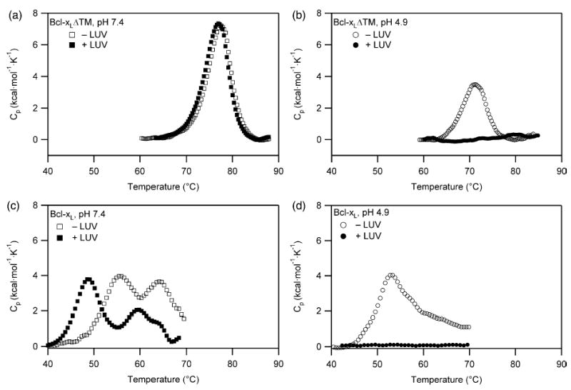
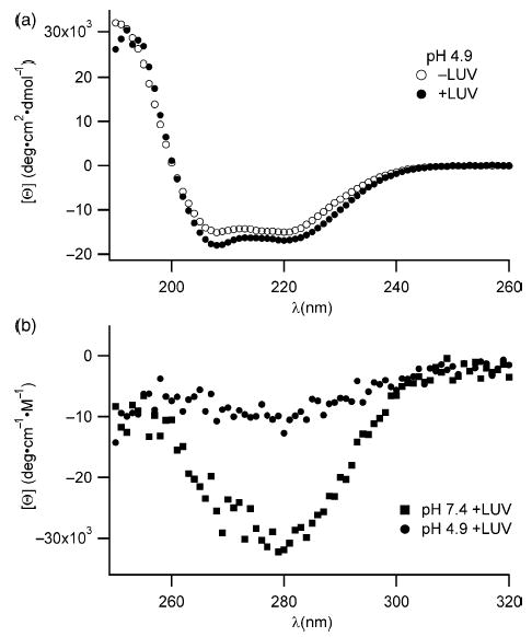
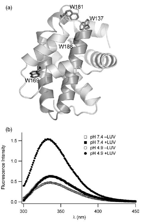
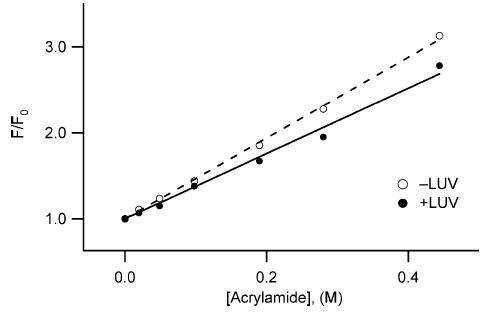
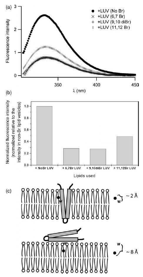
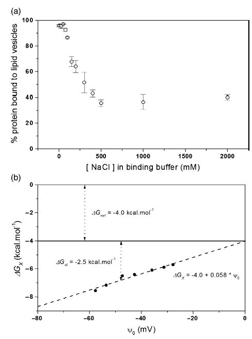
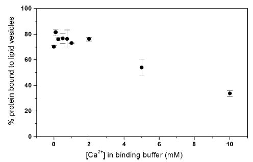
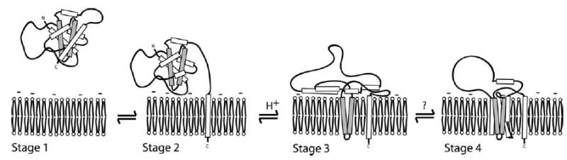
Signals from different cellular networks are integrated at the mitochondria in the regulation of apoptosis. This integration is controlled by the Bcl-2 proteins, many of which change localization from the cytosol to the mitochondrial outer membrane in this regulation. For Bcl-xL, this change in localization reflects the ability to undergo a conformational change from a solution to integral membrane conformation. To characterize this conformational change, structural and thermodynamic measurements were performed in the absence and presence of lipid vesicles with Bcl-xL. A pH-dependent model is proposed for the solution to membrane conformational change that consists of three stable conformations: a solution conformation, a conformation similar to the solution conformation but anchored to the membrane by its C-terminal transmembrane domain, and a membrane conformation that is fully associated with the membrane. This model predicts that the solution to membrane conformational change is independent of the C-terminal transmembrane domain, which is experimentally demonstrated. The conformational change is associated with changes in secondary and, especially, tertiary structure of the protein, as measured by far and near-UV circular dichroism spectroscopy, respectively. Membrane insertion was distinguished from peripheral association with the membrane by quenching of intrinsic tryptophan fluorescence by acrylamide and brominated lipids. For the cytosolic domain, the free energy of insertion (DeltaG degrees x) into lipid vesicles was determined to be -6.5 kcal mol(-1) at pH 4.9 by vesicle binding experiments. To test whether electrostatic interactions were significant to this process, the salt dependence of this conformational change was measured and analyzed in terms of Gouy-Chapman theory to estimate an electrostatic contribution of DeltaG degrees el approximately -2.5 kcal mol(-1) and a non-electrostatic contribution of DeltaG degrees nel approximately -4.0 kcal mol(-1) to the free energy of insertion, DeltaG degrees x. Calcium, which blocks ion channel activity of Bcl-xL, did not affect the solution to membrane conformational change more than predicted by these electrostatic considerations. The lipid cardiolipin, that is enriched at mitochondrial contact sites and reported to be important for the localization of Bcl-2 proteins, did not affect the solution to membrane conformational change of the cytosolic domain, suggesting that this lipid is not involved in the localization of Bcl-xL in vivo. Collectively, these data suggest the solution to membrane conformational change is controlled by an electrostatic mechanism. Given the distinct biological activities of these conformations, the possibility that this conformational change might be a regulatory checkpoint for apoptosis is discussed.


Figures

Figure 1
Figure 1
The Bcl-xL solution to membrane conformational change is pH-dependent involving at least three distinct conformations. (a) For Bcl-xLΔTM at pH 7.4, no association between protein and lipid vesicles is demonstrated by the absence of a change in the thermal unfolding transition of the protein upon addition of lipid vesicles in a 1:200 ratio. (b) By contrast, at pH 4.9 the thermal unfolding transition of Bcl-xLΔTM disappears suggesting a strong association with lipid vesicles upon addition. (c) For Bcl-xL at pH 7.4 in the absence of lipid vesicles, the C-terminal transmembrane domain of Bcl-xL unfolds first followed by the global unfolding of the protein. In the presence of lipid vesicles, an additional thermal transition is observed that is attributed to the unfolding of the cytosolic domain that is anchored to the lipid vesicle by its C-terminal transmembrane domain (d) For Bcl-xL at pH 4.9 in the presence of lipid vesicles, the thermal unfolding transition of the protein disappears, suggesting a strong association with lipid vesicles upon addition similar to Bcl-xLΔTM.
Figure 2
Figure 2
Changes in the secondary and tertiary structure of Bcl-xLΔTM upon association with lipid vesicles suggest a gross conformational change. (a) The far-UV CD signal of the protein is significantly decreased only in the presence of lipid vesicles at pH 4.9 and not at pH 7.4, suggesting an increase in α-helical structure upon association with lipid vesicles. (b) The near-UV CD signal arising from the aromatic residues disappears only in the presence of lipid vesicles at pH 4.9 and not at pH 7.4, suggesting a loss in side-chain packing around aromatic residues in the hydrophobic core of the protein upon association with lipid vesicles.
Figure 3
Figure 3
A pH and lipid vesicle dependent conformation change of Bcl-xLΔTM is confirmed by tryptophan fluorescence emission spectra. (a) The solution conformation of the cytosolic domain of Bcl-xL reveals a hydrophobic α-helical hairpin (shaded) surrounded by a sheath of amphiphilic α-helices (1MAZ.pdb). Six tryptophan residues served as spectroscopic probes of membrane insertion; four are highlighted. A large unstructured loop (residues 26–83) containing two tryptophan residues is omitted for clarity but this loop is present in our constructs. (b) At pH 7.4, tryptophan fluorescence emission does not change upon addition of lipid vesicles consistent with no association between protein and lipid vesicles. However, at pH 4.9, tryptophan fluorescence emission dramatic increases with a blue shift in λmax upon association with lipid vesicles, suggesting a net shift in the tryptophan residues to a more non-polar environment upon association with lipid vesicles at pH 4.9.
Figure 4
Figure 4
Solvent accessibility of tryptophan residues is reduced in the membrane conformation of Bcl-xLΔTM. Intrinsic tryptophan fluorescence quenching of Bcl-xLΔTM, in solution and in the presence of lipid vesicles at pH 5.0, was measured as a function of increasing concentrations of acrylamide, a membrane impermeable quencher. The data fit well to the Stern–Volmer relation with a Stern–Volmer quenching constant, KSV, estimated from the slope to be 4.73 M−1 for Bcl-xLΔTM in solution (○), and 3.90 M−1 in the presence of lipid vesicles (•). The correlation coefficients were 0.99 for the linear fits. The data suggest a net increase in protection of tryptophan residues from solvent exposure upon interaction with lipid vesicles.
Figure 5
Figure 5
The membrane conformation of Bcl-xLΔTM is deeply inserted into the membrane bilayer. (a) Fluorescence emission spectra were collected in the presence of lipid vesicles brominated at different positions along the fatty acyl chain. Emission spectra were collected at pH 5.0 in the presence of 150 mM NaCl with excitation at 295 nm. (b) Data from fluorescence quenching experiments normalized to the λmax of the fluorescence emission from protein in non-brominated lipid vesicles. (c) The quenching of intrinsic tryptophan fluorescence by phospholipids brominated at different points along the fatty acyl chain is distance dependent. Such data can distinguish between the insertion of the hydrophobic helical hairpin into the bilayer and the peripheral association of the hairpin with the membrane surface using the parallax method., Our data are consistent with insertion.
Figure 6
Figure 6
Anionic lipid dependence suggests an electrostatic component to the solution to membrane conformational change of Bcl-xLΔTM. A strong association of protein with lipid vesicles is only observed in the presence of lipid vesicles comprised of anionic lipids, because the thermal unfolding transition of the protein disappears only in the presence of lipid vesicles containing the negatively charged DOPG and not in the presence of lipid vesicles containing 100% of the net neutrally charged DOPC vesicles.
Figure 7
Figure 7
Electrostatic contribution of membrane insertion is confirmed by the salt dependence of Bcl-xLΔTM association with lipid vesicles at pH 5.0. (a) Protein binding to lipid vesicles as a function of increasing concentration of NaCl was measured by a sedimentation assay. (b) The free energy of membrane insertion, ΔGox, arises from electrostatic and non-electrostatic contributions as discussed in the text. The y-intercept estimates the non-electrostatic contribution, ΔGoel, of ~ −4.0 kcal mol−1 and is independent of pH. The slope indicates the effective charge on the protein that is modulated by electrostatic interactions, zeff, of ~2.3. Under conditions with buffer and 150mM NaCl, I=0.2, the free energy contribution from electrostatics, ΔGoel is about −2.5 kcal mol−1.
Figure 8
Figure 8
Calcium does not specifically influence the interaction of Bcl-xLΔTM with lipid vesicles. Protein binding to lipid vesicles at calcium concentrations ranging from 0 mM to 10 mM was not affected more than anticipated from electrostatic considerations, suggesting no specific role for calcium in the membrane insertion process.
Figure 9
Figure 9
A hypothetical model for the solution to membrane conformational change of Bcl-xL. Bcl-xL is localized to the cytosol and organellar membranes and translocates to the mitochondria during apoptosis. Protein localized in the cytosol presumably has the C-terminal transmembrane domain sequestered into the BH3 binding pocket of the protein in a conformation similar to Bax (stage 1). Protein localized to the membrane is anchored by the C-terminal transmembrane domain with the cytosolic domain of Bcl-xL poised to receive signals (stage 2). Insertion of the cytosolic domain via the hydrophobic helical hairpin into the membrane bilayer interior is caused by acidification or some other signal (stage 3). In the last step, a fully inserted membrane conformation is assembled by either oligomer formation via the helical hydrophobic hairpin (not shown), or by full insertion of the amphiphilic α-helices to span the membrane bilayer with their hydrophilic faces lining the lumen of the ion channel (stage 4). The results presented here support the first two stages of the model and that the transition to stage 3 is controlled by an electrostatic mechanism. Our data cannot distinguish between stages 3 and 4.

Similar articles

Cited by

References

    1. Hengartner MO. The biochemistry of apoptosis. Nature. 2000;407:770–776. - PubMed
    1. Kuwana T, Newmeyer DD. Bcl-2-family proteins and the role of mitochondria in apoptosis. Curr Opin Cell Biol. 2003;15:691–699. - PubMed
    1. Green DR, Reed JC. Mitochondria and apoptosis. Science. 1998;281:1309–1312. - PubMed
    1. Adams JM, Cory S. The Bcl-2 protein family: arbiters of cell survival. Science. 1998;281:1322–1326. - PubMed
    1. Gross A, McDonnell JM, Korsmeyer SJ. BCL-2 family members and the mitochondria in apoptosis. Genes Dev. 1999;13:1899–1911. - PubMed

Publication types