. 2006 May 9;103(19):7444-9.
doi: 10.1073/pnas.0602402103. Epub 2006 May 1.

Loss of p53 impedes the antileukemic response to BCR-ABL inhibition

Affiliations

Loss of p53 impedes the antileukemic response to BCR-ABL inhibition

Hans-Guido Wendel et al. Proc Natl Acad Sci U S A. .

Abstract

Targeted cancer therapies exploit the continued dependence of cancer cells on oncogenic mutations. Such agents can have remarkable activity against some cancers, although antitumor responses are often heterogeneous, and resistance remains a clinical problem. To gain insight into factors that influence the action of a prototypical targeted drug, we studied the action of imatinib (STI-571, Gleevec) against murine cells and leukemias expressing BCR-ABL, an imatinib target and the initiating oncogene for human chronic myelogenous leukemia (CML). We show that the tumor suppressor p53 is selectively activated by imatinib in BCR-ABL-expressing cells as a result of BCR-ABL kinase inhibition. Inactivation of p53, which can accompany disease progression in human CML, impedes the response to imatinib in vitro and in vivo without preventing BCR-ABL kinase inhibition. Concordantly, p53 mutations are associated with progression to imatinib resistance in some human CMLs. Our results identify p53 as a determinant of the response to oncogene inhibition and suggest one way in which resistance to targeted therapy can emerge during the course of tumor evolution.


Conflict of interest statement

Conflict of interest statement: No conflicts declared.

Figures

Fig. 1.
Fig. 1.
p53 modulates sensitivity to imatinib in vitro. (a) Immunoblot analysis of lysates prepared from Ba/F3 cells stably transduced with empty vector, BCR-ABL (p210), or mutant BCR-ABL (p210/T315I) treated with imatinib for 8 h as indicated were probed for p53, phosphorylated; and total levels of p56Dok-2 protein (P-p56 and p56, respectively), and tubulin (tub) as loading control. (b) Lysates of imatinib-treated Ba/F3/p210 cells were probed with antibodies against p53, total and phosphorylated (Ser-473) Akt (Akt and P-Akt), and total and phosphorylated (Ser-166) Mdm2 (Mdm2 and P-Mdm2), with tub as loading control. (c) Immunoblot of Ba/F3/p210 cells lysates expressing either an RNAi vector targeting p53 (p53D) or control vector (Vector) treated as indicated and probed for p53, phosphorylated (P-p56) and total (p56) p56 protein, and tub. (d) In vitro competition assay. Populations of Ba/F3 cells stably expressing either BCR-ABL (p210; Upper) or the T315I mutant (p210/T315I; Lower) were partially transduced with an RNAi vector against p53 (p53D) and propagated in the presence or absence of 1 μM imatinib for 1 week and then subjected to flow cytometry to determine the fraction of cells containing the RNAi vector (high GFP expression).
Fig. 2.
Fig. 2.
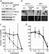
BCR-ABL sensitization of primary HSCs to imatinib depends on p53. (a) Immunoblot analysis of p53+/+ HSCs expressing BCR-ABL before therapy (Untr) or 8 h after treatment with different doses of imatinib, as indicated. Lysates were immunoblotted for p53, total and phosphorylated (Ser-473) Akt (Akt and P-Akt), phosphorylated (Ser-166) Mdm2 (P-Mdm2), and tubulin (tub). (b) Representative microphotographs of colonies formed by p53+/+ or p53−/− HSCs in methylcellulose untreated or treated with 1 μM imatinib (Lower) fluorescence detection of GFP expression in BCR-ABL-transformed colonies. (c and d) Results of methylcellulose colony-formation assays, p53+/+ (circles) and p53−/− (squares) HSC expressing BCR-ABL (c) or control (d) were incubated with imatinib at the indicated concentrations and colony-forming units counted after 10 days (mean ± SD, n = 7; P = 0.016 and P = 0.4 for IC50 (p53+/+ vs. p53−/−) in c and d, respectively).
Fig. 3.
Fig. 3.
BCR-ABL induced leukemia in vivo. (a) Schematic of the generation of mice harboring leukemias of defined p53 status; MIG-p210: MSCV-p210-IRES-GFP. (b) Representative microphotographs of blood smears illustrating the resulting pathologies. Most animals develop a CML-like myeloproliferative disease (Left), whereas some have acute leukemias (Right). (c) Latency to leukemia onset after transplantation (day 0) of BCR-ABL-transduced HSCs of these genotypes: p53+/+ (black, n = 28), p53+/− (blue, n = 57), and p53−/− (red, n = 30); P = 0.0008 (p53+/+ vs. p53+/−); P = 0.0005 (p53+/+ vs. p53−/−); P = 0.18 (p53+/− vs. p53−/−). (Inset) PCR to detect loss of heterozygosity in the p53 locus. N, the knockout allele; W, the wild-type allele. Lane 1, p53+/+ control; lane 2, p53+/− control; lane 3–6, CML samples derived from p53+/− HSCs.
Fig. 4.
Fig. 4.
p53 and targeted therapy in murine CML. (a) Representative immunohistochemical stains to assess p53 expression in the spleens of healthy vs. leukemic mice treated as indicated. (b) Fluorescence imaging of a cohort of p53+/+ and p53−/− leukemia-bearing mice killed at various times after imatinib treatment. Representative examples are shown. (c) Kaplan–Meier plot detailing survival times of leukemic mice grouped by genotype upon imatinib treatment; imatinib was started at the onset of leukemia (day 0), and a green bar indicates the treatment interval. Leukemias are derived from BCR-ABL-transduced p53+/+ HSCs (black, n = 15), p53+/− HSCs (blue, n = 24), or p53−/− (red, n = 10); P = 0.0002 (p53+/+ vs. p53+/−); P < 0.0001 (p53+/+ vs. p53−/−); P = 0.08 (p53+/− vs. p53−/−). (d) Bone marrow lysates of p53+/+ (+) or p53−/− (−) leukemias were prepared from untreated animals (Untr) or at various times after a single treatment with imatinib and subjected to immunoblotting with antibodies against phosphorylated and total p56 (P-p56, p56) and tubulin (tub).

References

    1. Sawyers C. Nature. 2004;432:294–297. - PubMed
    1. Weinstein I. B. Science. 2002;297:63–64. - PubMed
    1. Jonkers J., Berns A. Cancer Cell. 2004;6:535–538. - PubMed
    1. Gorre M. E., Mohammed M., Ellwood K., Hsu N., Paquette R., Rao P. N., Sawyers C. L. Science. 2001;293:876–880. - PubMed
    1. Shah N. P., Tran C., Lee F. Y., Chen P., Norris D., Sawyers C. L. Science. 2004;305:399–401. - PubMed

Publication types

MeSH terms