. 2006 May;168(5):1619-30.
doi: 10.2353/ajpath.2006.050924.

Gangliosides trigger inflammatory responses via TLR4 in brain glia

Affiliations

Gangliosides trigger inflammatory responses via TLR4 in brain glia

Ilo Jou et al. Am J Pathol. 2006 May.

Abstract

Gangliosides participate in various cellular events of the central nervous system and have been closely implicated in many neuronal diseases. However, the precise molecular mechanisms underlying the pathological activity of gangliosides are poorly understood. Here we report that toll-like receptor 4 (TLR4) may mediate the ganglioside-triggered inflammation in glia, brain resident immune cells. Gangliosides rapidly altered the cell surface expression of TLR4 in microglia and astrocytes within 3 hours. Using TLR4-specific siRNA and a dominant-negative TLR4 gene, we clearly demonstrate the functional importance of TLR4 in ganglioside-triggered activation of glia. Inhibition of TLR4 expression by TLR4-siRNA suppressed nuclear factor (NF)-kappaB-binding activity, NF-kappaB-dependent luciferase activity, and transcription of inflammatory cytokines after exposure to gangliosides. Transient transfection of dominant-negative TLR4 also attenuated NF-kappaB-binding activity and interleukin-6 promoter activity. In contrast, these activities were slightly elevated in cells with wild-type TLR4. In addition, CD14 was required for ganglioside-triggered activation of glia, and lipid raft formation may be associated with ganglioside-stimulated signal propagation. Taken together, these results suggest that TLR4 may provide an explanation for the pathological ability of gangliosides to cause inflammatory conditions in the brain.


Figures
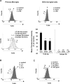
Figure 1
Figure 1
Gangliosides rapidly modulate cell surface expression of TLR4 in microglia. Rat primary microglia and BV2 microglial cells (A) were treated with 50 μg/ml of brain ganglioside mixture (Gmix) or 100 ng/ml of LPS for 18 hours, and the TLR4 surface expression level was analyzed by flow cytometric analysis using phycoerythrin-conjugated TLR4/MD2(MTS510) antibody. Data shown are representative of at least four independent experiments. The MFI was analyzed by CellQuest software (BD Biosciences), and the change in MFI of cells incubated with TLR4 antibody was calculated for LPS- or Gmix-treated cells and untreated cells after subtraction of the MFI obtained with the isotype control antibody. The MFI values are mean ± SD of three independent experiments. *P < 0.01 when compared with control samples. B: Primary microglia were treated with 50 μg/ml of Gmix for the indicated times, and TLR4 surface protein expression was determined by FACS analysis. C: Primary microglia were treated with the indicated concentrations of Gmix for 18 hours. Data shown are representative of at least four independent experiments.
Figure 2
Figure 2
Sialic acid residue is essential for ganglioside-modulated TLR4 expression. A: Microglia were stimulated with GM1, GD1a, GT1b, or Gmix for 12 hours, and TLR4 expression was analyzed at the cell surface protein levels. B: Microglia were treated with the indicated concentrations of GT1b for 12 hours, after which expression of TLR4 and IL-1β was determined by FACS and RT-PCR analysis, respectively. C: TLR4 cell surface expression levels were determined in cells with 25 μg/ml of Gmix or asialo GM1. Data shown are representative of at least three independent experiments.
Figure 3
Figure 3
Polymyxin B, a LPS scavenger, does not affect the ganglioside-dependent expression of TLR4 and cytokine. A: Primary microglia were stimulated with Gmix in the presence or the absence of 10 μg/ml of polymyxin B, after which TLR4 expression was analyzed at the cell surface protein level. B: Primary microglia were pretreated with polymyxin B for 1 hour, and stimulated with Gmix or LPS for 3 hours. mRNA expression of TNF-α and IL-1β was detected using a RT-PCR-based assay.
Figure 4
Figure 4
Gangliosides modulate TLR4 expression also in primary astrocytes. A: Rat primary astrocytes were treated with 50 μg/ml of Gmix or 100 ng/ml of LPS for 18 hours, and the TLR4 surface expression levels were analyzed by flow cytometric analysis. B: Rat primary astrocytes were stimulated with 25 μg/ml of GM1, GD1a, or GT1b, after which TLR4 expressions were analyzed at cell surface protein levels.
Figure 5
Figure 5
siRNA-mediated suppression of TLR4 reduces ganglioside-induced transcription of inflammatory cytokines. A: Rat primary astrocytes were transfected with a TLR4-specific 21-bp siRNA duplex or a nonsilencing control siRNA (Scramble 1 duplex, Dharmacon) using oligofectamine. Transcript levels of TLR4 were then detected by RT-PCR. Twenty-four hours after rat primary astrocytes were transfected with oligofectamine alone (mock), 20 pmol of Scramble 1 (Scr)-siRNA or 20 pmol of TLR4-siRNA, TLR4 cell surface expression levels were measured by FACS analysis. B: Rat primary astrocytes were transfected with either 20 pmol of TLR4-siRNA or 20 pmol of Scr control-siRNA, and the cells were treated with 50 μg/ml of brain ganglioside mixture (a), 100 ng/ml of LPS as a positive control (b), or 10 U/ml of IFN-γ as a negative control (c) for 3 hours. Total RNA was then extracted for RT-PCR analysis.
Figure 6
Figure 6
siRNA-mediated inhibition of TLR4 also suppresses ganglioside-induced NF-κB activity. A: Eighteen hours after transfection with TLR4-siRNA or Scramble control-siRNA in rat primary astrocytes, cells were transfected with luciferase reporter plasmids containing 5×NF-κB binding elements. After incubation for 24 hours, the cells were stimulated with 50 μg/ml of Gmix for 6 hours. Luciferase activity was then determined, and the result was normalized for transfection efficiency by comparing β-galactosidase activity. Values are mean ± SD of triplicates. Data are representative of three independent experiments. B: Eighteen hours after transfection with 20 pmol of control siRNA, 20 pmol of TLR4-siRNA, or oligofectamine alone (mock) in rat primary astrocytes, cells were serum-starved for 24 hours and then treated with 50 μg/ml of Gmix for 30 minutes in the presence of 2.5% serum. Nuclear extracts were then prepared and analyzed using a γ-32P-labled consensus NF-κB binding elements oligonucleotide probe.
Figure 7
Figure 7
Transient transfection of wild-type or mutant TLR4 genes affects the ganglioside-dependent nuclear factor binding activity to NF-κB binding elements as well as IL-6 promoter activity. A: Rat primary astrocytes were transiently transfected with wild-type TLR4 [pDisplay-Tlr4(wt)], dominant-negative TLR4 [pDisplay-Tlr4(P712H)], or vector alone, and their expressions were determined at the transcript and surface protein expression level using RT-PCR and FACS analysis. The sequences for mouse-specific TLR4 primer used in this study were (forward) 5′-GAACAAACAGCCTGAGAC-3′ and (reverse) 5′-GACTGGTCAAGCCAAGAA-3′. B: Rat primary astrocytes were transiently transfected with pDisplay-Tlr4(P712H) (a), pDisplay-Tlr4(wt) (b), or vector, and then incubated for 18 hours. The cells were serum-starved for 24 hours, and treated with 50 μg/ml of Gmix for the indicated times in the presence of 2.5% serum. Nuclear extracts were then prepared and analyzed using a γ-32P-labled consensus NF-κB binding elements oligonucleotide probe. C: Eighteen hours after transfection with luciferase reporter plasmids containing IL-6 promoter elements and pDisplay-Tlr4(wt), pDisplay-Tlr4(P712H), or control vector, cells were serum-starved for 24 hours and stimulated with 50 μg of Gmix for 6 hours. Luciferase activity was then determined, and the result was normalized for transfection efficiency by comparison with β-galactosidase activity. Values are mean ± SD of triplicates. Data shown are representative of at least three independent experiments.
Figure 8
Figure 8
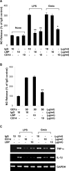
CD14 is required for ganglioside-induced inflammatory response. A: Primary microglia were preincubated with 10 μg/ml of anti-CD14 mAb, anti-LBP mAb, or isotype control mAb, and then the cells were treated with 50 μg/ml of Gmix or 100 ng/ml of LPS as a control for 24 hours. B: Primary microglia with 10 μg/ml of anti-CD14 mAb, anti-LBP mAb, or isotype control mAb were treated with 50 μg/ml of GD1a for 24 hours. The amount of NO produced was determined by measuring the amount of nitrite converted from NO in the media. Values are mean ± SD of triplicates. **P < 0.001 when compared with isotype mAb-treated control samples; *P < 0.005 when compared with isotype mAb-treated control samples. C: Cells were treated with 50 μg/ml of Gmix or 100 ng/ml of LPS for 3 hours. Total RNA was then extracted for RT-PCR analysis.
Figure 9
Figure 9
Ganglioside-induced NO release is significantly inhibited by raft-disrupting drugs, such as MβCD and filipin. Primary microglia were incubated with MβCD or filipin for 30 minutes and then stimulated with the ganglioside mixture for 24 hours. The amount of NO produced was determined by measuring the amount of nitrite converted from NO in the media. Values are mean ± SD of triplicates. *P < 0.001 when compared with Gmix alone-treated samples.
Figure 10
Figure 10
Schematic diagram depicting possible mechanisms for ganglioside-triggered inflammation in the brain. Brain injury causes damage to neuronal cells, which release gangliosides into the extracellular space. TLR4 and CD14 recognize these gangliosides within lipid rafts and activate downstream inflammatory signaling cascades in glia, leading to pathological conditions in the brain.

Similar articles

Cited by

References

    1. Ridet JL, Malhotra SK, Privat A, Gage FH. Reactive astrocytes: cellular and molecular cues to biological function. Trends Neurosci. 1997;20:570–577. - PubMed
    1. Matyszak MK. Inflammation in the CNS: balance between immunological privilege and immune responses. Prog Neurobiol. 1998;56:19–35. - PubMed
    1. Streit WJ, Walter SA, Pennell NA. Reactive microgliosis. Prog Neurobiol. 1999;57:563–581. - PubMed
    1. Aloisi F, Ria F, Adorini L. Regulation of T-cell responses by CNS antigen-presenting cells: different roles for microglia and astrocytes. Immunol Today. 2000;21:141–147. - PubMed
    1. Mutoh T, Tokuda A, Miyadai T, Hamagucci M, Fujiki N. Ganglioside GM1 binds to the Trk protein and regulates receptor function. Proc Natl Acad Sci USA. 1995;92:5087–5041. - PMC - PubMed

Publication types

MeSH terms

LinkOut - more resources