The myxoid/round cell liposarcoma fusion oncogene FUS-DDIT3 and the normal DDIT3 induce a liposarcoma phenotype in transfected human fibrosarcoma cells
- PMID: 16651630
- PMCID: PMC1606602
- DOI: 10.2353/ajpath.2006.050872
The myxoid/round cell liposarcoma fusion oncogene FUS-DDIT3 and the normal DDIT3 induce a liposarcoma phenotype in transfected human fibrosarcoma cells
Abstract
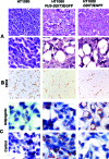
Myxoid/round cell liposarcoma (MLS/RCLS) is the most common subtype of liposarcoma. Most MLS/RCLS carry a t(12;16) translocation, resulting in a FUS-DDIT3 fusion gene. We investigated the role of the FUS-DDIT3 fusion in the development of MLS/RCLS in FUS-DDIT3- and DDIT3-transfected human HT1080 sarcoma cells. Cells expressing FUS-DDIT3 and DDIT3 grew as liposarcomas in severe combined immunodeficient mice and exhibited a capillary network morphology that was similar to networks of MLS/RCLS. Microarray-based comparison of HT1080, the transfected cells, and an MLS/RCLS-derived cell line showed that the FUS-DDIT3- and DDIT3-transfected variants shifted toward an MLS/RCLS-like expression pattern. DDIT3-transfected cells responded in vitro to adipogenic factors by accumulation of fat and transformation to a lipoblast-like morphology. In conclusion, because the fusion oncogene FUS-DDIT3 and the normal DDIT3 induce a liposarcoma phenotype when expressed in a primitive sarcoma cell line, MLS/RCLS may develop from cell types other than preadipocytes. This may explain the preferential occurrence of MLS/RCLS in nonadipose tissues. In addition, development of lipoblasts and the typical MLS/RCLS capillary network could be an effect of the DDIT3 transcription factor partner of the fusion oncogene.
Figures


References
-
- Fletcher CDM, Unni KK, Mertens F. Tumors of Soft Tissue and Bone. Lyon, France: IARC Press; 2000
-
- Åman P, Ron D, Mandahl N, Fioretos T, Heim S, Arheden K, Willen H, Rydholm A, Mitelman F. Rearrangement of the transcription factor gene CHOP in myxoid liposarcomas with t(12;16)(q13;p11). Genes Chromosomes Cancer. 1992;5:278–285. - PubMed
-
- Panagopoulos I, Åman P, Mandahl N, Mitelman F. Two distinct FUS breakpoint clusters in myxoid liposarcoma and acute myeloid leukemia with the translocations t(12;16) and t(16;21). Oncogene. 1995;11:1133–1137. - PubMed
-
- Crozat A, Åman P, Mandahl N, Ron D. Fusion of CHOP to a novel RNA-binding protein in human myxoid liposarcoma. Nature. 1993;363:640–644. - PubMed
Publication types
MeSH terms
Substances
LinkOut - more resources
Full Text Sources
Research Materials

