

Rapid development of salivary gland carcinomas upon conditional expression of K-ras driven by the cytokeratin 5 promoter
- PMID: 16651631
- PMCID: PMC1606594
- DOI: 10.2353/ajpath.2006.050847
Rapid development of salivary gland carcinomas upon conditional expression of K-ras driven by the cytokeratin 5 promoter
Abstract
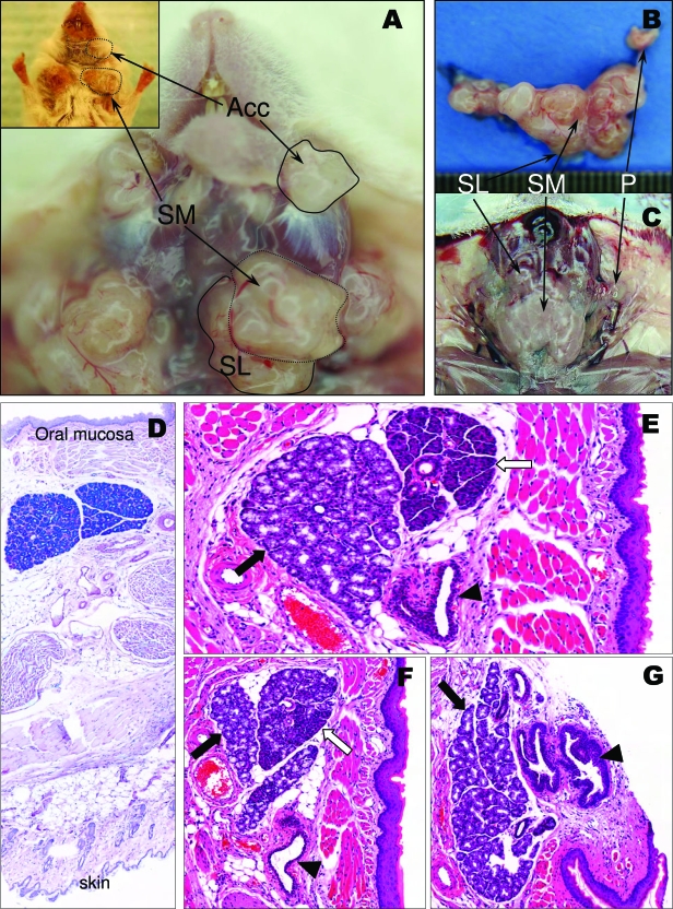
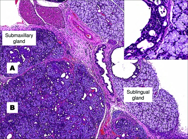
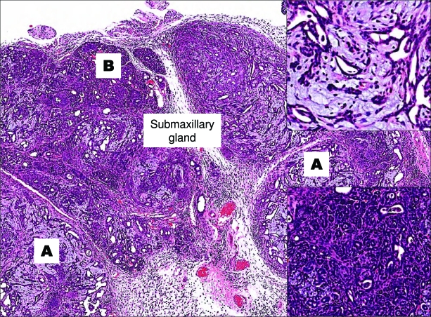
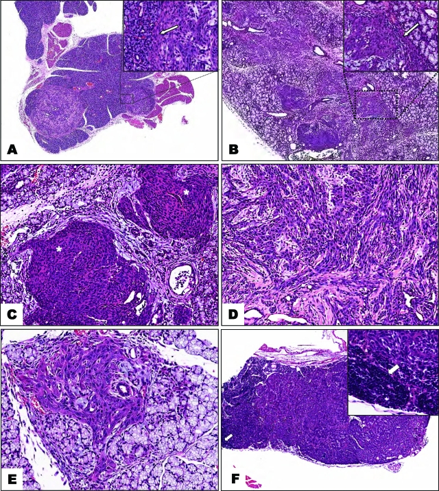
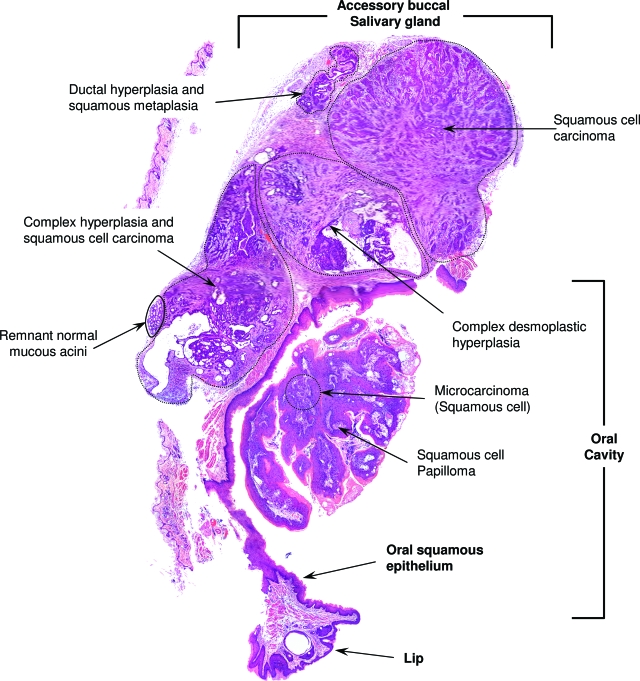
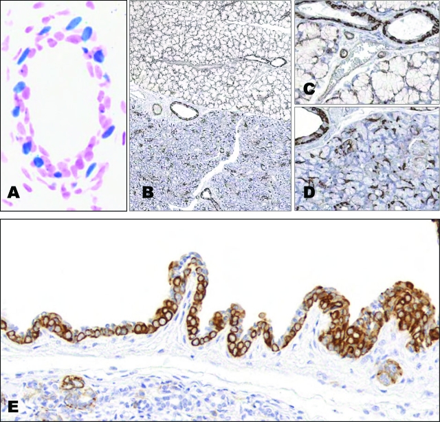
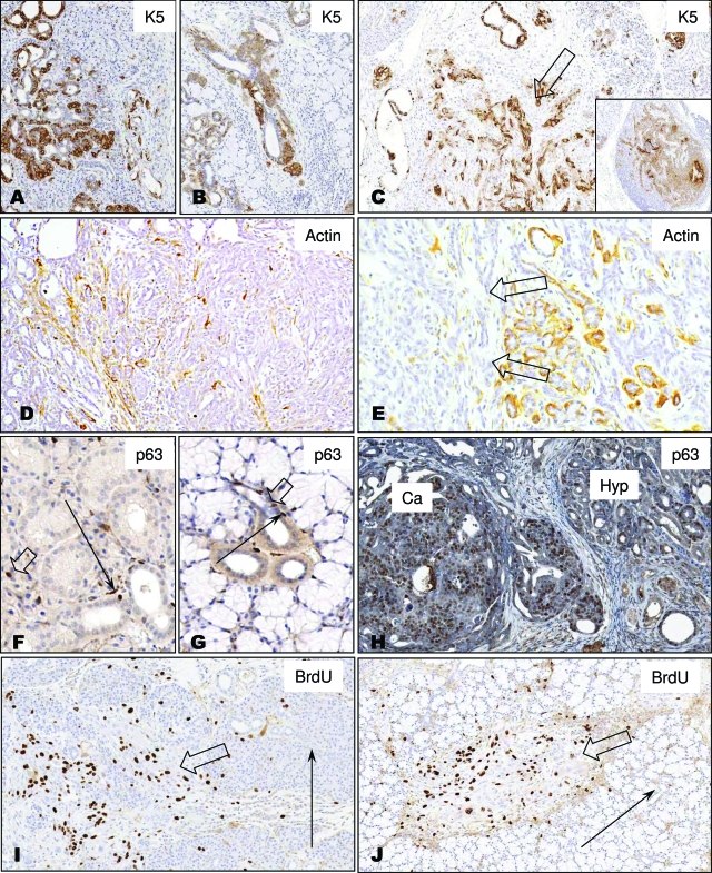
We have used a recently described model in which a ras oncogene is expressed in cytokeratin 5 (K5)-expressing cells on doxycycline administration to explore the effects of this oncogene in salivary glands of adult mice. Inducible expression of a mutated K-ras gene under the control of the K5 promoter led to the development of hyperplastic and dysplastic epithelial lesions and carcinomas, with an incidence of 100% and a minimum latency of a week. All major salivary glands were affected, as well as a set of previously undescribed buccal accessory salivary glands located on the apex of the masseter muscle, close to the oral angle. The tumors appear to arise from the cytokeratin 5-positive basal cell compartment. Myoepithelial cells participated in the hyperplasias but not in carcinomas, because the tumors are negative for smooth muscle actin. Carcinomas did not accumulate immunoreactive p53 but are positive for p63, as assayed by immunohistochemistry using an antibody against the N terminus of DeltaN p63, a splice variant of p63 that can inhibit p53 transcriptional activity. In this study, we provide evidence that the ras oncogene, targeted to a specifically sensitive cell compartment within the salivary glands, can trigger a series of event that are sufficient for full carcinogenesis.
Figures
Similar articles
-
Squamous/epidermoid differentiation in normal breast and salivary gland tissues and their corresponding tumors originate from p63/K5/14-positive progenitor cells.Virchows Arch. 2015 Jan;466(1):21-36. doi: 10.1007/s00428-014-1671-x. Epub 2014 Oct 26. Virchows Arch. 2015. PMID: 25344874
-
Expression and regulation of the ΔN and TAp63 isoforms in salivary gland tumorigenesis clinical and experimental findings.Am J Pathol. 2011 Jul;179(1):391-9. doi: 10.1016/j.ajpath.2011.03.037. Epub 2011 May 7. Am J Pathol. 2011. PMID: 21703418 Free PMC article.
-
Conditional expression of K-ras in an epithelial compartment that includes the stem cells is sufficient to promote squamous cell carcinogenesis.Cancer Res. 2004 Dec 15;64(24):8804-7. doi: 10.1158/0008-5472.CAN-04-2623. Cancer Res. 2004. PMID: 15604235
-
[New developments in molecular diagnostics of carcinomas of the salivary glands: "translocation carcinomas"].Cesk Patol. 2016;52(3):139-45. Cesk Patol. 2016. PMID: 27526014 Review. Czech.
-
Advances in salivary gland pathology.Histopathology. 2007 Jul;51(1):1-20. doi: 10.1111/j.1365-2559.2007.02719.x. Epub 2007 May 31. Histopathology. 2007. PMID: 17539914 Review.
Cited by
-
Immunocompromised and immunocompetent mouse models for head and neck squamous cell carcinoma.Onco Targets Ther. 2016 Jan 27;9:545-55. doi: 10.2147/OTT.S95633. eCollection 2016. Onco Targets Ther. 2016. PMID: 26869799 Free PMC article. Review.
-
GSTM1 and GSTT1 null polymorphisms and risk of salivary gland carcinoma.Int J Clin Exp Med. 2009;2(1):68-75. Epub 2009 Feb 25. Int J Clin Exp Med. 2009. PMID: 19436833 Free PMC article.
-
Immunolocalization patterns of cytokeratins during salivary acinar cell development in mice.J Mol Histol. 2018 Feb;49(1):1-15. doi: 10.1007/s10735-017-9742-3. Epub 2017 Nov 27. J Mol Histol. 2018. PMID: 29181608
-
Parasympathetic innervation maintains epithelial progenitor cells during salivary organogenesis.Science. 2010 Sep 24;329(5999):1645-7. doi: 10.1126/science.1192046. Science. 2010. PMID: 20929848 Free PMC article.
-
FLCN alteration drives metabolic reprogramming towards nucleotide synthesis and cyst formation in salivary gland.Biochem Biophys Res Commun. 2020 Feb 19;522(4):931-938. doi: 10.1016/j.bbrc.2019.11.184. Epub 2019 Dec 2. Biochem Biophys Res Commun. 2020. PMID: 31806376 Free PMC article.
References
-
- Speight PM, Barrett AW. Salivary gland tumours. Oral Dis. 2002;8:229–240. - PubMed
-
- Dardick I. New York: Igaku-Shoin Medical Publisher, Inc.; Color Atlas/Text of Salivary Gland Tumor Pathology. 1996
-
- Histological Typing of Salivary Gland Tumours. New York: TELOS, Springer-Verlag; World Health Organization International histological classification of tumours. 1991
-
- Ellis GL, Auclair PL. Atlas of tumor pathology. Washington, DC: Armed Forces Institute of Pathology Tumors of the salivary glands; 1996:p 31.
-
- Ide F, Suka N, Kitada M, Sakashita H, Kusama K, Ishikawa T. Skin and salivary gland carcinogenicity of 7,12-dimethylbenz[a]anthracene is equivalent in the presence or absence of aryl hydrocarbon receptor. Cancer Lett. 2004;214:35–41. - PubMed
Publication types
MeSH terms
Substances
Grants and funding
LinkOut - more resources
Full Text Sources
Medical
Molecular Biology Databases
Research Materials
Miscellaneous
