. 2006 May 1;20(9):1162-74.
doi: 10.1101/gad.1367206.

A mitotic topoisomerase II checkpoint in budding yeast is required for genome stability but acts independently of Pds1/securin

Affiliations

A mitotic topoisomerase II checkpoint in budding yeast is required for genome stability but acts independently of Pds1/securin

Catherine A Andrews et al. Genes Dev. .

Abstract

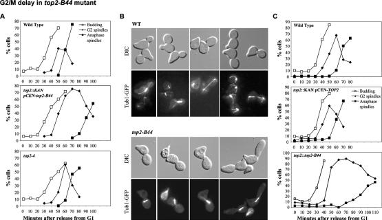
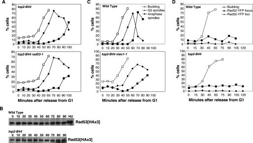
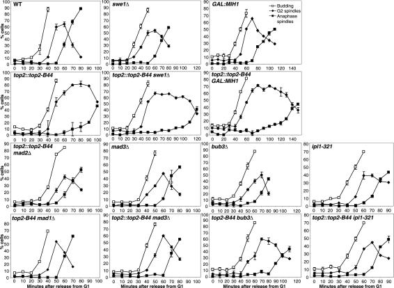
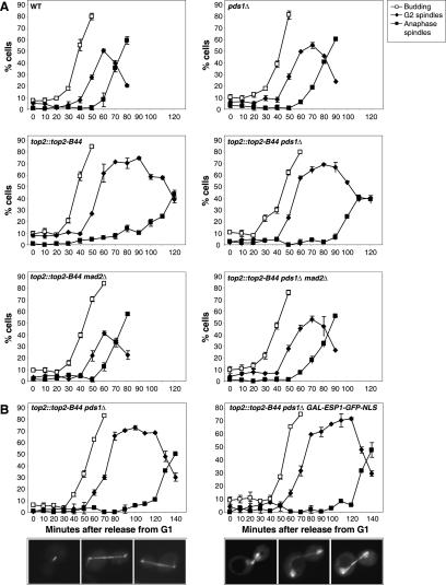
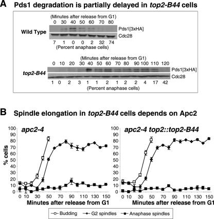
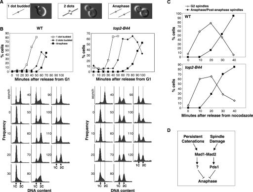
Topoisomerase II (Topo II) performs topological modifications on double-stranded DNA molecules that are essential for chromosome condensation, resolution, and segregation. In mammals, G2 and metaphase cell cycle delays induced by Topo II poisons have been proposed to be the result of checkpoint activation in response to the catenation state of DNA. However, the apparent lack of such controls in model organisms has excluded genetic proof that Topo II checkpoints exist and are separable from the conventional DNA damage checkpoint controls. But here, we define a Topo II-dependent G2/M checkpoint in a genetically amenable eukaryote, budding yeast, and demonstrate that this checkpoint enhances cell survival. Conversely, a lack of the checkpoint results in aneuploidy. Neither DNA damage-responsive pathways nor Pds1/securin are needed for this checkpoint. Unusually, spindle assembly checkpoint components are required for the Topo II checkpoint, but checkpoint activation is not the result of failed chromosome biorientation or a lack of spindle tension. Thus, compromised Topo II function activates a yeast checkpoint system that operates by a novel mechanism.


Figures

Figure 1.
Figure 1.
top2-B44 mutant cells delay in G2/M. Cell cycle analysis of wild-type versus top2 mutants at 32°C, released from mating pheromone-induced G1 synchrony. Samples were processed for FACScan analysis of DNA content (not shown) and cytology (budding and spindle morphology was scored). (A) Cell cycle progression after release from G1 in wild-type versus top2∷KAN pCEN-top2-B44 and top2-4. (B) Photomicrographs showing G2/M delay in top2∷KAN pCEN-top2-B44 cells; wild-type cells with large buds (top; photos taken 70 min after release from G1) have elongated or disassembled spindles, while top2-B44 cells with large buds often contain short G2 spindles (photos taken 80 or 90 min after release from G1). (C) Cell cycle progression after release from G1 in wild-type versus a top2-null strain containing pCEN-TOP2(TRP1) (top two graphs) and in top2∷top2-B44 cells (bottom graph).
Figure 2.
Figure 2.
G2/M delay in top2-B44 is DNA damage checkpoint-independent. (A,C) Cell cycle analysis of double mutants of top2∷KAN pCEN-top2-B44 combined with DNA damage checkpoint mutants, performed as described in Figure 1 after G1 synchrony. (A) Cell cycle progression after release from G1 in top2-B44 rad53-1. (C) Cell cycle progression after release from G1 in top2-B44 mec1-1 sml1Δ. (B) Western blot showing Rad53 phosphorylation shift after hydroxyurea (HU) treatment, but no shift in wild-type or top2-B44 cells progressing through the cell cycle at 32°C, released from mating pheromone-induced G1 synchrony. (D) Rad52 foci (a measure of the presence of DNA breaks) in wild-type or top2-B44 cells progressing through the cell cycle at 32°C, released from mating pheromone-induced G1 synchrony. Foci = cells with more than one fluorescent dot; focus = cells with one fluorescent dot.
Figure 3.
Figure 3.
G2/M delay in top2-B44 depends on spindle checkpoint proteins, but not the Cdc28 kinase Swe1. Cell cycle analysis of double mutants of top2∷top2-B44 combined with a swe1-null or spindle assembly checkpoint mutants, performed as described in Figure 1 after G1 synchrony. In the case of the GAL1-MIH1 and top2∷top2-B44 GAL1-MIH1 strains, the cells were synchronized in G1in medium containing raffinose, then released into the cell cycle in the presence of galactose to induce overexpression of MIH1. For each time point, at least 200 cells were scored. The averages of several counts are plotted and the error bars show standard deviations. Derived from these data, approximate lengths of the G2/M period are listed for these stains in Supplementary Table 1.
Figure 4.
Figure 4.
G2/M delay in top2-B44 is not enforced by Pds1/Securin. (A) Cell cycle analysis of double and triple mutants of top2∷top2-B44 combined with pds1Δ or pds1Δ mad2Δ. Since the pds1Δ cells are temperature-sensitive slightly above 28°C, the G1 synchrony was performed at 26°C, then upon release from G1 the temperature was shifted up to 28°C. (B) Cell cycle analysis of top2∷top2-B44 pds1Δ in combination with GAL1-ESP1-GFP-NLS (Jensen et al. 2001). Cells were synchronized in medium containing raffinose at 26°C, then upon release from G1 the temperature was shifted up to 28°C and galactose was added to induce expression of Esp1-GFP-NLS. (Note that the cell cycle progresses slightly slower in medium with raffinose/galactose as the carbon source.) As previously described (Jensen et al. 2001), accumulation of Esp1-GFP-NLS in the nucleus occurred efficiently in the majority of cells; photomicrographs show examples of G2/M and anaphase cells with nuclear Esp1 (right) and cells not expressing Esp1-GFP (left) for comparison.
Figure 5.
Figure 5.
Spindle elongation in top2-B44 cells depends on the APC. (A) Timing of Pds1-3xHA degradation in top2-B44 cells. Cells were released from G1 synchrony as described in Figure 1 and samples were taken for scoring budding and spindle morphologies, as well as for preparing whole-cell protein extracts for Western blots. Pds1 was detected using an anti-HA antibody, and as a loading control, Cdc28 was detected using the PSTAIRE antibody. Percent anaphase cells at each time point is indicated beneath the Western blots. (B) Cell cycle analysis of top2∷top2-B44 in combination with the apc2-4 mutation. Cells were synchronized in G1 at 26°C, then upon release from G1 the temperature was shifted up to 32°C, the nonpermissive temperature for the apc2-4 mutation. Budding and spindle morphologies were scored as described in Figure 1.
Figure 6.
Figure 6.
Chromosomes biorient properly in top2-B44 cells. (A) Schematic representation (cartoons) and photomicrographs describing a chromosome biorientation assay in budding yeast. (Left) A locus at CEN4 is tagged with a fluorescent signal (see Materials and Methods) yielding a single fluorescent dot in small budded cells. (Middle) Two closely apposed fluorescent spots are seen once the CEN4 locus has been replicated and has undergone the typical precocious separation (Goshima and Yanagida 2000) seen when the chromosome becomes bioriented, and is under spindle tension. (Right) During anaphase, the spots are segregated to the mother and daughter cells. (B) Cell cycle analysis of wild-type and top2-B44 mutants after release from G1 synchrony. (Bottom) Cell morphologies were scored as depicted in A, and samples were processed for FACScan analysis of DNA content (time points are shown at the side of each histogram plot and the vertical lines indicate approximate position of 1C and 2C DNA contents). (C) Cell cycle analysis of wild-type versus top2-B44 mutants at 32°C. Cells were released from mating pheromone-induced G1 synchrony into medium containing nocodazole, then released from the nocodazole after 120 min. Samples were processed for cytology and spindle morphology was scored. (D) Model showing the proposed relationship between the Topo II checkpoint and the spindle assembly checkpoint in yeast. We suggest that the Topo II checkpoint is activated until decatenation reactions have been performed adequately to allow a successful mitosis. In this case, anaphase is inhibited independently of Pds1/securin.

References

    1. Adachi Y., Luke M., Laemmli U.K. Chromosome assembly in vitro: Topoisomerase II is required for condensation. Cell. 1991;64:137–148. - PubMed
    1. Adachi N., Iiizumi S., So S., Koyama H. Genetic evidence for involvement of two distinct nonhomologous end-joining pathways in repair of topoisomerase II-mediated DNA damage. Biochem. Biophys. Res. Commun. 2004;318:856–861. - PubMed
    1. Bachant J., Alcasabas A., Blat Y., Kleckner N., Elledge S.J. The SUMO-1 isopeptidase Smt4 is linked to centromeric cohesion through SUMO-1 modification of DNA topoisomerase II. Mol. Cell. 2002;9:1169–1182. - PubMed
    1. Charron M., Hancock R. DNA topoisomerase II is required for formation of mitotic chromosomes in Chinese hamster ovary cells: Studies using the inhibitor 4′-demethylepipodophyllotoxin 9-(4,6-O-thenylidene-β-D-glucopyranoside). Biochemistry. 1990;29:9531–9537. - PubMed
    1. Clarke D.J., Giménez-Abián J.F. Checkpoints controlling mitosis. Bioessays. 2000;22:351–363. - PubMed

Publication types

MeSH terms