. 2006 May 1;20(9):1187-202.
doi: 10.1101/gad.1407906.

SOX2 is a dose-dependent regulator of retinal neural progenitor competence

Affiliations

SOX2 is a dose-dependent regulator of retinal neural progenitor competence

Olena V Taranova et al. Genes Dev. .

Abstract

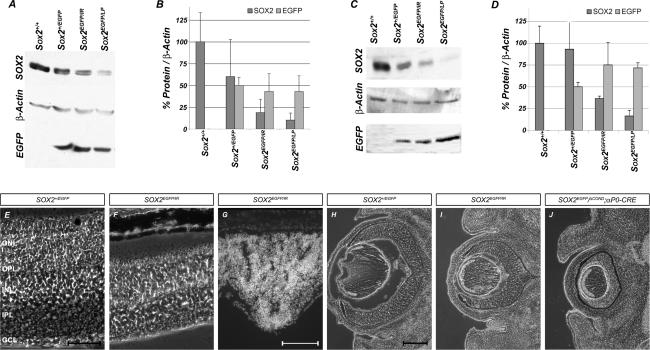
Approximately 10% of humans with anophthalmia (absent eye) or severe microphthalmia (small eye) show haploid insufficiency due to mutations in SOX2, a SOXB1-HMG box transcription factor. However, at present, the molecular or cellular mechanisms responsible for these conditions are poorly understood. Here, we directly assessed the requirement for SOX2 during eye development by generating a gene-dosage allelic series of Sox2 mutations in the mouse. The Sox2 mutant mice display a range of eye phenotypes consistent with human syndromes and the severity of these phenotypes directly relates to the levels of SOX2 expression found in progenitor cells of the neural retina. Retinal progenitor cells with conditionally ablated Sox2 lose competence to both proliferate and terminally differentiate. In contrast, in Sox2 hypomorphic/null mice, a reduction of SOX2 expression to <40% of normal causes variable microphthalmia as a result of aberrant neural progenitor differentiation. Furthermore, we provide genetic and molecular evidence that SOX2 activity, in a concentration-dependent manner, plays a key role in the regulation of the NOTCH1 signaling pathway in retinal progenitor cells. Collectively, these results show that precise regulation of SOX2 dosage is critical for temporal and spatial regulation of retinal progenitor cell differentiation and provide a cellular and molecular model for understanding how hypomorphic levels of SOX2 cause retinal defects in humans.


Figures

Figure 1.
Figure 1.
SOX2 defines progenitor cell population in the retina. SOX2 expression was evaluated using Sox2+/EGFP (green) mice (Ellis et al. 2004) and specific antibodies (red). (A,E,B,F) At E15.5, Sox2-EGFP fluorescence, SOX2, and PCNA are coexpressed in the RBL. (C,G) SOX1 expression is restricted to lens cells. Sox2EGFP is down-regulated in differentiating neurons, as marked by βTUBULINIII (D,H) NEUROFILAMENT (I,L; inset in L; arrow in I); RHODOPSIN (J,M; inset in M; arrow in J); and PKC (K,N; inset in N). Sox2-EGFP expression was maintained in a subpopulation of amacrine cells identified by ISLET1 (O,R; inset in R) and CALRETININ (P,S; inset in S), and Müller glia stained for CRALBP (Q,T; inset in T). (ONL) outer nuclear layer; (OPL); outer plexiform layer (INL); inner nuclear layer (IPL); inner plexiform layer (GCL) ganglion cell layer. Bars: A–H, 200 μm; I–T, 50 μm; I–T (insets), 20 μm.
Figure 2.
Figure 2.
Generation of allelic series of mouse Sox2 locus. (A) Targeting vector introduced two loxP sites flanking the Sox2 promoter and mRNA coding regions. Homologous recombination of this vector at the Sox2 genomic locus in ES cells resulted in the generation of two alleles. Complete insertion of the vector generated the Sox2COND allele, while partial incorporation of the 3′ part of this vector generated the Sox2LP allele, which contains only the 3′ insertion, but not the 5′ loxP site. CRE-driven recombination in Sox2+/COND;ACTB-Cre mice (in ACTB mice CRE recombinase is expressed in the female germline) resulted in deletion of the Sox2 promoter and mRNA coding region as well as the Neo cassette, generating a null allele (Sox2ΔCOND). (B) The DNA recombination events were confirmed by Southern blot analysis of mouse tail DNA. (C) In targeting vector B, the 3′ UTR of Sox2 was replaced by internal ribosome entry site (IRES) and dsRED2 coding sequence followed by a Neo cassette flanked with two loxP sites. The incorporation of the insertion into Sox2 locus generated the Sox2IR allele. The restriction enzymes used were AvrII (A), NheI (N), EcoRI (RI), SalI (S), SpeI (Sp), EcoRV (V). (D) Segregation of the alleles in the mouse lines was confirmed by Southern blotting with the P1 after EcoRI digest. (E) Quantification of pups and embryos of each genotype recovered from indicated breedings.
Figure 3.
Figure 3.
Analysis of Sox2IR and Sox2LP hypomorphic alleles. (A,C) Immunoblot of protein extracts from wild-type and Sox2 mutant embryos; E14.5 brains and eyes, respectively, were developed with antibodies against SOX2. β-ACTIN antibody was used as a loading quantity control and an antibody against EGFP was used to control for the amount of putative SOX2 expressing cells in the sample. Note that the expression level of EGFP from the Sox2 locus is preserved throughout. (B,D) Quantification of the immunoblot results. SOX2 expression in Sox2+/+ was taken as baseline (100%), and EGFP expression in Sox2+/EGFP was designated as 50%. N = 2 for each genotype. (E–G) Compared with the Sox2 heterozygous mouse (Sox2+/EGFP; E), the compound null hypomorphic adult (Sox2EGFP/IR; F) shows a significant reduction in the thickness of the retina. By E14.5, the null hypomorph exhibits distinct rosette structures (Sox2EGFP/IR; G). (H–J) Comparison of eye morphology of E14.5 Sox2+/EGFP, Sox2EGFP/IR, and Sox2EGFP/ΔCOND;αP0-CRE mice. Bars: E,F, 50 μm; G, 100 μm; H–J, 200 μm.
Figure 5.
Figure 5.
Decrease in SOX2 levels leads to hypoplasia of the optic nerve and loss of retinal ganglion cells. Ventral side of 3-mo-old Sox2+/EGFP (A) and Sox2EGFP/IR (E) brains. (E) Note the absence of developed optic nerve (arrow) in Sox2EGFP/IR. Immunostained sections through the retina of adult Sox2+/EGFP (B–D,I–L) and Sox2EGFP/IR (F–H,M–P) mice. Compared with the Sox2+/EGFP, Sox2EGFP/IR mice show ∼95% reduction in the number of cells immunostained for NEUROFILAMENT (B,F, arrows), Brn3b (C,G, arrows), and ISLET1 (D,H, arrows), and a ∼60%–70% reduction in cells stained with CALRETININ (I,M, arrowhead) in the ganglion cell layer (GCL). In contrast, interneurons demonstrate a relatively normal distribution, as shown by staining for NEUROFILAMENT (B,F, top arrowheads), ISLET1 (D,H), CALRETININ (I,M), and PKC (J,N). (I,M) Note the significant reduction in size of inner plexiform synaptic layer (IPL) as visualized by CALRETININ staining (arrowheads). (K,O) No cell loss was apparent in outer nuclear layer (ONL) formed by photoreceptor cell bodies, as confirmed by RHODOPSIN staining of the outer segments (arrowheads). (L,P) We noted an up-regulation of GFAP expression in central retina of the Sox2EGFP/IR mice as compared with the Sox2+/EGFP mice (insets, gross morphology). Bars: A,E, 2 μm; B–D,F–P, 100 μm; L,P (insets), 200 μm.
Figure 6.
Figure 6.
Reduction of SOX2 expression disrupts retinal laminar morphology and impairs differentiation of RGCs. Immunostained sections through the retina of P20 Sox2+/EGFP (A–C) and Sox2EGFP/IR (D–F) mice. Compared with the Sox2+/EGFP (A–C), Sox2EGFP/IR (D–F), mice show disruption of retinal cell layers with rosette structure formation. The rosette structures contain rod cells (F, RHODOPSIN, arrow) surrounded by horizontal cells (E, NEUROFILAMENT, arrow) and amacrine cells (D, CALRETININ, arrow). Representative horizontal sections of E15.5 Sox2+/EGFP (G,H) and Sox2EGFP/IR (L,M) littermates immunostained for Brn3b and βTUBULINIII. Both embryos show the presence of GCL; however, the differentiating neurites in the compound null hypomorphic retinas (L,M) do not form a distinguishable fiber layer, and in the majority of cases fail to enter the optic nerve; βTUBULINIII-positive cell bodies can be identified in the neuroblast layer (M, arrow). Compared with Sox2+/EGFP (I), these regions in Sox2EGFP/IR (N) retinas contain fewer progenitor cells as marked by PCNA staining. Anterograde labeling of the E14.5 retina with Cy3-dextran confirmed the loss of axonal projections from the retina in compound null hypomorphic retina (O, dotted outline), compared with the Sox2 heterozygote (J, dotted outline). (K) Quantitative analysis of cells immunoreactive for activated CASPASE3. Cells were counted from horizontal section of E12, E14, and E15 mouse embryos; n = 4 at each age. (P) Representative section of E14 Sox2EGFP/IR retina illustrating CASPASE-3 staining in GCL. Bars, A–F, 50 μm; G–I,L–N, 200 μm; J,D, 300 μm; P, 50 μm.
Figure 7.
Figure 7.
Expression of molecular regulators of RGC differentiation in Sox2 hypomorphic mice. In situ hybridization on sections from E14 Sox2+/EGFP (A–C,G–I) and Sox2EGFP/IR (D–F,J–L) embryos with probes for transcription factors involved in RGC differentiation. (A,D) Math5. (B,E) NeuroD1. (C,F) Gli1. (H,K) Notch1. Hes-5 (I,L). (G,J) Immunostaining for PAX6. Bars, A–J, 200 μm.
Figure 8.
Figure 8.
SOX2 binds IVS13 cis elements in the Notch1 gene. (A) Whole-cell extracts from ES cells and Control STO cells (which lack SOX2) were incubated with SOX2 antibody. SOX2 was efficiently immunoprecipitated (IPed) from only the ES cell extracts. (B) Schematic representation of standard ChIP PCR assay. PCR primers flanking the SOX2 consensus cis elements were used in a standard ChIP PCR assay with DNA template IPed from ES cells, E14.5 CNS, or MEFs as a control. Sonicated DNA was volumetrically divided in half to ensure equivalent input templates, and ChIP was conducted with either a SOX2 or nonspecific IgG as a control antibody. (C) The Notch1 IVS13 locus was IPed as a direct function of SOX2 binding in both ES cells and E14.5 CNS, but not from MEFs (which lack SOX2). Known SOX2 cis elements positioned in well-characterized enhancers of Fgf4 and Nestin were used as positive controls to demonstrate efficient ChIP using the SOX2 antibody. (D) Probe sequence used for DNase I footprinting analysis. Numbers indicate the nucleotide positions of the SOX2 consensus sites within Notch1. The underlined sequence represents the area of the footprint pattern. Red boxes indicate bases in the SOX2 consensus sites that were mutated (+26988: TG to CC, and +27001: TGT to GCA). (E) DNase I footprinting demonstrates the binding of proteins that produce interference patterns that localize specifically to SOX2 consensus sites within the Notch1 IVS13. Numbered arrows on the left indicate the location of the SOX2 consensus sites in the probe. The schematic diagram on the right represents the approximate locations of SOX2 binding to the probe. (F, left schematic) Luciferase reporter gene constructs were generated that contained the intact 150-bp IVS13 DNA fragment or the same DNA fragment in which either the +26988 or +27001 SOX2 consensus sites were mutated. (Right graph) Transient transfections in P19 cells indicate that the 150-bp IVS13 of Notch1 has the ability to enhance transcription of the reporter gene by approximately threefold, and that the +26988 and +27001 SOX2 consensus sites are required for transcriptional enhancement.
None

Similar articles

Cited by

References

    1. Ahmad I., Dooley C.M., Polk D.L. Delta-1 is a regulator of neurogenesis in the vertebrate retina. Dev. Biol. 1997;185:92–103. - PubMed
    1. Austin C.P., Feldman D.E., Ida J.A., Jr., Cepko C.L. Vertebrate retinal ganglion cells are selected from competent progenitors by the action of Notch. Development. 1995;121:3637–3650. - PubMed
    1. Avilion A.A., Nicolis S.K., Pevny L.H., Perez L., Vivian N., Lovell-Badge R. Multipotent cell lineages in early mouse development depend on SOX2 function. Genes & Dev. 2003;17:126–140. - PMC - PubMed
    1. Blackshaw S., Harpavat S., Trimarchi J., Cai L., Huang H., Kuo W.P., Weber G., Lee K., Fraioli R.E., Cho S.H., et al. Genomic analysis of mouse retinal development. PLoS Biol. 2004;2:E247. - PMC - PubMed
    1. Bylund M., Andersson E., Novitch B.G., Muhr J. Vertebrate neurogenesis is counteracted by Sox1-3 activity. Nat. Neurosci. 2003;6:1162–1168. - PubMed

Publication types

MeSH terms