Comparative Study
. 2006 May;16(5):567-75.
doi: 10.1101/gr.4769606.

A genome-wide distribution of 8-oxoguanine correlates with the preferred regions for recombination and single nucleotide polymorphism in the human genome

Affiliations
Comparative Study

A genome-wide distribution of 8-oxoguanine correlates with the preferred regions for recombination and single nucleotide polymorphism in the human genome

Mizuki Ohno et al. Genome Res. 2006 May.

Abstract

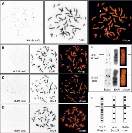
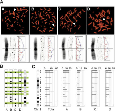
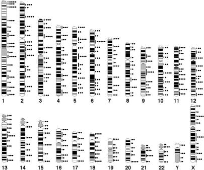
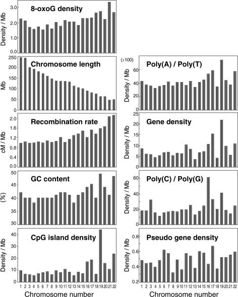
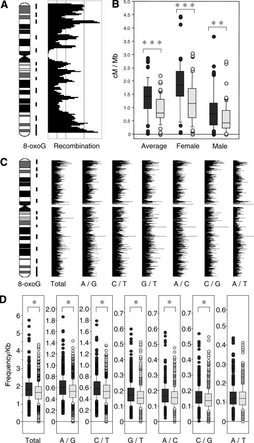
8-Oxoguanine (8-oxoG), a major spontaneous form of oxidative DNA damage, is considered to be a natural cause of genomic diversity in organisms because of its mutagenic potential. The steady-state level of 8-oxoG in the nuclear genome of a human cell has been estimated to be several residues per 10(6) guanines. In the present study, to clarify the genome-wide distribution of 8-oxoG in the steady state, we performed fluorescence in situ detection of 8-oxoG on human metaphase chromosomes using a monoclonal antibody. Multiple dot-like signals were observed on each metaphase chromosome. We then mapped the position of the signal at megabase resolution referring to the cytogenetically identified chromosomal band, and demonstrated that 8-oxoG is unevenly distributed in the normal human genome and that the distribution pattern is conserved among different individuals. Moreover, we found that regions with a high frequency of recombination and single nucleotide polymorphisms (SNPs) are preferentially located within chromosomal regions with a high density of 8-oxoG. Our findings suggest that 8-oxoG is one of the main causes of frequent recombinations and SNPs in the human genome, which largely contribute to the genomic diversity in human beings.


Figures

Figure 1.
Figure 1.
In situ detection of 8-oxoG on human metaphase chromosomes. (A) Immunological detection of 8-oxoG. The arrowhead indicates one example of a twin dot on sister chromatids. (B) Immunological detection of 8-oxoG after treatment with MutM. (C) MutM-dependent in situ end-labeling. (D) MutM (−) negative control for in situ end-labeling. (E) Signal patterns on chromosome 7: comparison between immunological detection (top) and MutM-sensitive sites (bottom). Lines indicate centromere. The monochrome images of signal (left) and DAPI (middle) are digitally enhanced and positively inverted. For a merged image, signal and DAPI images are pseudo-colored with green and red, respectively (right). (F) Signal positions on chromosome 7 are indicated as filled circles on corresponding regions in the ideogram of the G-banding pattern at the 850-band resolution obtained from the NCBI ftp site. The line indicates centromeres.
Figure 2.
Figure 2.
Genome-wide distribution of 8-oxoG is conserved among individuals. (A, top) Immunological detection of 8-oxoG on metaphase chromosomes from four individuals (A–D, green: immuno-fluorescence signal, red: DAPI). Arrowheads indicate a single chromosome 1. (A, bottom) High-magnification image of chromosome 1 (arrow, top). A positively inverted monochrome image of DAPI (left) and signal (middle) is presented. The fluorescence intensity of DAPI (blue) and signal (red) are plotted on the profile (vertical axis: chromosome length, horizontal axis: fluorescence intensity). (B) The position of each signal detected on chromosome 1 is indicated by a filled circle on the corresponding region in the ideogram. The regions detected in more than three chromosomal copies of the four individuals are colored. (C) Total scoring for 8-oxoG from 10 copies of chromosome 1 of the four subjects are represented as bar plots with the chromosome length on the y-axis and the 8-oxoG score on the x-axis, max = 20 points, see Methods). The vertical lines on the right of the ideogram indicate mapped regions. A bar indicating frequency per region was plotted at the center of the corresponding region.
Figure 3.
Figure 3.
Comprehensive mapping of 8-oxoG. The positions at which 8-oxoG was detected are indicated as vertical lines on the right of the ideogram. The total 8-oxoG score for each region was graded from zero to five and indicated as filled circles.
Figure 4.
Figure 4.
Bar plots of 8-oxoG density and eight predictors. The recombination rate (sex-averaged) was obtained from the deCODE study (Kong et al. 2002). The GC content and CpG island density were obtained from Venter et al. (2001). Gene density, pseudo gene density, and chromosomal length were obtained from the Ensembl Genome browser (http://www.ensembl.org/Homo_sapiens/). Poly (A or T) and (C or G) fractions were obtained from Rohrer et al. (2002).
Figure 5.
Figure 5.
Comparison of 8-oxoG distribution on chromosome 11 and recombination rate or SNP frequency. (A) Regions with a high density of 8-oxoG are indicated by vertical lines on the right of the ideogram. The sex-averaged recombination rate (deCODE) was calculated with a 3-Mb window in 1-Mb steps and indicated as a bar plot with the chromosome lengths on the y-axis and recombination rates on the x-axis. (B) Box-whisker plot of recombination rates in chromosome 11 within regions with a high density of 8-oxoG (gray) and regions without a high density of 8-oxoG (light gray). (Mann-Whitney U-test, **P < 0.01, ***P < 0.0001). The center line within the box gives the median. The top and bottom boxes show the 75 and 25 percentiles, respectively. The top and bottom of the whisker show the 5 and 95 percentiles, respectively. (C) Bar plots of SNP frequencies. The SNP frequency for each base substitution was calculated with a 200-Kb window and in 50-Kb steps. The substitution spectra are indicated by two letters separated with a slash. For example, A/G means that A and G were recorded in a single position, and does not suggest the direction of the substitution. The regions with a high density of 8-oxoG are indicated by vertical lines on the right of the ideogram of chromosome 11. (D) Box-whisker plots of SNP frequencies in chromosome 11 regions with a high density of 8-oxoG (gray) and in regions without a high density of 8-oxoG (light gray) (Mann-Whitney U-test, *P < 0.05). Centrometic regions were excluded from the calculation.

References

    1. Aburatani H., Hippo Y., Ishida T., Takashima R., Matsuba C., Kodama T., Takao M., Yasui A., Yamamoto K., Asano M., Hippo Y., Ishida T., Takashima R., Matsuba C., Kodama T., Takao M., Yasui A., Yamamoto K., Asano M., Ishida T., Takashima R., Matsuba C., Kodama T., Takao M., Yasui A., Yamamoto K., Asano M., Takashima R., Matsuba C., Kodama T., Takao M., Yasui A., Yamamoto K., Asano M., Matsuba C., Kodama T., Takao M., Yasui A., Yamamoto K., Asano M., Kodama T., Takao M., Yasui A., Yamamoto K., Asano M., Takao M., Yasui A., Yamamoto K., Asano M., Yasui A., Yamamoto K., Asano M., Yamamoto K., Asano M., Asano M. Cloning and characterization of mammalian 8-hydroxyguanine-specific DNA glycosylase/apurinic, apyrimidinic lyase, a functional mutM homologue. Cancer Res. 1997;57:2151–2156. - PubMed
    1. Aquadro C.F., Bauer Dumont V., Reed F.A., Bauer Dumont V., Reed F.A., Reed F.A. Genome-wide variation in the human and fruitfly: A comparison. Curr. Opin. Genet. Dev. 2001;11:627–634. - PubMed
    1. Arai T., Kelly V.P., Komoro K., Minowa O., Noda T., Nishimura S., Kelly V.P., Komoro K., Minowa O., Noda T., Nishimura S., Komoro K., Minowa O., Noda T., Nishimura S., Minowa O., Noda T., Nishimura S., Noda T., Nishimura S., Nishimura S. Cell proliferation in liver of Mmh/Ogg1-deficient mice enhances mutation frequency because of the presence of 8-hydroxyguanine in DNA. Cancer Res. 2003;63:4287–4292. - PubMed
    1. Barch M.J. The ACT cytogenetics laboratory manual. Raven Press; New York.: 1991.
    1. Barnes D.E., Lindahl T., Lindahl T. Repair and genetic consequences of endogenous DNA base damage in mammalian cells. Annu. Rev. Genet. 2004;38:445–476. - PubMed

Publication types

LinkOut - more resources