. 2006 May 9;45(18):5896-906.
doi: 10.1021/bi052619p.

Extrinsic interactions dominate helical propensity in coupled binding and folding of the lactose repressor protein hinge helix

Affiliations

Extrinsic interactions dominate helical propensity in coupled binding and folding of the lactose repressor protein hinge helix

Hongli Zhan et al. Biochemistry. .

Abstract

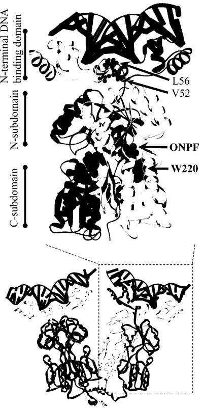
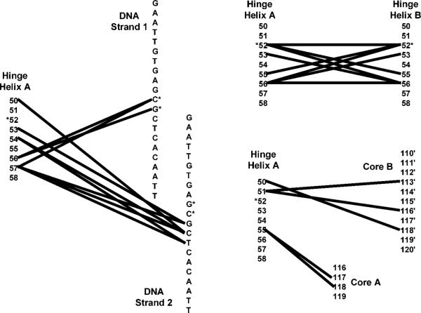
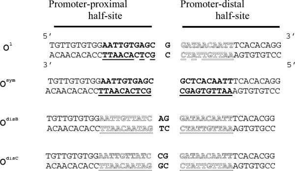
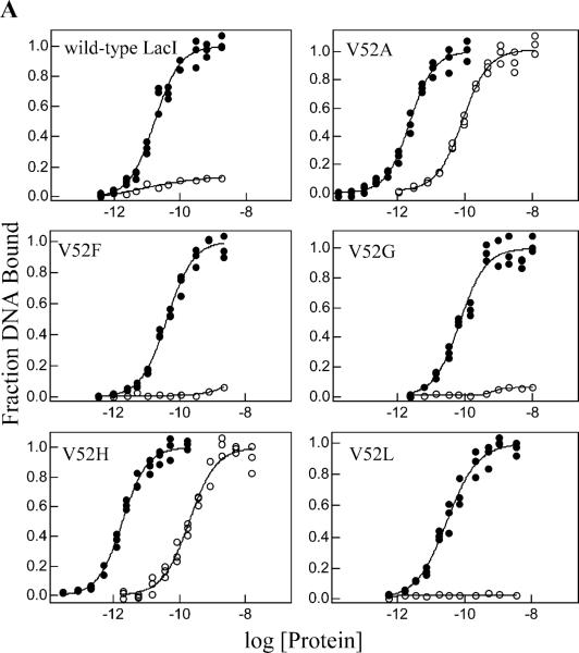
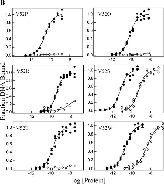
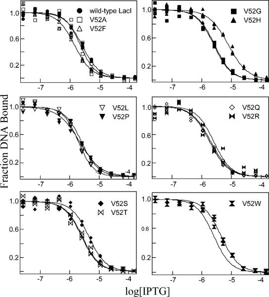
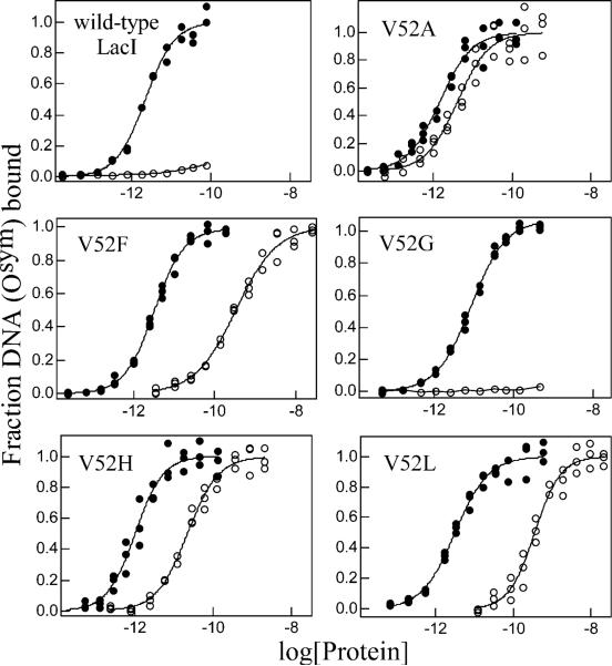
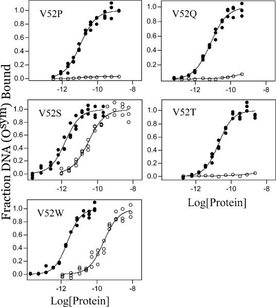
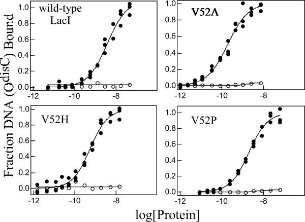
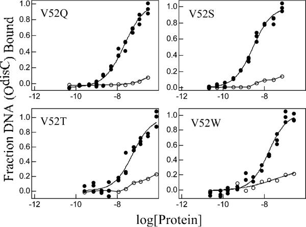
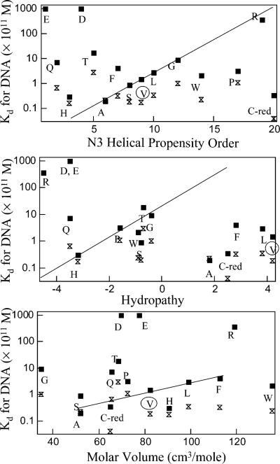
A significant number of eukaryotic regulatory proteins are predicted to have disordered regions. Many of these proteins bind DNA, which may serve as a template for protein folding. Similar behavior is seen in the prokaryotic LacI/GalR family of proteins that couple hinge-helix folding with DNA binding. These hinge regions form short alpha-helices when bound to DNA but appear to be disordered in other states. An intriguing question is whether and to what degree intrinsic helix propensity contributes to the function of these proteins. In addition to its interaction with operator DNA, the LacI hinge helix interacts with the hinge helix of the homodimer partner as well as to the surface of the inducer-binding domain. To explore the hierarchy of these interactions, we made a series of substitutions in the LacI hinge helix at position 52, the only site in the helix that does not interact with DNA and/or the inducer-binding domain. The substitutions at V52 have significant effects on operator binding affinity and specificity, and several substitutions also impair functional communication with the inducer-binding domain. Results suggest that helical propensity of amino acids in the hinge region alone does not dominate function; helix-helix packing interactions appear to also contribute. Further, the data demonstrate that variation in operator sequence can overcome side chain effects on hinge-helix folding and/or hinge-hinge interactions. Thus, this system provides a direct example whereby an extrinsic interaction (DNA binding) guides internal events that influence folding and functionality.


Figures

Figure 1
Figure 1
(A). Structure of LacI dimer bound to DNA (top, black ladder) and anti-inducer, ONPF (middle, gray spacefilling). One monomer is in dark gray, the other light gray. The hinge helix side chains of L56, which intercalate into the minor groove of DNA (24), are shown as a gray ball/stick (top), and those of V52 are black ball/stick. W220 (middle, spacefill) is shown in black. The coordinates are from Protein Data Bank file 1efa (15). The protein is composed of the N-terminal DNA binding domain, the core domain (each monomer comprising N- and C-subdomains with an inducer-binding site between them), and the C-terminal tetramerization domain (not present in the dimeric structure shown). Below: The structure of tetrameric LacI bound to Osym DNA (shown as black ladder on the top of each dimer), with two monomers of right-hand dimer colored as black and light gray, respectively, and two monomers in the left-hand dimer colored to correspond with the structural domains described above. (B). Interactions between the hinge helix of a single monomer (“Hinge Helix A”) and other regions in the LacI•DNA complex. Columns of numbers represent residues of the protein; DNA basepairs of Osym are represented by columns of letters. Lines between any pair of numbers/letters indicate an interaction occurs between them. For clarity, the interactions of hinge helix A are separated onto three networks, which were made using the structure of 1efa (15) and the program RESMAP (25, 68). The interactions between 55 and 117/118 are “long” hydrogen bonds μ slightly above 3.5 Å but below 3.6 and 3.7 Å, respectively. The position of V52 and the central basepairs of Osym are indicated by asterisks. Note that position 52 is the only residue in the hinge helix that does not interact with DNA or the core inducer-binding domain.
Figure 1
Figure 1
(A). Structure of LacI dimer bound to DNA (top, black ladder) and anti-inducer, ONPF (middle, gray spacefilling). One monomer is in dark gray, the other light gray. The hinge helix side chains of L56, which intercalate into the minor groove of DNA (24), are shown as a gray ball/stick (top), and those of V52 are black ball/stick. W220 (middle, spacefill) is shown in black. The coordinates are from Protein Data Bank file 1efa (15). The protein is composed of the N-terminal DNA binding domain, the core domain (each monomer comprising N- and C-subdomains with an inducer-binding site between them), and the C-terminal tetramerization domain (not present in the dimeric structure shown). Below: The structure of tetrameric LacI bound to Osym DNA (shown as black ladder on the top of each dimer), with two monomers of right-hand dimer colored as black and light gray, respectively, and two monomers in the left-hand dimer colored to correspond with the structural domains described above. (B). Interactions between the hinge helix of a single monomer (“Hinge Helix A”) and other regions in the LacI•DNA complex. Columns of numbers represent residues of the protein; DNA basepairs of Osym are represented by columns of letters. Lines between any pair of numbers/letters indicate an interaction occurs between them. For clarity, the interactions of hinge helix A are separated onto three networks, which were made using the structure of 1efa (15) and the program RESMAP (25, 68). The interactions between 55 and 117/118 are “long” hydrogen bonds μ slightly above 3.5 Å but below 3.6 and 3.7 Å, respectively. The position of V52 and the central basepairs of Osym are indicated by asterisks. Note that position 52 is the only residue in the hinge helix that does not interact with DNA or the core inducer-binding domain.
Figure 2
Figure 2
Sequence of the variant operators used for the DNA binding studies. The 40 base pair sequences are shown. The region of the O1 operator protected by LacI from DNase footprinting is highlighted in bold and outlined letters (83). Symmetric sites within each operator are underlined and are shown in the sequences in similar print to the O1 sequence. The two half-sites are labeled as proximal and distal based on the natural operator, O1.
Figure 3
Figure 3
Operator O1 binding and inducibility of wild-type LacI and V52 mutants. Experiments were performed in buffer containing 0.01 M Tris-HCl, pH 7.4, 0.15 M KCl, 0.3 mM DTT, 0.1 mM EDTA, 5% DMSO in the absence (closed circles) or presence (open circles) of IPTG. The experiments were conducted as described in Materials and Methods with operator concentration below 1.5 × 10⁻¹³ M for V52A and V52H and below 1.5 × 10⁻¹² M for other proteins. IPTG concentration was 1 mM. Data shown are for single determinations (triplicate points), and the results from multiple determinations are summarized in Tables 1 and 2. (A) Wild-type LacI, V52A, V52F, V52G, V52H, and V52L; (B) V52P, V52Q, V52R, V52S, V52T, and V52W. Note that V52A, V52H, V52S, and V52W retain significant affinity in the presence of inducer.
Figure 3
Figure 3
Operator O1 binding and inducibility of wild-type LacI and V52 mutants. Experiments were performed in buffer containing 0.01 M Tris-HCl, pH 7.4, 0.15 M KCl, 0.3 mM DTT, 0.1 mM EDTA, 5% DMSO in the absence (closed circles) or presence (open circles) of IPTG. The experiments were conducted as described in Materials and Methods with operator concentration below 1.5 × 10⁻¹³ M for V52A and V52H and below 1.5 × 10⁻¹² M for other proteins. IPTG concentration was 1 mM. Data shown are for single determinations (triplicate points), and the results from multiple determinations are summarized in Tables 1 and 2. (A) Wild-type LacI, V52A, V52F, V52G, V52H, and V52L; (B) V52P, V52Q, V52R, V52S, V52T, and V52W. Note that V52A, V52H, V52S, and V52W retain significant affinity in the presence of inducer.
Figure 4
Figure 4
Operator release by IPTG of wild-type protein and V52 variants. The buffer was 0.01 M Tris-HCl, pH 7.4, 0.15 M KCl, 0.3 mM DTT, 0.1 mM EDTA, and 5% DMSO, and various concentrations of IPTG were added. In the experiments shown, operator concentration was 1.5 × 10⁻¹² M, and protein concentrations were as follows: wild-type, 2.4 × 10⁻¹⁰ M; V52A, 1 × 10⁻¹¹ M; V52F, 2.4 × 10⁻¹⁰ M; V52R, 5 × 10⁻⁹ M; V52G, 3.5 × 10⁻⁹ M; V52H, 2.1 × 10⁻¹¹ M; V52L, 2.1 × 10⁻¹⁰ M; V52P, 1.5 × 10⁻¹⁰ M; V52Q, 5 × 10⁻¹⁰ M; V52S, 2.4 × 10⁻¹⁰ M; V52T, 3.5 × 10⁻⁹ M; V52W, 2.4 × 10⁻¹⁰ M. Data for a single experiment are shown (triplicate points), and the results from multiple measurements are summarized in Table 3. Dotted lines correspond to the wild-type data and are provided for comparison.
Figure 5
Figure 5
Osym binding and inducibility of wild-type LacI and V52 mutants. Buffer was 0.01 M Tris-HCl, pH 7.4, 0.15 M KCl, 0.3 mM DTT, 0.1 mM EDTA, 5% DMSO in the absence (closed circles) or presence (open circles) of IPTG. The experiments were conducted as described in Materials and Methods with Osym concentration below 1.5 × 10⁻¹³ M in the absence of inducer. In the presence of 1 mM IPTG, operator concentration was below 1.5 × 10⁻¹³ M for V52A and below 1.5 × 10⁻¹² M for other proteins. Data are shown for single determinations (triplicate points), and the results from multiple determinations are summarized in Table 2. (A) Wild-type LacI, V52A, V52F, V52G, V52H, and V52L; (B) V52P, V52Q, V52S, V52T, and V52W. Note that V52A, V52F, V52H, V52L, V52S, and V52W show high affinity Osym binding in the presence of a saturating inducer.
Figure 5
Figure 5
Osym binding and inducibility of wild-type LacI and V52 mutants. Buffer was 0.01 M Tris-HCl, pH 7.4, 0.15 M KCl, 0.3 mM DTT, 0.1 mM EDTA, 5% DMSO in the absence (closed circles) or presence (open circles) of IPTG. The experiments were conducted as described in Materials and Methods with Osym concentration below 1.5 × 10⁻¹³ M in the absence of inducer. In the presence of 1 mM IPTG, operator concentration was below 1.5 × 10⁻¹³ M for V52A and below 1.5 × 10⁻¹² M for other proteins. Data are shown for single determinations (triplicate points), and the results from multiple determinations are summarized in Table 2. (A) Wild-type LacI, V52A, V52F, V52G, V52H, and V52L; (B) V52P, V52Q, V52S, V52T, and V52W. Note that V52A, V52F, V52H, V52L, V52S, and V52W show high affinity Osym binding in the presence of a saturating inducer.
Figure 6
Figure 6
OdisC binding and inducibility of wild-type LacI and V52 mutants. Buffer was 0.01 M Tris-HCl, pH 7.4, 0.15 M KCl, 0.3 mM DTT, 0.1 mM EDTA, 5% DMSO in the absence (closed circles) or presence (open circles) of IPTG. OdisC concentration was below 1.5 × 10⁻¹² M. When present, IPTG concentration was 1 mM. Data are shown for single determinations (triplicate points), and the results from multiple determinations are summarized in Table 4. (A) Wild-type LacI, V52A, V52H, and V52P; (B) V52Q, V52S, V52T, and V52W.
Figure 6
Figure 6
OdisC binding and inducibility of wild-type LacI and V52 mutants. Buffer was 0.01 M Tris-HCl, pH 7.4, 0.15 M KCl, 0.3 mM DTT, 0.1 mM EDTA, 5% DMSO in the absence (closed circles) or presence (open circles) of IPTG. OdisC concentration was below 1.5 × 10⁻¹² M. When present, IPTG concentration was 1 mM. Data are shown for single determinations (triplicate points), and the results from multiple determinations are summarized in Table 4. (A) Wild-type LacI, V52A, V52H, and V52P; (B) V52Q, V52S, V52T, and V52W.
Figure 7
Figure 7
Correlation between DNA binding affinities (O1-squares and Osym-bowties) of V52 mutants and N3 helical propensity order (top) (69-73), hydropathy (middle) (75), and molar volume (bottom) (74). For helical propensity versus Kd, a trend is noted with a line but has many obvious outliers. No trend is seen for hydropathy versus Kd, but data suggest a correlation between affinity and molar volume for about one-half of the substitutions. We predict that this may reflect enhanced hinge•hinge packing interactions.

References

    1. Gilbert W, Müller-Hill B. Isolation of the lac repressor. Proc. Natl. Acad. Sci. U. S. A. 1966;56:1891–1898. - PMC - PubMed
    1. Matthews KS, Nichols JC. Lactose repressor protein: Functional properties and structure. Prog. Nucleic Acid Res. Mol. Biol. 1998;58:127–164. - PubMed
    1. Straney DC, Crothers DM. Effect of drug-DNA interactions upon transcription initiation at the lac promoter. Biochemistry. 1987;26:1987–1995. - PubMed
    1. Lin S-Y, Riggs AD. A comparison of lac repressor binding to operator and to nonoperator DNA. Biochem. Biophys. Res. Commun. 1975;62:704–710. - PubMed
    1. Jobe A, Bourgeois S. lac repressor-operator interaction. VI. The natural inducer of the lac operon. J. Mol. Biol. 1972;69:397–408. - PubMed

Publication types