Review
. 2006:57:837-58.
doi: 10.1146/annurev.arplant.56.032604.144208.

Phytochrome structure and signaling mechanisms

Affiliations
Review

Phytochrome structure and signaling mechanisms

Nathan C Rockwell et al. Annu Rev Plant Biol. 2006.

Abstract

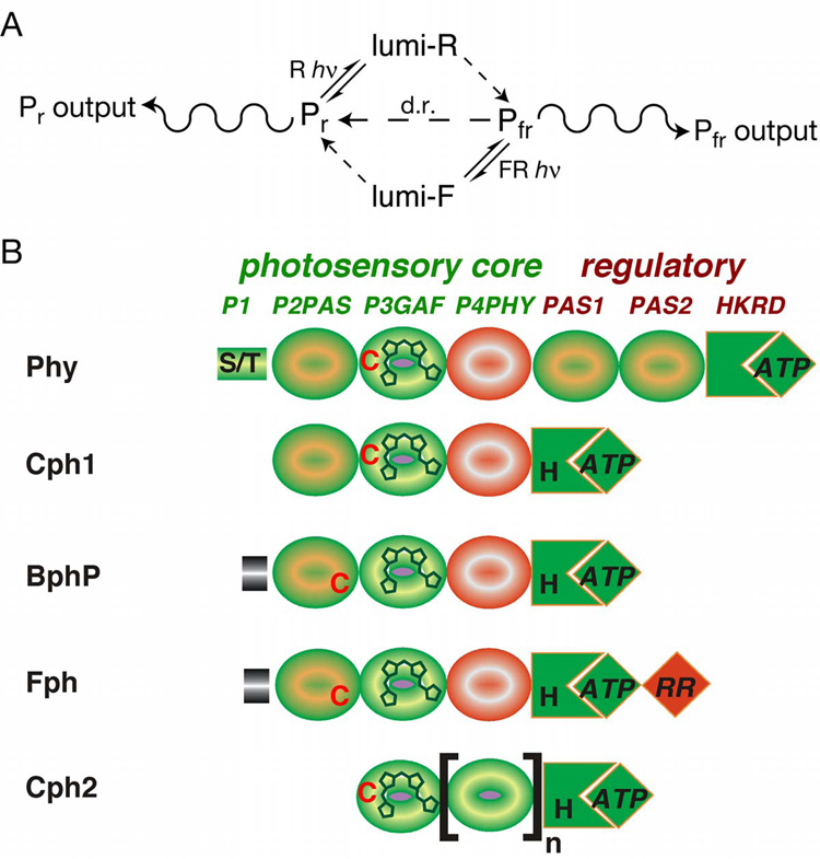
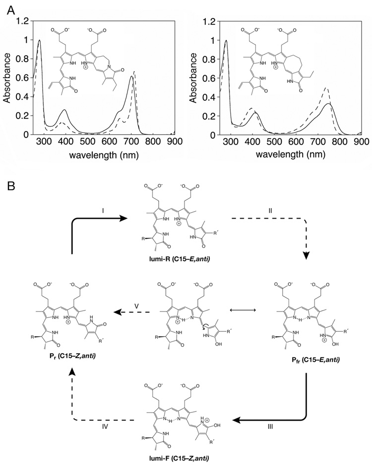
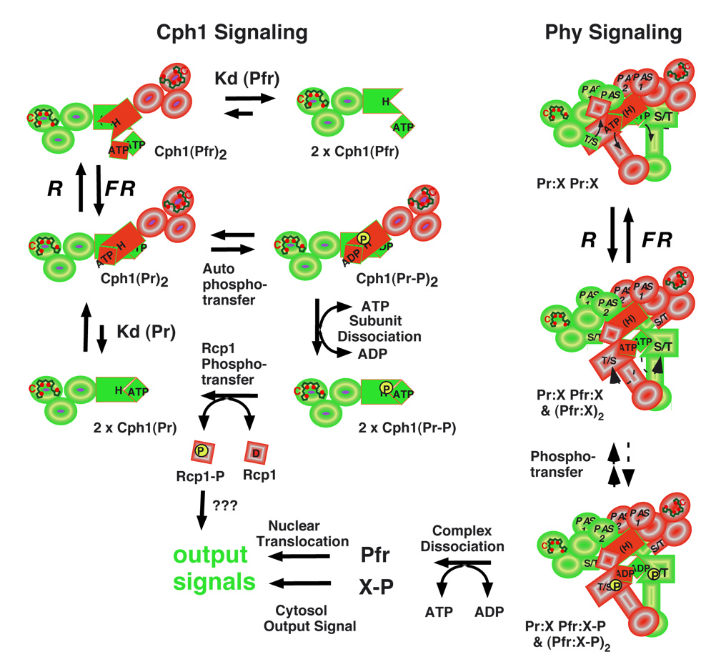
Phytochromes are a widespread family of red/far-red responsive photoreceptors first discovered in plants, where they constitute one of the three main classes of photomorphogenesis regulators. All phytochromes utilize covalently attached bilin chromophores that enable photoconversion between red-absorbing (P(r)) and far-red-absorbing (P(fr)) forms. Phytochromes are thus photoswitchable photosensors; canonical phytochromes have a conserved N-terminal photosensory core and a C-terminal regulatory region, which typically includes a histidine-kinase-related domain. The discovery of new bacterial and cyanobacterial members of the phytochrome family within the last decade has greatly aided biochemical and structural characterization of this family, with the first crystal structure of a bacteriophytochrome photosensory core appearing in 2005. This structure and other recent biochemical studies have provided exciting new insights into the structure of phytochrome, the photoconversion process that is central to light sensing, and the mechanism of signal transfer by this important family of photoreceptors.


Figures

Figure 1
Figure 1. Domain structure and chromophores of phytochromes
(a) The phytochrome photocycle. Illumination of Pr phytochrome with red light (R) produces lumi-R as the primary photoproduct. This is subsequently converted to Pfr via multiple light-independent steps. Pfr can be converted into Pr either by illumination with far-red light (FR), producing lumi-F and then Pr via subsequent thermal steps, or by an entirely thermal process known as dark reversion (d.r., center). The ratio between Pr and Pfr (and hence between the two physiological outputs) is thus determined by the light environment and by the rate of dark reversion. (b) Domain architecture of the extended phytochrome family. The five classes of phytochromes possess an N-terminal photosensory core region and typically share regulatory output domains related to those found on two component histidine kinases (HKRD). The P3/GAF domain is associated with the bilin chromophore and is highly conserved. All phytochromes except those found in the Cph2 subfamily share P2/PAS domains, while P4/PHY photosensory domains are specific to phytochromes and are thought to have folds similar to GAF domains (69). Plant phytochromes (Phys) possess two additional PAS domains within the regulatory region. Fungal phytochromes (Fphs) have a domain structure similar to those of the cyanobacterial phytochrome 1 (Cph1) and bacteriophytochrome (BphP) families, except for an additional C-terminal response regulator receiver domain (RR) extension and variable N-terminal extensions.
Figure 2
Figure 2. Chromophore structure and sssembly
(a) The structures of the bilin chromophores utilized by known phytochromes are shown. Left, phycocyanobiliin (PCB) and phytochromobilin (PΦB) chromophores share a reduced A ring and differ only at the C18 side chain. These chromophores are utilized by plant and algal Phys and cyanobacterial Cph1s and Cph2s. Right, the BphPs and Fphs instead utilize biliverdin (BV) as chromophore. All chromophores are shown in the C5–Z,syn C10–Z,syn C15–Z,anti configuration adopted in the Pr state (103). (b) Conformations of PCB thought to be present during the assembly reaction with Cph1 are shown (6). The cyclic, porphyrin-like C15–Z,syn species (left) is the most stable in solution at neutral pH and initially binds to apoCph1. After binding, the B/C ring system becomes protonated, driving adoption of a C15–Z,anti conformation (right) which is characterized by enhanced, red-shifted long wavelength absorbance. This species then becomes covalently attached to Cys259 of Cph1 to give the Pr structure shown in (a). BV is bound to a different Cys upstream of the P2/PAS domain of BphPs (58, 103).
Figure 3
Figure 3. Conservation of the PAS and GAF domains of phytochromes
The 2.5 Å crystal structure of DrBphP P2/PAS and P3/GAF domains (PDB code 1ZTU: (103)) is shown with bound BV chromophore covalently attached to Cys24 (bronze) colored by domains (top left), similarity (top right), gaps (bottom left), and known alleles of plant Phys (bottom right). The DrBphP structure colored by domains (top left) uses the following color scheme: PAS, blue; GAF, red; N-terminal knot interface, green; GAF insert knot interface, purple; N-terminus, grey. The DrBphP structure colored by similarity (top right) uses a normalized BLOSUM62 matrix (38) and the alignment of 122 phytochromes presented in Supplemental Figure 1. A continuous color scale is used, ranging from dark blue (100% conserved) to bright red (variable). The DrBphP structure colored by length of gaps (bottom left) uses the alignment in Supplemental Figure 1. A continuous color scale ranges from light blue (no gaps) to bright red (gaps ≥ 5 amino acids long), with a gap defined as a position where any phytochrome has insertions relative to DrBphP. The DrBphP structure colored by the location of alleles in plant phytochromes (bottom right) shows alleles that have been reported within the PAS/GAF domains of DrBphP against a grey background (see Supplemental Table 2). Loss-of-function alleles are colored red, gain-of-function alleles are colored blue, positions with multiple phenotypes are colored yellow, and silent alleles are colored green. Figure 3 and Figure 4 were prepared using VMD (40), Tachyon (96), STRIDE (28) and homolmapper (N. C. R. and J. C. L., unpublished results). (b) Stereoview of the conserved trefoil knot at the interface between the PAS and GAF domains by residues 27–43 (green, upstream of the PAS domain and the first beta strand of the PAS domain) and 227–257 (purple). Ile35 (blue) is at the center of the knot.
Figure 4
Figure 4. Chromophore-protein interactions in DrBphP are conserved
(a) Interaction of the buried carboxylate side chains of BV (bronze) with DrBphP (103). The B ring carboxylate interacts with the conserved Arg254, which is part of the trefoil knot, while the C ring carboxylate interacts with conserved Ser272 and His260. All protein residues within 3.5 Å of the carboxylate oxygens are shown colored by similarity as in Figure 2. Secondary structure elements are shown in transparent grey for residues 214–218, 254–262, and 271–275 for reference. (b) Environment of the C10 bridging carbon (bronze sphere). This atom is held in place by the B and C rings of biliverdin along with the conserved Asp207, Ile208, Tyr216, His260, and Pro209 (shown as sticks colored by atom type and as solvent-accessible surface, with the surface colored by similarity as in Figure 2). (c) Environment of the D ring. Residues within 5 Å of the chromophore D ring and/or C15 methine bridge are shown as sticks and surface as in part (b).
Figure 5
Figure 5. Chemical delineation of the phytochrome photocycle
(a) Structures and spectra for synthetic, sterically locked bilins (44) assembled with the bacteriophytochrome Agp1 from Agrobacterium tumefaciens. (left) Spectra for the C15–Z,anti locked bilin (dashed) and Pr biliverdin (solid) adducts. (b) Spectra for the C15–E,anti locked bilin (dashed) and Pfr biliverdin (solid) adducts. Spectra in (a) and (b) are courtesy of Drs. Tilman Lamparter and Katsuhiko Inomata. (c) Proposed photocycle for phytochromes utilizing PCB or PΦB. The Pr conformation is assigned based on the crystal structure of DrBphP, the locked bilin data presented in part (a), and the known stereochemistry of the 3 stereocenters in these molecules. Illumination with red light triggers photoisomerization about the C15–C16 double bond (I) to give the C15–E,anti primary photoproduct lumi-R, which is subsequently converted to Pfr in several light-independent steps (II). As discussed in the text, the proposed Pfr is hypothetical but would account for the observed instability of the Pfr chromopeptide, the red-shifted Pfr absorbance maximum, and the observed Pfr dark reversion. Illumination of Pfr with far-red light (III) triggers the reverse photoisomerization to yield the C15–Z,anti lumi-F primary photoproduct, which is subsequently converted to Pr in a series of light-independent steps (IV). Dark reversion would proceed through the Pfr resonance form with single-bond character about C15–C16, which would readily undergo thermal rotation about this bond and then convert to Pr(V).
Figure 6
Figure 6. Hypothetical signaling mechanisms for prokaryote and eukaryote phytochromes
Homodimerization of the prokaryote phytochrome (Cph1) is dynamic and light-dependent (upper left) since autophosphorylation is favored by the formation of homodimers in the Pr form (Cph1(Pr)2) and inhibited by conversion to Pfr (Cph1(Pfr)2) which dissociates to an inactive monomer ((Cph1(Pfr)). Exchange of bound ADP with ATP, a process that promotes dissociation of the phosphorylated Pr dimer (Cph1(Pr-P)2) by inhibiting reassociation of the phosphorylated Pr monomer (Cph1(Pr-P)), stimulates histidine to aspartate phosphotransfer to Cph1's substrate Rcp1. The dephosphorylated Pr monomer (Cph1(Pr)) reassociates to form the active homodimer (Cph1(Pr)2). Eukaryote phytochromes (Phys) are obligate homodimers that are associated with a cytosolic anchoring protein X in an ATP-dependent protein complex (upper right). Photoconversion yields a Pr-Pfr heterodimer/Pfr-Pfr homodimer mixture (Pr:X Pfr:X & (Pfr:X)2) which results in activation of the Ser/Thr kinase activity and the stimulation of phosphotransfer to anchoring protein X. The exchange of bound ADP with ATP favors dissociation of the Pfr:X complexes, enabling free Pfr to move to the nucleus and phosphorylated X to mediate a cytosolic output signal.

Similar articles

Cited by

References

    1. Andel F, Lagarias JC, Mathies RA. Resonance Raman analysis of chromophore structure in the lumi-R photoproduct of phytochrome. Biochem. 1996;35:15997–16008. - PubMed
    1. Andel F, Murphy JT, Haas JA, McDowell MT, van der Hoef I, et al. Probing the photoreaction mechanism of phytochrome through analysis of resonance Raman vibrational spectra of recombinant analogues. Biochem. 2000;39:2667–2676. - PubMed
    1. Batschauer A, editor. Photoreceptors and Light Signaling. Vols. 3. Cambridge, UK: Royal Society of Chemistry; 2003. 388 pp.
    1. Bhoo SH, Davis SJ, Walker J, Karniol B, Vierstra RD. Bacteriophytochromes are photochromic histidine kinases using a biliverdin chromophore. Nature. 2001;414:776–779. - PubMed
    1. Blumenstein A, Vienken K, Tasler R, Purschwitz J, Veith D, et al. The Aspergillus nidulans phytochrome FphA represses sexual development in red light. Curr. Biol. 2005;15:1833–1838. - PubMed

Publication types