. 2006 Jul 1;397(1):223-31.
doi: 10.1042/BJ20060220.

Functional interactions of the SPAK/OSR1 kinases with their upstream activator WNK1 and downstream substrate NKCC1

Affiliations

Functional interactions of the SPAK/OSR1 kinases with their upstream activator WNK1 and downstream substrate NKCC1

Alberto C Vitari et al. Biochem J. .

Abstract

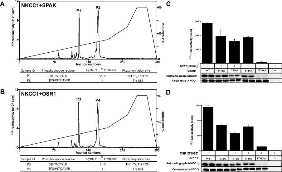
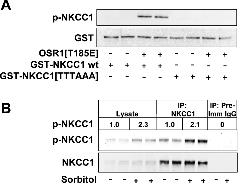
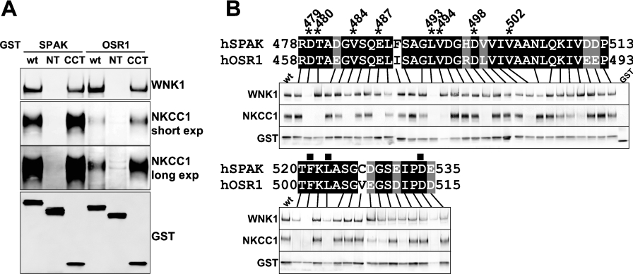
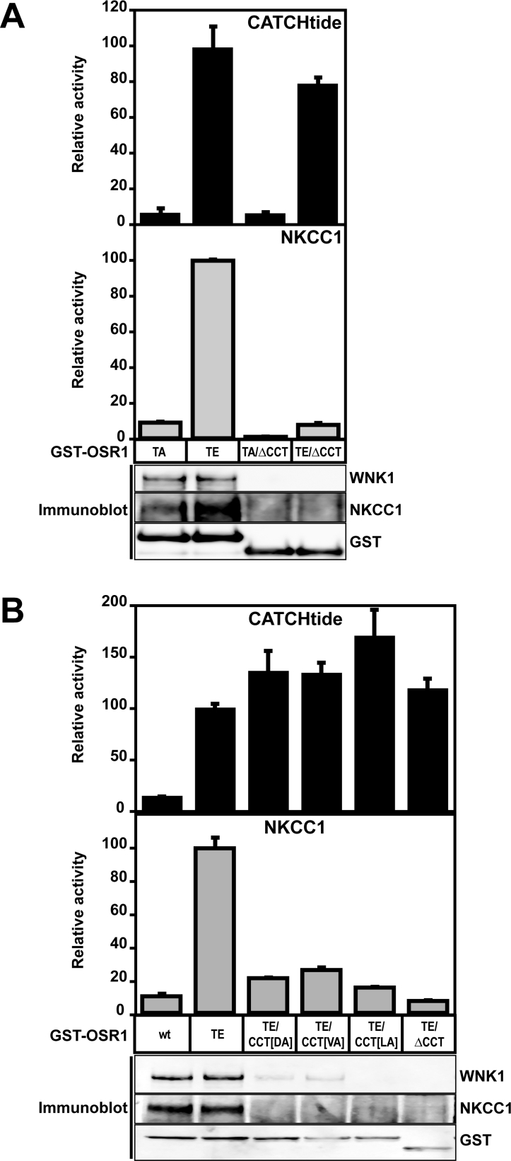
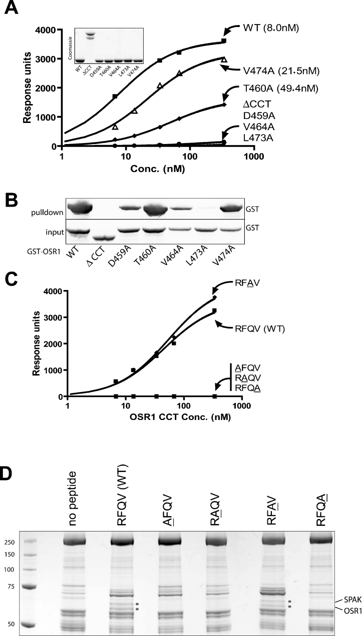
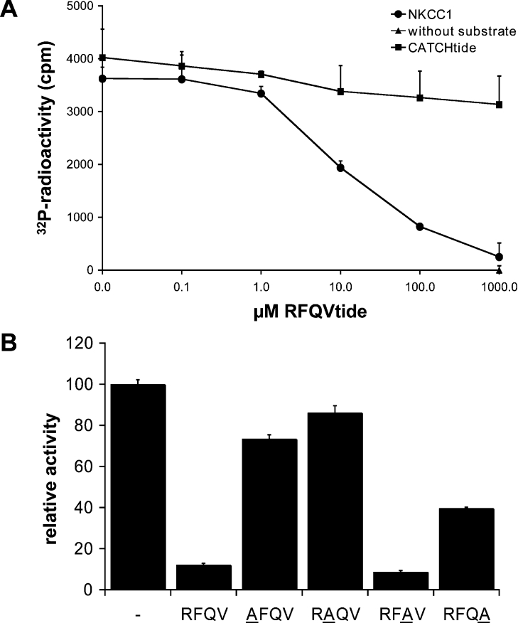
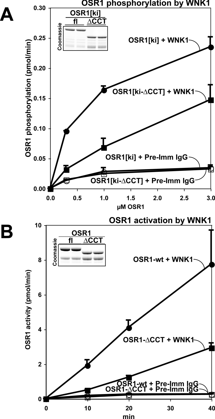
The SPAK (STE20/SPS1-related proline/alanine-rich kinase) and OSR1 (oxidative stress-responsive kinase-1) kinases interact and phosphorylate NKCC1 (Na+-K+-2Cl- co-transporter-1), leading to its activation. Recent studies indicated that SPAK and OSR1 are phosphorylated and activated by the WNK1 [with no K (lysine) protein kinase-1] and WNK4, genes mutated in humans affected by Gordon's hypertension syndrome. In the present study, we have identified three residues in NKCC1 (Thr175/Thr179/Thr184 in shark or Thr203/Thr207/Thr212 in human) that are phosphorylated by SPAK and OSR1, and have developed a peptide substrate, CATCHtide (cation chloride co-transporter peptide substrate), to assess SPAK and OSR1 activity. Exposure of HEK-293 (human embryonic kidney) cells to osmotic stress, which leads to phosphorylation and activation of NKCC1, increased phosphorylation of NKCC1 at the sites targeted by SPAK/OSR1. The residues on NKCC1, phosphorylated by SPAK/OSR1, are conserved in other cation co-transporters, such as the Na+-Cl- co-transporter, the target of thiazide drugs that lower blood pressure in humans with Gordon's syndrome. Furthermore, we characterize the properties of a 92-residue CCT (conserved C-terminal) domain on SPAK and OSR1 that interacts with an RFXV (Arg-Phe-Xaa-Val) motif present in the substrate NKCC1 and its activators WNK1/WNK4. A peptide containing the RFXV motif interacts with nanomolar affinity with the CCT domains of SPAK/OSR1 and can be utilized to affinity-purify SPAK and OSR1 from cell extracts. Mutation of the arginine, phenylalanine or valine residue within this peptide abolishes binding to SPAK/OSR1. We have identified specific residues within the CCT domain that are required for interaction with the RFXV motif and have demonstrated that mutation of these in OSR1 inhibited phosphorylation of NKCC1, but not of CATCHtide which does not possess an RFXV motif. We establish that an intact CCT domain is required for WNK1 to efficiently phosphorylate and activate OSR1. These data establish that the CCT domain functions as a multipurpose docking site, enabling SPAK/OSR1 to interact with substrates (NKCC1) and activators (WNK1/WNK4).


Figures

Figure 1
Figure 1. Sites on NKCC1 phosphorylated by SPAK and OSR1
(A) Shark NKCC1-(1–260) was phosphorylated by wild-type SPAK as described in the Materials and methods section. The 32P-labelled NKCC1 was isolated by electrophoresis on a polyacrylamide gel and digested with trypsin, and the resulting peptides were chromatographed on a C18 column equilibrated in 0.1% (v/v) trifluoroacetic acid in water. The column was developed with a linear acetonitrile gradient (diagonal line) at a flow rate of 0.2 ml/min, and fractions (0.1 ml each) were collected. The peak fractions containing the major 32P-labelled peptides are marked as P1 and P2. The indicated peptides were analysed by MALDI–TOF/TOF MS as described in the Materials and methods section. The site of phosphorylation within each peptide was determined by solid-phase Edman sequencing in which 32P radioactivity was measured after each cycle of Edman degradation. The cycle number in which 32P radioactivity was released is indicated. The deduced amino acid sequences of P1 and P2 are indicated in which the phosphorylated residues are underlined. (B) As in (A), except that wild-type OSR1 was employed in place of SPAK, and the major 32P-labelled peptides are marked as P3 and P4. (C) Active [T233E]SPAK was incubated with bacterially expressed wild-type NKCC1-(1–260) or the indicated mutants in the presence of Mg-[γ-32P]ATP. After incubation, the samples were analysed by PAGE and phosphorylation of NKCC1 protein substrate was determined by scintillation counting. NKCC1-TTTAAA corresponds to a mutant in which Thr175, Thr179 and Thr184 were all mutated to alanine. (D) As in (C), except that active [T185E]OSR1 was employed in place of [T233E]SPAK.
Figure 2
Figure 2. Phosphorylation of endogenous NKCC1
(A) The indicated forms of GST–NKCC1-(1–260) were incubated with or without constitutively active [T185E]OSR1 in the presence of MgATP and immunoblotted with anti-phospho-NKCC1 antibody (p-NKCC1) or an anti-GST antibody. (B) HEK-293 cells were incubated with or without 0.5 M sorbitol for 30 min and then lysed. Cell lysates (40 μg), or immunoprecipitations (IP) undertaken from 2 mg of lysate with NKCC1 or pre-immune (Pre-Imm) IgG, were analysed by immunoblotting with the anti-phospho-NKCC1 antibody (p-NKCC1) or an antibody raised against NKCC1-(1–260). Similar results were obtained in three separate experiments. Quantification of the phospho-NKCC1 immunoblot was undertaken using the Li-Cor Odyssey® IR imaging system. Results are presented relative to the mean value observed in unstimulated cells, which was given a value of 1.0. OSR1[T185E], GST–[T185E]OSR1; GST-NKCC1 wt, GST–NKCC1-(1–260); GST-NKCC1[TTTAAA], GST–[T175A/T179A/T184A]NKCC1-(1–260).
Figure 3
Figure 3. Generation of a peptide substrate for OSR1
(A) Multiple sequence alignment of the indicated fragments of cation–chloride co-transporters encompassing the NKCC1 SPAK and OSR1 sites of phosphorylation, performed using the program T-Coffee [18]. The alignment was graphically represented with BOXSHADE version 3.21 at http://www.ch.embnet.org/software/BOX_form.html using default parameters. Identical residues are highlighted in black and similar residues are in grey. The region of NKCC1 that encompasses CATCHtide is marked with a continuous line, the sites phosphorylated by SPAK and OSR1 are marked with a closed inverted triangle, and the previously identified residues on NKCC1 phosphorylated in cells [13] are marked with closed square. h, human; s, shark. (B) Constitutively active GST–[T185E]OSR1 was incubated with the indicated amount of CATCHtide in presence of Mg-[γ-32P]ATP and incorporation of [32P]phosphate into the substrate was determined. Each assay was carried out in triplicate and the results are means±S.D. Similar results were obtained in at least three experiments.
Figure 4
Figure 4. Analysis of the interaction of SPAK and OSR1 with WNK1 and NKCC1
(A) HEK-293 cells were transfected with constructs encoding the indicated GST-fusion proteins. At 36 h post-transfection, the GST-fusion proteins were affinity-purified on glutathione–Sepharose beads that were immunoblotted with anti-GST, anti-WNK1 or anti-NKCC1 antibody to detect endogenously associated WNK1 and NKCC1. SPAK wt, full-length GST–SPAK (547 amino acids long); SPAK NT, GST–SPAK-(1–455); SPAK CCT, GST–SPAK-(449–547); OSR1 wt, GST–OSR1 (527 amino acids long); OSR1 NT, GST–OSR1-(1–435); OSR1 CCT, GST–OSR1-(429–527); exp, exposure. (B) HEK-293 cells were transfected with constructs encoding the isolated CCT domain of SPAK [GST–SPAK-(449–547)] or the mutants in which the indicated residues had been mutated to alanine. Binding of NKCC1 and WNK1 to these was assessed as above. Residues marked with an asterisk represent mutations that markedly affected binding to NKCC1 and/or WNK1, whereas residues marked with a closed square, although they affected binding to NKCC1 and WNK1, were also found to markedly decrease expression of the isolated CCT domain as well as the full-length protein (results not shown). wt, wild-type.
Figure 5
Figure 5. Role of the CCT domain in enabling OSR1 to phosphorylate NKCC1 and CATCHtide
(A and B) HEK-293 cells were transfected with constructs encoding the indicated mutants of OSR1 as GST-fusion proteins. At 36 h post transfection, the fusion proteins were affinity-purified and incubated with CATCHtide (upper histograms) or sNKCC1-(1–260) (lower histograms) as substrates in the presence of Mg-[γ-32P]ATP, and phosphorylation of these substrates was determined as described in the Materials and methods section. The results are presented relative to the kinase activity observed with GST–[T185E]OSR1, which was given a value of 100. The purified GST–OSR1 proteins were also immunoblotted with indicated antibodies. (A) TA, GST–[T185A]OSR1; TE, GST–[T185E]OSR1; TA/ΔCCT, GST–[T185A]OSR1-ΔCCT; TE/ΔCCT, GST–[T185E]OSR1-ΔCCT. (B) wt, GST–OSR1; TE, GST–[T185E]OSR1; TE/CCT[DA], GST–[T185E/D459A]OSR1; TE/CCT[VA], GST–[T185E/V464A]OSR1; TE/CCT[LA], GST–[T185E/L473A]OSR1, TE/ΔCCT, GST-[T185E]OSR1-ΔCCT.
Figure 6
Figure 6. Analysis of the binding properties of the CCT domain
(A) The binding of bacterially expressed isolated forms of the OSR1 CCT domain [WT; OSR1-(429–527)] or OSR1-ΔCCT (residues 1–435) to a biotinylated RFQV motif-containing peptide derived from WNK4 (biotin–SEEGKPQLVGRFQVTSSK) was analysed using BiaCore as described in the Materials and methods section. The analysis was performed over a range of protein concentrations (6.8–340 nM) and the response level at steady state was plotted against the log of the protein concentration. The Kd values were calculated by fitting the data to the equation y=Bmax·x/(Kd+x) using GraphPad 4 software, which describes the binding of a ligand to a receptor that follows the law of mass action. Bmax is the maximal binding and Kd is the concentration of ligand required to reach half-maximal binding, whereas x and y correspond to the protein concentration and the response units respectively. The inset shows the similar level of the recombinant proteins employed for this analysis. (B) HEK-293 cells were transfected with GST-tagged wild-type (WT) OSR1 or the indicated mutants. At 36 h post-transfection, cells were lysed and subjected to affinity-purification with the biotinylated RFQV peptide as described in the Materials and methods section. The affinity-purified samples (termed pulldown) were immunoblotted with an anti-GST antibody to detect interaction of GST-fusion proteins with the peptide. The cell lysates before affinity-purification (termed input) were also subjected to immunoblotting. (C) As in (A), except that binding of the isolated OSR1 CCT domain to the indicated biotinylated peptides was determined. The peptide sequences employed are RFQV, biotin–SEEGKPQLVGRFQVTSSK; AFQV, biotin–SEEGKPQLVGAFQVTSSK; RAQV, biotin–SEEGKPQLVGRAQVTSSK; RFAV, biotin–SEEGKPQLVGRFAVTSSK; RFQA, biotin–SEEGKPQLVGRFQATSSK. Mutated residues are underlined. (D) Non-transfected HEK-293 cell lysates were incubated with the biotinylated peptides described in (C) and subjected to affinity-purification with streptavidin–Sepharose. The purified samples were analysed on a Coomassie Blue-stained SDS/PAGE gel. The bands marked with an asterisk were subjected to MS fingerprinting and their identity was confirmed as endogenously expressed SPAK and OSR1. Molecular-masses are given in kDa.
Figure 7
Figure 7. Effects of RFQV motif-containing peptide on OSR1 kinase activity
(A) GST–[T185E]OSR1 was incubated with either NKCC1-(1–260) (5 μM) or CATCHtide (10 μM) or alone in the presence of Mg-[γ-32P]ATP and the indicated concentration of RFQV motif-containing peptide. After incubation, the phosphorylation of substrates was determined. Results are means±S.D. for a triplicate experiment. Similar results were obtained in two separate experiments. (B) As in (A), except that samples were incubated with 1 mM of the indicated peptide whose sequence is the same as that described in the legend to Figure 6, except that they were not biotinylated.
Figure 8
Figure 8. Role of the CCT domain in regulating OSR1 activation by WNK1
(A) WNK1 immunoprecipitated from HEK-293 cell lysate (closed symbols) or a control immunoprecipitation performed with pre-immune (Pre-Imm) IgG (open symbols) was incubated in the presence of Mg-[γ-32P]ATP and the indicated concentration of full-length kinase-inactive OSR1[ki] or kinase-inactive OSR1-[ki-ΔCCT] purified from E. coli. After incubation, phosphorylation of OSR1 substrate was determined, and results are means±S.D. for duplicate experiments. The insert shows a Coomassie Blue-stained SDS/PAGE gel of the purified proteins quantified using a Li-Cor Odyssey® IR imaging system to ensure that a similar molar concentration of full-length OSR1[ki] and mutant OSR1[ki-ΔCCT] were employed in the assay. OSR1[ki] or fl, full-length [D164A]OSR1; OSR1[ki-ΔCCT] or ΔCCT, [D164A]OSR1-ΔCCT. (B) Full-length GST-OSR1 or GST–OSR1-ΔCCT purified from E. coli were incubated with either WNK1 immunoprecipitated from HEK-293 cell lysate (closed symbols) or with a control immunoprecipitation performed with pre-immune (Pre-Imm) IgG antibody (open symbols), in the presence of CATCHtide and Mg-[γ-32P]ATP. After the indicated time of incubation, phosphorylation of CATCHtide was determined and results are mean±S.D. activities for triplicate experiments. The inset shows a Coomassie Blue-stained SDS/PAGE gel of the purified GST-fusion proteins quantified using a Li-Cor Odyssey® IR imaging system to ensure that a similar molar concentration of wild-type OSR1 and mutant OSR1-ΔCCT were employed in the activity assay. Recombinant OSR1 migrates as a doublet band with the upper band migrating with the expected molecular mass. We presume that the lower band is a proteolytic fragment containing the kinase domain and lacking the C-terminal portion, as these enzymes were expressed as N-terminal GST-fusion proteins. OSR1-wt or fl, wild-type GST–OSR1; OSR1-ΔCCT or ΔCCT, GST–OSR1-ΔCCT.

Similar articles

Cited by

References

    1. Piechotta K., Lu J., Delpire E. Cation chloride cotransporters interact with the stress-related kinases Ste20-related proline-alanine-rich kinase (SPAK) and oxidative stress response 1 (OSR1) J. Biol. Chem. 2002;277:50812–50819. - PubMed
    1. Dowd B. F., Forbush B. PASK (proline-alanine-rich STE20-related kinase), a regulatory kinase of the Na–K–Cl cotransporter (NKCC1) J. Biol. Chem. 2003;278:27347–27353. - PubMed
    1. Gagnon K. B., England R., Delpire E. Volume sensitivity of cation–Cl− cotransporters is modulated by the interaction of two kinases: Ste20-related prolinealanine-rich kinase and WNK4. Am. J. Physiol. Cell Physiol. 2006;290:C134–C142. - PubMed
    1. Piechotta K., Garbarini N., England R., Delpire E. Characterization of the interaction of the stress kinase SPAK with the Na+–K+–2Cl− cotransporter in the nervous system: evidence for a scaffolding role of the kinase. J. Biol. Chem. 2003;278:52848–52856. - PubMed
    1. Moriguchi T., Urushiyama S., Hisamoto N., Iemura S., Uchida S., Natsume T., Matsumoto K., Shibuya H. WNK1 regulates phosphorylation of cation–chloride-coupled cotransporters via the STE20-related kinases, SPAK and OSR1. J. Biol. Chem. 2005;280:42685–42693. - PubMed

Publication types

MeSH terms