. 2006 May 9;103(19):7315-20.
doi: 10.1073/pnas.0510033103. Epub 2006 May 2.

Growth hormone promotes skeletal muscle cell fusion independent of insulin-like growth factor 1 up-regulation

Affiliations

Growth hormone promotes skeletal muscle cell fusion independent of insulin-like growth factor 1 up-regulation

Athanassia Sotiropoulos et al. Proc Natl Acad Sci U S A. .

Abstract

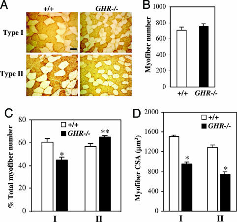
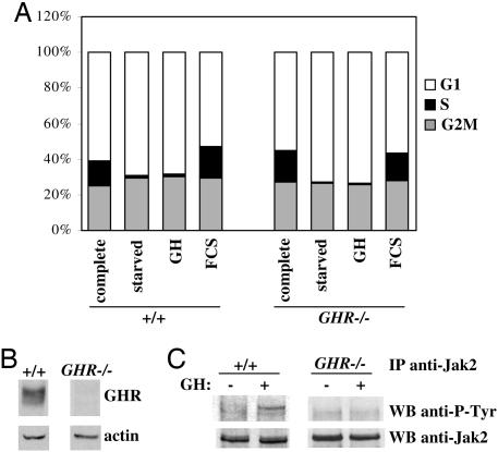
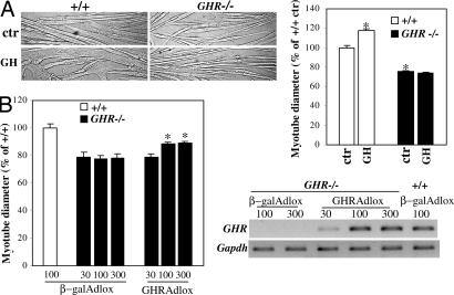
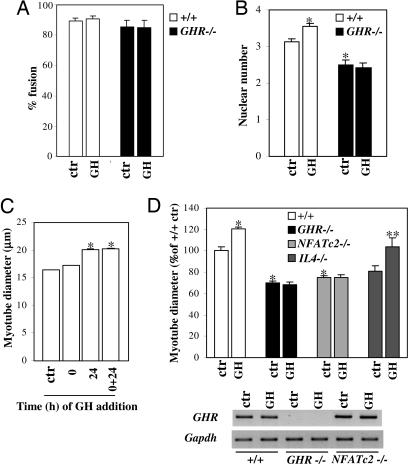
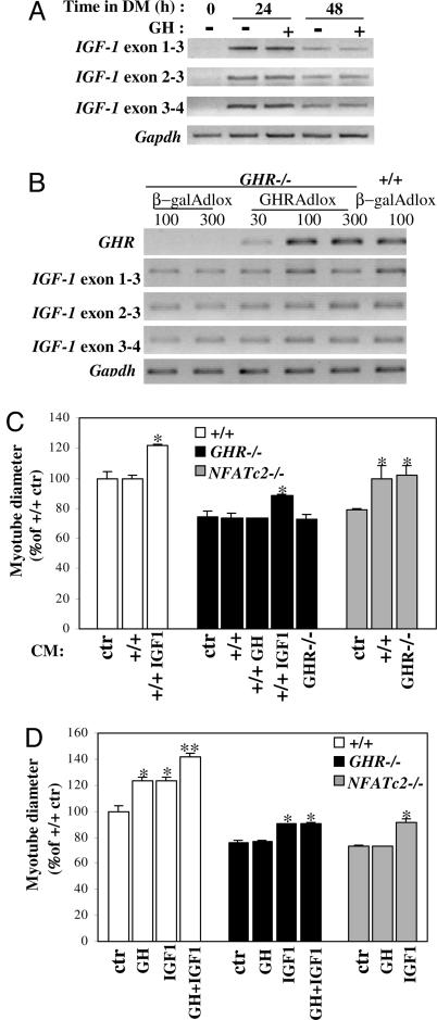
Growth hormone (GH) participates in the postnatal regulation of skeletal muscle growth, although the mechanism of action is unclear. Here we show that the mass of skeletal muscles lacking GH receptors is reduced because of a decrease in myofiber size with normal myofiber number. GH signaling controls the size of the differentiated myotubes in a cell-autonomous manner while having no effect on size, proliferation, and differentiation of the myoblast precursor cells. The GH hypertrophic action leads to an increased myonuclear number, indicating that GH facilitates fusion of myoblasts with nascent myotubes. NFATc2, a transcription factor regulating this phase of fusion, is required for GH action because GH is unable to induce hypertrophy of NFATc2-/- myotubes. Finally, we provide three lines of evidence suggesting that GH facilitates cell fusion independent of insulin-like growth factor 1 (IGF-1) up-regulation. First, GH does not regulate IGF-1 expression in myotubes; second, GH action is not mediated by a secreted factor in conditioned medium; third, GH and IGF-1 hypertrophic effects are additive and rely on different signaling pathways. Taken together, these data unravel a specific function of GH in the control of cell fusion, an essential process for muscle growth.


Conflict of interest statement

Conflict of interest statement: No conflicts declared.

Figures

Fig. 1.
Fig. 1.
Reduced muscle size in GHR−/− soleus muscles is due to a decrease in myofiber CSA. (A) Representative sections of wild-type (+/+) and GHR−/− soleus immunostained with anti-type I and anti-type II MyHC antibodies. (Scale bars: 50 μm in Upper Left.) (B) Myofiber number of +/+ and GHR−/− soleus. (C) Fiber type distribution in +/+ and GHR−/− soleus. (D) The CSA of both type I and type II MyHC-expressing myofibers. Data are means ± SEM of four mice per genotype. ∗, P < 0.005; ∗∗, P < 0.05, vs. +/+.
Fig. 2.
Fig. 2.
GH signaling does not affect myoblast proliferation and size. (A) Cell-cycle distribution of wild-type and GHR−/− cells grown in complete medium, starved or stimulated with 300 ng/ml GH or with 20% FCS, were analyzed by flow cytometry after propidium iodide staining. Percentages of cells in G1-phase, in S-phase, and in G2M-phase from one representative experiment are presented. (B) Analysis of GHR expression by immunoblot in myoblasts. Actin expression was used as a loading control. (C) Jak2 phosphorylation in cells starved and stimulated with GH. Immunoprecipitated proteins with anti-Jak2 were analyzed by immunoblot with anti-phosphotyrosine (P-Tyr) and anti-Jak2 antibodies.
Fig. 3.
Fig. 3.
GH signaling increases myotube size but does not affect myogenesis. (A) Myoblasts were induced to differentiate for 48 h in the presence of GH, and myotube diameter was measured and expressed as percentage change over control wild-type (+/+) cells. Histograms are means ± SEM of at least eight experiments from three independent cell cultures. ∗, P < 0.005, vs. +/+ control. Bright-field images of myotubes from +/+ and GHR−/− cells are shown. (B) GHR−/− myoblasts were transduced with GHR (GHRAdlox) or β-galAdlox adenoviruses at 30, 100, and 300 multiplicities of infection and induced to differentiate for 48 h. Myotube diameter was measured and is expressed as percentage change over +/+ cells transduced by β-galAdlox. Histograms are means ± SEM of four experiments on two independent cultures. ∗, P < 0.005, vs. GHR−/− transduced by β-galAdlox. Expression of GHR mRNA was determined by semiquantitative RT-PCR analysis. Gapdh expression was used as a control.
Fig. 4.
Fig. 4.
GH facilitates muscle cell fusion. (A) Wild-type (+/+) and GHR−/− myoblasts were induced to differentiate and treated with GH for 48 h. After staining of nuclei, the fusion index was calculated. Histograms are means ± SEM of four experiments on two independent cultures. (B) The experiment was performed as in A, and the nuclear number was counted. Data are means ± SEM of five experiments on two independent cultures. ∗, P < 0.05, vs. +/+ control. (C) Cells were differentiated and treated with GH at 0 h, 24 h, or both 0 and 24 h. Myotube diameter was measured after 48 h. Data are means ± SEM of three experiments. ∗, P < 0.005, vs. +/+ control. (D) Myoblasts of the indicated genotype were differentiated in the presence of GH. After 48 h, myotube diameter was measured and is expressed as percentage change over +/+ control. Histograms are means ± SEM of at least three independent experiments. ∗, P < 0.005, vs. +/+ control; ∗∗, P < 0.05, vs. IL4−/− control. Expression of endogenous GHR mRNA was determined by semiquantitative RT-PCR analysis (Lower). Gapdh expression was used as a control.
Fig. 5.
Fig. 5.
GH hypertrophic action is independent of IGF-1. (A) Myoblasts were induced to differentiate in the presence of GH. RNA was extracted at the indicated time, and expression of IGF-1 transcripts was studied by semiquantitative RT-PCR analysis. Primers designed to detect transcripts regulated by promoter 1 (exons 1 and 3) or promoter 2 (exons 2 and 3) and primers detecting all IGF-1 isoforms were used (exons 3 and 4). Gapdh expression was used as a control. (B) GHR−/− myoblasts were transduced with GHRAdlox or β-galAdlox adenoviruses at 30, 100, and 300 multiplicities of infection and induced to differentiate. After 48 h, expression of IGF-1 transcripts was analyzed by semiquantitative RT-PCR as in A. The experiments were repeated at least twice on two independent cultures with similar results. (C) Myoblasts of the indicated genotype were induced to differentiate in DM or in +/+ (with or without GH or IGF-1-R3) or GHR−/− CM for 48 h, and myotube diameter was measured. Histograms are means ± SEM of three independent experiments. ∗, P < 0.05, vs. +/+, GHR−/−, or NFATc2−/− control. (D) Myoblasts were induced to differentiate for 48 h in the presence of GH, IGF-1-R3, or both GH and IGF-1-R3, and myotube diameter was measured. Histograms are means ± SEM of three independent experiments. ∗, P < 0.005, vs. +/+, GHR−/−, or NFATc2−/− control; ∗∗, P < 0.005, vs. +/+ treated by GH or IGF-1.

Similar articles

Cited by

References

    1. Florini J. R., Ewton D. Z., Coolican S. A. Endocr. Rev. 1996;17:481–517. - PubMed
    1. Hoffman A. R., Kuntze J. E., Baptista J., Baum H. B., Baumann G. P., Biller B. M., Clark R. V., Cook D., Inzucchi S. E., Kleinberg D., et al. J. Clin. Endocrinol. Metab. 2004;89:2048–2056. - PubMed
    1. McNurlan M. A., Garlick P. J., Steigbigel R. T., Decristofaro K. A., Frost R. A., Lang C. H., Johnson R. W., Santasier A. M., Cabahug C. J., Fuhrer J., et al. J. Clin. Invest. 1997;100:2125–2132. - PMC - PubMed
    1. Le Roith D., Bondy C., Yakar S., Liu J. L., Butler A. Endocr. Rev. 2001;22:53–74. - PubMed
    1. Shavlakadze T., Winn N., Rosenthal N., Grounds M. D. Growth Horm. IGF Res. 2005;15:4–18. - PubMed

Publication types

MeSH terms

LinkOut - more resources