Review
. 2006 May;7(5):490-5.
doi: 10.1038/sj.embor.7400682.

T-cell antigen-receptor stoichiometry: pre-clustering for sensitivity

Affiliations
Review

T-cell antigen-receptor stoichiometry: pre-clustering for sensitivity

Balbino Alarcón et al. EMBO Rep. 2006 May.

Abstract

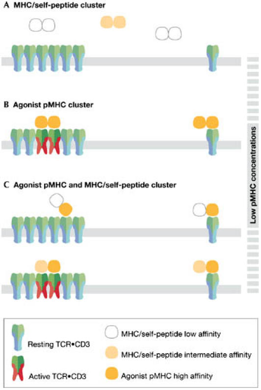
The T-cell antigen receptor (TCR x CD3) is a multi-subunit complex that is responsible for triggering an adaptive immune response. It shows high specificity and sensitivity, while having a low affinity for the ligand. Furthermore, T cells respond to antigen over a wide concentration range. The stoichiometry and architecture of TCR x CD3 in the membrane have been under intense scrutiny because they might be the key to explaining its paradoxical properties. This review highlights new evidence that TCR x CD3 is found on intact unstimulated T cells in a monovalent form (one ligand-binding site per receptor) as well as in several distinct multivalent forms. This is in contrast to the TCR x CD3 stoichiometries determined by several biochemical means; however, these data can be explained by the effects of different detergents on the integrity of the receptor. Here, we discuss a model in which the multivalent receptors are important for the detection of low concentrations of ligand and therefore confer sensitivity, whereas the co-expressed monovalent TCR x CD3s allow a wide dynamic range.


Figures

Figure 1
Figure 1
The T-cell antigen receptor (TCR•CD3) complex consists of the αβ, γε, δε and ζζ dimers. Schematic pictures of the individual components of the TCR•CD3 complex are shown. The variable immunoglobulin domains of TCRαβ (V) bind to the ligand, whereas the cytoplasmic tails of the CD3 subunits (γε and δε) and TCRζζ interact with cytosolic-signalling proteins. Disulphide bridges are shown by thick black lines. Potential charges in the transmembrane regions are also depicted.
Figure 2
Figure 2
Arrangement of the T-cell antigen receptor (TCR•CD3) complexes on the cell surface before stimulation. The old model shows monovalent receptors with a stoichiometry of αβγεδεζζ distributed evenly on the plasma membrane. The new model has monovalent receptors and a variety of multivalent receptors that co-exist in the intact membrane of unstimulated T cells, and that have been detected by fluorescence resonance-energy transfer and electron microscopy. Due to the lack of certainty, the stoichiometries of the monovalent and multivalent TCR•CD3s are not shown. The monovalent complexes, however, might have the αβγεδεζζ stoichiometry. The multivalent TCR•CD3s might be present in a different lipid context (dark grey). The cell surface also contains large areas that are devoid of any TCR•CD3. TCRαβ is shown in green and CD3 is shown in various shades of blue/turquoise.
Figure 3
Figure 3
Influence of different detergents on the integrity of the multivalent T-cell antigen receptor (TCR•CD3) complex. The detergents Brij96, Brij98, lubrol and octylglucoside keep the multivalent TCR•CD3 intact. Digitonin disrupts the multivalent complexes, resulting in the αβγεδεζζ stoichiometry. In contrast to murine TCR•CD3, the human receptor is disassembled into its dimeric constituents by nonidet P-40 (NP40) and TritonX-100 (TX-100; San Jose et al, 1998). Sodium dodecyl sulphate (SDS) treatment leads to unfolding of the subunits and subsequent destruction of all non-covalent interactions.
Figure 4
Figure 4
Multivalent T-cell antigen receptors (TCR•CD3s) have higher avidity towards the ligand than monovalent TCR•CD3s. (A) Major histocompatibility complex (MHC)/self-peptide clusters (white or yellow) do not bind with sufficient avidity to either TCR•CD3 form and thus do not activate T cells. (B) Clusters of MHC containing two agonistic peptides bind with sufficient avidity to the multivalent, but not the monovalent, TCR•CD3. Consequently, at low pMHC concentrations, only multivalent TCR•CD3s are activated. (C) In clusters with one antigenic pMHC (orange) and MHC/self-peptides of intermediate affinity (yellow), the average affinity of both interactions will determine whether the multivalent TCR•CD3s can be bound with a sufficiently high avidity for T-cell activation. If the affinity of the MHC/self-peptide is low (upper panel), then TCR•CD3 triggering does not occur. If the affinity of the MHC/self-peptide is above a certain threshold, then, in combination with the agonist pMHC, the avidity might be high enough to trigger multivalent TCR•CD3 (lower panel). This avidity is not sufficient to cluster and activate two monovalent TCR•CD3s. Thus, small amounts of agonistic pMHC presented by an antigen-presenting cell might preferentially trigger multivalent TCR•CD3.
Figure 5
Figure 5
Multivalent T-cell antigen receptors (TCR•CD3s) allow spreading of the signal within the multimer. (A) At low agonist–major histocompatibility complex peptide (pMHC) concentrations, the multimeric TCR•CD3 complexes become preferentially activated. This might be due to their higher avidity for multimeric antigenic pMHC (orange). TCRαβs of multivalent TCR•CD3s that are not engaged by antigenic pMHC can bind to MHC/self-peptide (white) and thereby become activated. By amplifying the effect of a few pMHC-binding events to neighbouring receptors within a TCR•CD3 multimer, the T cell might be able to respond to low pMHC doses. (B) At medium agonist–pMHC concentrations, the multimers are saturated. (C) At high agonist–pMHC concentrations, monovalent TCR•CD3s begin to be activated. This allows the T cell to sense high doses of antigen when the multivalent receptors are saturated. (D) Only at high ligand concentrations, when all multivalent and monovalent receptors are engaged, is the T-cell response saturated. APC, antigen-presenting cell.

Similar articles

Cited by

References

    1. Alarcón B, Gil D, Delgado P, Schamel WWA (2003) Initiation of TCR signaling: regulation within CD3 dimers. Immunol Rev 191: 38–46 - PubMed
    1. Anderson HA, Hiltbold EM, Roche PA (2000) Concentration of MHC class II molecules in lipid rafts facilitates antigen presentation. Nat Immunol 1: 156–162 - PubMed
    1. Bachmann MF, Speiser DE, Zakarian A, Ohashi PS (1998) Inhibition of TCR triggering by a spectrum of altered peptide ligands suggests the mechanism for TCR antagonism. Eur J Immunol 28: 3110–3119 - PubMed
    1. Blumberg RS, Alarcón B, Sancho J, McDermott FV, Lopez P, Breitmeyer J, Terhorst C (1990) Assembly and function of the T cell antigen receptor. Requirement of either the lysine or arginine residues in the transmembrane region of the α chain. J Biol Chem 265: 14036–14043 - PubMed
    1. Blumberg RS, Ley S, Sancho J, Lonberg N, Lacy E, McDermott F, Schad V, Greenstein JL, Terhorst C (1990) Structure of the T-cell antigen receptor: evidence for two CD3ε subunits in the T-cell receptor–CD3 complex. Proc Natl Acad Sci USA 87: 7220–7224 - PMC - PubMed

Publication types

MeSH terms

Substances