Review
. 2006 May;116(5):1159-66.
doi: 10.1172/JCI28547.

T cell control in autoimmune bullous skin disorders

Affiliations
Review

T cell control in autoimmune bullous skin disorders

Michael Hertl et al. J Clin Invest. 2006 May.

Abstract

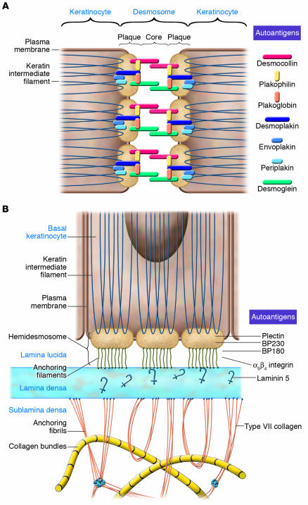
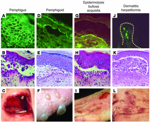
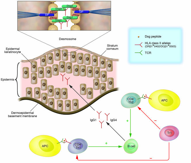
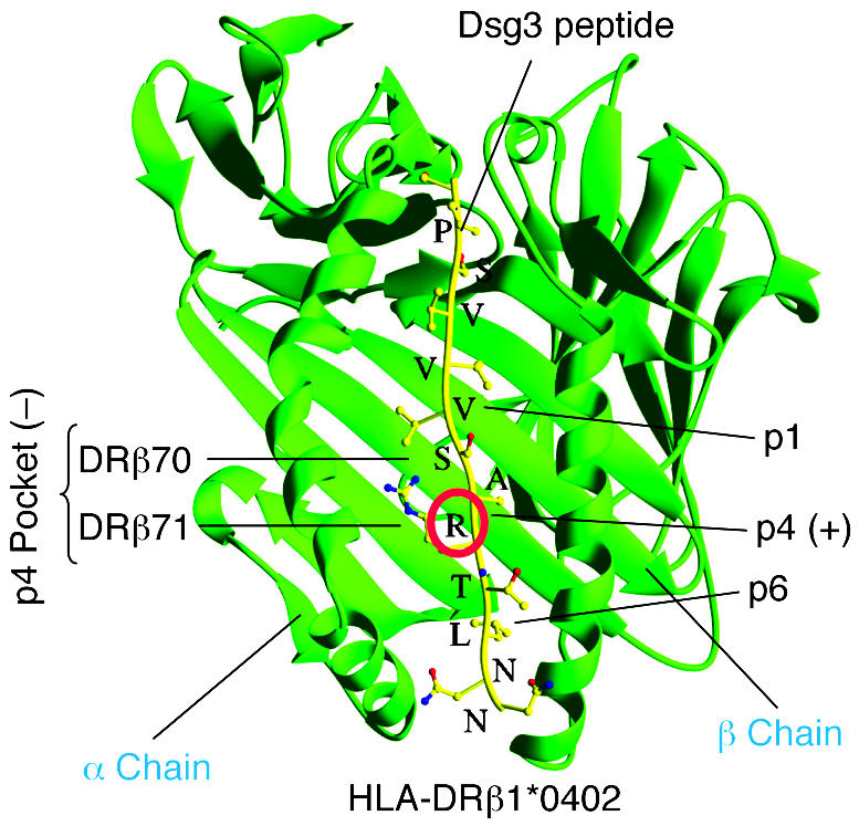
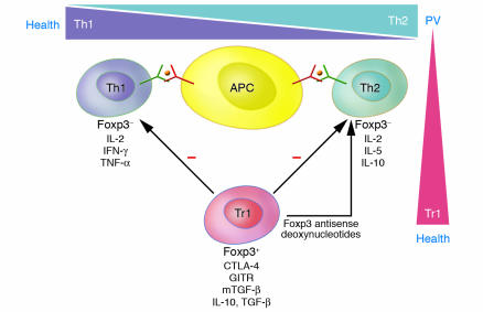
Autoimmune bullous disorders are a group of severe skin diseases characterized clinically by blisters and erosions of skin and/or mucous membranes. A hallmark of these disorders is the presence of IgG and occasionally IgA autoantibodies that target distinct adhesion structures of the epidermis, dermoepidermal basement membrane, and anchoring fibrils of the dermis. This Review focuses on the potential role of autoreactive T cells in the pathogenesis of these disorders. Pemphigus vulgaris (PV) and bullous pemphigoid (BP) are the best-characterized bullous disorders with regard to pathogenesis and T cell involvement. Activation of autoreactive T cells in PV and BP is restricted by distinct HLA class II alleles that are prevalent in individuals with these disorders. Autoreactive T cells are not only present in patients but can also be detected in healthy individuals. Recently, a subset of autoreactive T cells with remarkable regulatory function was identified in healthy individuals and to a much lesser extent in patients with PV, suggesting that the occurrence of autoimmune bullous disorders may be linked to a dysfunction of Tregs.


Figures

Figure 1
Figure 1. Autoantigens of human autoimmune bullous skin disorders and their role in epidermal and dermoepidermal adhesion.
(A) Desmosomes. The integrity of epidermal cell cohesion is largely dependent on desmosomes, plaque-like intercellular adhesion structures that connect transmembranous adhesion molecules such as the desmogleins and desmocollins with keratins of the cytoskeleton through interaction with intracellular components of the desmosomal plaque such as desmoplakin, plakoglobin, plakophilin, envoplakin, and periplakin. In addition to Dsg3 and Dsg1, which are autoantigens in pemphigus, all the other aforementioned components of desmosomes have been identified as autoantigens of different clinical variants of pemphigus. (B) Hemidesmosomes and components of the dermoepidermal basement membrane. Basal keratinocytes adhere to the basement membrane zone by the interaction of cytoplasmic and transmembranous components of hemidesmosomes, such as BP230 and BP180 and α6β4 integrin, with ligands such as laminin 5 located in the lamina lucida and lamina densa of the dermoepidermal basement membrane zone. The intracellular hemidesmosomal components BP230 and plectin are linked to keratins of the cytoskeleton and interact with the cytoplasmic domains of BP180 and α6β4 integrin, which in turn interact with laminin 5 via their ectodomains. Type VII collagen is the major component of anchoring fibrils, which link the basement membrane to interstitial dermal collagen fibers by direct interaction with laminin 5 in the lamina densa of the basement membrane. Figure modified from ref. .
Figure 2
Figure 2. Immunological and clinical characteristics of autoimmune bullous skin disorders.
(AC) Pemphigus is characterized by the presence of IgG (and occasionally IgA) specific for desmosomal target antigens (visualized as an intercellular staining pattern by direct immunofluorescence; A) resulting in a loss of intraepidermal adhesion (as shown by histopathology; B) and blisters and/or erosions of the mucous membranes and skin (C). (DF) In the pemphigoids, including linear IgA bullous dermatosis, IgG (or IgA) autoantibodies bind to antigens of hemidesmosomes and the lamina lucida of the dermoepidermal junction (D), resulting in a loss of subepidermal adhesion (E) and tense blisters (F). (GI) Epidermolysis bullosa acquisita is associated with IgG (and sometimes IgA) binding to the anchoring fibrils underneath the lamina densa of the dermoepidermal junction (G), resulting in a subepidermal loss of adhesion (H) and tense blisters with a tendency toward scarring and milia formation (I). (JL) Dermatitis herpetiformis is associated with deposits in the papillary dermis (dotted line indicates the dermoepidermal junction) of IgA reactive with epidermal transglutaminase (J), a subepidermal loss of adhesion (K), and herpetiform blisters or pruritic papules (L). Magnification, ×100 (A, B, D, E, G, H, J, and K).
Figure 3
Figure 3. T cell involvement in the immune pathogenesis of PV.
PV is the prototype of an autoantibody-mediated immunobullous skin disorder and is characterized by a loss of intraepidermal adhesion primarily caused by autoantibodies belonging to the IgG4 and, to a lesser extent, IgG1 subclasses specific for Dsg3 and Dsg1, components of the desmosomal adhesion complex of epidermal keratinocytes that are connected to the keratin cytoskeleton through interaction with the intracellular plaque proteins plakoglobin (PKG) and desmoplakin (DP; inset). IgG production by autoreactive B cells is presumably regulated by Dsg3- and Dsg1-reactive Th1 and Th2 cells. The Dsg3-reactive Th cells recognize epitopes of the Dsg3 ectodomain in association with the HLA class II alleles HLA-DRβ1*0402 and HLA-DQβ1*0503 presented by APCs including dendritic cells and B cells. Dsg3-reactive Th cells that recognize identical epitopes are also found in healthy carriers of the aforementioned PV-associated HLA class II alleles. Thus Tregs may be critical in maintaining peripheral B cell tolerance to Dsg3. This contention is supported by the finding that Dsg3-reactive Tr1s are more frequently detected in healthy individuals than in patients with PV.
Figure 4
Figure 4. Algorithm for HLA class II–Dsg3 peptide interaction in PV.
Shown is the physical interaction of DRβ1*0402 with a representative Dsg3 peptide. HLA-DRβ1*0402 differs from other HLA-DR4 molecules by the presence of a negative charge at amino acid residue 71 (DRβ71) of the β chain, which is a critical binding motif for T cell peptides. Several T cell epitopes of Dsg3, the major autoantigen of PV, have been identified that carry a positively charged amino acid (mostly lysine, K, or arginine, R) at relative position 4 (p4; red circle), which serves as an anchor motif to the negatively charged p4 pocket formed by residues DRβ70 and DRβ71 of DRβ1*0402. Thus DRβ1*0402 shapes the fine specificity of the T cell autoimmune response against a limited set of Dsg3 epitopes that fulfill the binding criteria of this specific HLA class II molecule.
Figure 5
Figure 5. Putative T cell network in PV and health.
Dsg3- and Dsg1-reactive Th1 and Th2 cells are present in patients with PV and healthy carriers of PV-associated HLA class II alleles, which recognize identical epitopes of the Dsg3 ectodomain presented by APCs such as dendritic cells and B cells. There is a predominance of Dsg3-reactive Th2 cells in PV and of autoreactive Th1 cells in healthy individuals. In addition, Tr1s specific for Dsg3 are present at higher frequencies in healthy individuals than in PV patients. These Tr1s express the phenotypic markers glucocorticoid-induced TNF receptor (GITR), membrane-bound TGF-β (mTGF-β), and cytotoxic T cell antigen–4 (CTLA-4) and inhibit the activation of Dsg3-specific autoreactive Th1 and Th2 cells via the secretion of IL-10 and TGF-β and the uptake of exogenous IL-2 produced by the Th cells. Inactivation of the regulatory transcription factor Foxp3 by antisense deoxynucleotides converts Tr1s into a Th2-like population with regard to cytokine profile, loss of inhibitory function on Th cells, and the ability to develop a proliferative response to Dsg3. Thus an imbalance of the putative relationship between autoreactive Th and Tr1 cells may be critical in the pathogenesis of PV. Hence Dsg-specific Tr1s may be critical for the maintenance of tolerance against Dsg3 and Dsg1 in health and the restoration of tolerance against these autoantigens in remittent PV.

References

    1. Beutner E.H., Jordon R.E., Chorzelski T.P. The immunopathology of pemphigus and bullous pemphigoid. J. Invest. Dermatol. 1968;51:63–80. - PubMed
    1. Yancey K.B., Egan C.A. Pemphigoid: clinical, histologic, immunopathologic, and therapeutic considerations. JAMA. 2000;284:350–356. - PubMed
    1. Stanley J.R. Pemphigus and pemphigoid as paradigms of organ-specific, autoantibody-mediated diseases. J. Clin. Invest. 1989;83:1443–1448. - PMC - PubMed
    1. Hertl M. Humoral and cellular autoimmunity in autoimmune bullous skin disorders. Int. Arch. Allergy Immunol. 2000;122:91–100. - PubMed
    1. Borradori L., Sonnenberg A. Hemidesmosomes: roles in adhesion, signaling and human diseases. Curr. Opin. Cell Biol. 1996;8:647–656. - PubMed

Publication types