Review
. 2006 May;116(5):1167-73.
doi: 10.1172/JCI28607.

Prophylactic human papillomavirus vaccines

Affiliations
Review

Prophylactic human papillomavirus vaccines

Douglas R Lowy et al. J Clin Invest. 2006 May.

Abstract

Human papillomavirus (HPV) infection causes virtually all cases of cervical cancer, the second most common cause of death from cancer among women worldwide. This Review examines prophylactic HPV subunit vaccines based on the ability of the viral L1 capsid protein to form virus-like particles (VLPs) that induce high levels of neutralizing antibodies. Following preclinical research by laboratories in the nonprofit sector, Merck and GlaxoSmithKline are developing commercial versions of the vaccine. Both vaccines target HPV16 and HPV18, which account for approximately 70% of cervical cancer. The Merck vaccine also targets HPV6 and HPV11, which account for approximately 90% of external genital warts. The vaccines have an excellent safety profile, are highly immunogenic, and have conferred complete type-specific protection against persistent infection and associated lesions in fully vaccinated women. Unresolved issues include the most critical groups to vaccinate and when the vaccine's cost may be low enough for widespread implementation in the developing world, where 80% of cervical cancer occurs.


Figures

Figure 1
Figure 1. Papillomavirus life cycle.
To establish infection, the virus must infect basal epithelial cells that are long lived or have stem cell–like properties. Microtrauma to the suprabasal epidermal cells probably enables the virus to infect the cell within the basal layer. The viral genome maintains itself as an episome in basal cells, where the viral genes are poorly expressed. Viral replication takes place in suprabasal layers and is tied to the epidermal differentiation process. The presence of the virus causes morphological abnormalities in the epithelium, including papillomatosis, parakeratosis, and koilocytosis. Progeny virus is released in desquamated cells.
Figure 2
Figure 2. Progression from a benign cervical lesion to invasive cervical cancer.
Infection by oncogenic HPV types, especially HPV16, may directly cause a benign condylomatous lesion, low-grade dysplasia, or sometimes even an early high-grade lesion. Carcinoma in situ rarely occurs until several years after infection. It results from the combined effects of HPV genes, particularly those encoding E6 and E7, which are the 2 viral oncoproteins that are preferentially retained and expressed in cervical cancers; integration of the viral DNA into the host DNA; and a series of genetic and epigenetic changes in cellular genes. HSIL, high-grade squamous intraepithelial lesion; LSIL, low-grade squamous intraepithelial lesion.
Figure 3
Figure 3. Relationship among incidences of cervical HPV infection, precancer, and cancer.
The HPV curve emphasizes the high incidence of infection that develops soon after women initiate sexual activity and subsequent lower incidence because a high proportion of infections are self-limited. The precancer incidence curve follows several years behind the HPV incidence curve and is substantially lower than that of HPV incidence, as there is generally a delay between the acquisition of HPV infection and precancer development, and only a subset of infected women develop precancers. The cancer incidence curve follows several years behind the precancer curve, reflecting the relatively long interval between precancer and progression to invasive cancer. As women approach 40 years of age, the incidence of cancer begins to approach the incidence of precancer. Figure modified with permission from the New England Journal of Medicine (53).
Figure 4
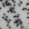
Figure 4. Electron micrograph of HPV16 L1 VLPs.
Original magnification, ×14,500. Image courtesy of Yuk-Ying Susana Pang (Laboratory of Cellular Oncology, National Cancer Institute).

References

    1. Pisani P., Parkin D.M., Munoz N., Ferlay J. Cancer and infection: estimates of the attributable fraction in 1990. Cancer Epidemiol. Biomarkers Prev. 1997;6:387–400. - PubMed
    1. Lowy D.R., Howley P.H. In Fields virology . D.M. Knipe and P.H. Howley, editors. Lippincott, Williams, & Wilkins.; Philadelphia, Pennsylvania, USA.: 2001. Papillomaviruses. pp. 2231–2264.
    1. Zur Hausen H. Papillomaviruses and cancer: from basic studies to clinical application. Nat. Rev. Cancer. 2002;2:342–350. - PubMed
    1. Bosch F.X., Schiffman M., Solomon D. editors.Future directions in epidemiologic and preventive research on human papillomaviruses and cancer. J. Natl. Cancer Inst. Monogr. 2003;31:131. - PubMed
    1. Frisch M. On the etiology of anal squamous carcinoma. Dan. Med. Bull. 2002;49:194–209. - PubMed

Publication types