Review
. 2006 May;116(5):1174-86.
doi: 10.1172/JCI28553.

Frontiers in pruritus research: scratching the brain for more effective itch therapy

Affiliations
Review

Frontiers in pruritus research: scratching the brain for more effective itch therapy

Ralf Paus et al. J Clin Invest. 2006 May.

Abstract

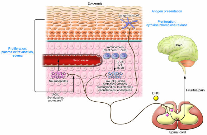
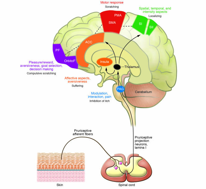
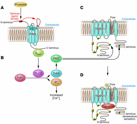
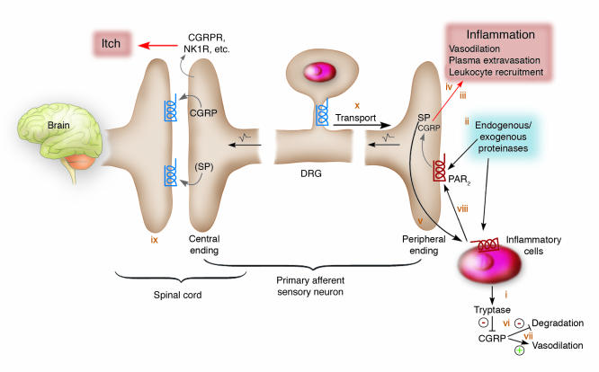
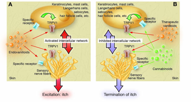
This Review highlights selected frontiers in pruritus research and focuses on recently attained insights into the neurophysiological, neuroimmunological, and neuroendocrine mechanisms underlying skin-derived itch (pruritogenic pruritus), which may affect future antipruritic strategies. Special attention is paid to newly identified itch-specific neuronal pathways in the spinothalamic tract that are distinct from pain pathways and to CNS regions that process peripheral pruritogenic stimuli. In addition, the relation between itch and pain is discussed, with emphasis on how the intimate contacts between these closely related yet distinct sensory phenomena may be exploited therapeutically. Furthermore, newly identified or unduly neglected intracutaneous itch mediators (e.g., endovanilloids, proteases, cannabinoids, opioids, neurotrophins, and cytokines) and relevant receptors (e.g., vanilloid receptor channels and proteinase-activated, cannabinoid, opioid, cytokine, and new histamine receptors) are discussed. In summarizing promising new avenues for managing itch more effectively, we advocate therapeutic approaches that strive for the combination of peripherally active antiinflammatory agents with drugs that counteract chronic central itch sensitization.


Figures

Figure 1
Figure 1. Table 2 and neurophysiological pathways activated during pruritus (pruritogenic itch).
Exogenous or endogenous mediators stimulate specific subtypes of peripheral nerve endings of primary afferent neurons (pruriceptors). High-affinity receptors for pruritogenic mediators transmit the stimulus via intracellular signaling from the periphery to the dorsal root ganglia (DRG) and the spinal cord. Within the spinal cord, itch signals can be modulated (see It is the brain that itches, not the skin). From lamina I, a specific area within the dorsal horn of the spinal cord, the signal is transmitted to the CNS after crossing to the contralateral side (see Figure 3). Activation of specific areas in the CNS results in the perception of itch, leading to discomfort and a scratch response. Additionally, the associated peripheral axon reflex may lead to the release of mast cell–stimulating neuropeptides (e.g., SP), thereby amplifying pruritus via release of histamine, tryptase, and TNF-α, for example (see Table 1). This figure does not consider the interaction between pain and itch fibers on the spinal cord level (see The enigmatic neurophysiology of itch is becoming increasingly understood). Figure modified with permission from The Journal of Investigative Dermatology (5).
Figure 2
Figure 2. Central processing of pruritus.
Pruriceptive primary afferent nerve fibers from the skin activate spinal neurons in lamina I of the dorsal horn, which project to the thalamus. Direct excitatory connections from the thalamus include anterior cingulate cortex (ACC), insular cortex (Insula), and primary and secondary somatosensory cortices (SI, SII). The putative function of brain areas activated in central imaging studies of itch are summarized. SMA, supplementary motor area; PMA, premotor area; PF, prefrontal cortex; OrbitoF, orbitofrontal cortex; PAG, periaqueductal gray.
Figure 3
Figure 3. Pruritogenic receptors interact synergistically, thereby amplifying itch or pain.
(A) Activation of PAR2 leads to binding of G proteins (Gαq/11), followed by (B) stimulation of the intracellular PKC pathway and mobilization of intracellular [Ca2+] via phopholipase Cβ (PLCβ), diacylglycerol (DAG), and inositol triphosphate (IP3). PIP2, phosphatidylinositol 4,5-biphosphate. This results in (C) sensitization of TRPV1 by phosphorylation of the intracellular C terminus (heterologous sensitization). Sensitization of TRPV1 leads to (D) a lowered threshold for capsaicin binding or temperature, i.e., stimulation of TRPV1. This mechanism affects release of neuropeptides (activation of mast cell degranulation) from nerve terminals of sensory nerves as well as stimulation of activation potentials (transmission of pain and pruritus to the spinal cord).
Figure 4
Figure 4. PARs play a key role in pruritus during neurogenic inflammation.
(i) Tryptase released from degranulated mast cells activates PAR2 at the plasma membrane of sensory nerve endings. (ii) Activation of PAR2 by tryptase, trypsins, kallikreins, or probably exogenous proteinases (bacteria, house dust mite) stimulates the release of calcitonin gene–related peptide (CGRP) and tachykinins, e.g., SP, from sensory nerve endings. (iii) CGRP interacts with the CGRP1 receptor to induce arteriolar dilation and hyperaemia. (iv) SP interacts with the neurokinin-1 receptor (NK1R) on endothelial cells of postcapillary venules to cause gap formation and plasma extravasation. Hyperaemia and plasma extravasation cause edema during inflammation. (v) SP may stimulate degranulation of mast cells, providing a positive-feedback mechanism. (vi) Tryptase degrades CGRP and terminates its effects. (vii) CGRP inhibits SP degradation by neutral endopeptidase and also enhances SP release, thereby amplifying its effects. (viii) Mediators from mast cells and other inflammatory cells stimulate the release of vasoactive peptides from sensory nerves and also sensitize nerves. (ix) At the spinal cord level, PAR2-induced intracellular Ca2+ mobilization leads to release of CGRP (and SP) from central nerve endings, thereby activating CGRP receptor (CGRPR) and NK1R to transit itch responses to the central nervous system. (x) During inflammation, PAR2 may be peripherally transported, thereby increasing receptor density and stimulation. Figure modified with permission from Nature Medicine (64, 125).
Figure 5
Figure 5. The putative role of TRPV1 signaling in the pathogenesis and therapy of itch.
(A) Pruritogenic endovanilloids either directly or indirectly (via their own cognate receptors) activate and/or sensitize TRPV1 expressed on sensory neurons and nonneuronal cell types, which augments the bidirectional intercellular network to initiate itch. (B) Repeatedly applied topical vanilloids (such as capsaicin) may desensitize neuronal and nonneuronal TRPV1–mediated signaling so as to counteract the pruritogenic intercellular network and hence terminate itch. Note that certain cannabinoids (via acting either on specific CB receptors or directly on TRPV1) may augment the efficacy of vanilloid therapy.

References

    1. Greaves M.W., Khalifa N. Itch: more than skin deep. Int. Arch. Allergy Immunol. 2004;135:166–172. - PubMed
    1. Graham D.T., Goodell H., Wolff H.G. Neural mechanisms involved in itch, itchy skin, and tickle sensations. J. Clin. Invest. 1951;30:37–49. - PMC - PubMed
    1. Yosipovitch G., Greaves M.W., Schmelz M. Itch. Lancet. 2003;361:690–694. - PubMed
    1. Biro T., et al. How best to fight that nasty itch - from new insights into the neuroimmunological, neuroendocrine, and neurophysiological bases of pruritus to novel therapeutic approaches. Exp. Dermatol. 2005;14:225–240. - PubMed
    1. Steinhoff M., et al. Neurophysiological, neuroimmunological and neuroendocrine basis of pruritus. J. Invest. Dermatol. 2006 In press. - PubMed

Publication types