. 2006 May 3:7:104.
doi: 10.1186/1471-2164-7-104.

Four genomic islands that mark post-1995 pandemic Vibrio parahaemolyticus isolates

Affiliations

Four genomic islands that mark post-1995 pandemic Vibrio parahaemolyticus isolates

Catherine C Hurley et al. BMC Genomics. .

Abstract

Background: Vibrio parahaemolyticus is an aquatic, halophilic, Gram-negative bacterium, first discovered in 1950 in Japan during a food-poisoning outbreak. Infections resulting from consumption of V. parahaemolyticus have increased globally in the last 10 years leading to the bacterium's classification as a newly emerging pathogen. In 1996 the first appearance of a pandemic V. parahaemolyticus clone occurred, a new O3:K6 serotype strain that has now been identified worldwide as a major cause of seafood-borne gastroenteritis.

Results: We examined the sequenced genome of V. parahaemolyticus RIMD2210633, an O3:K6 serotype strain isolated in Japan in 1996, by bioinformatic analyses to uncover genomic islands (GIs) that may play a role in the emergence and pathogenesis of pandemic strains. We identified 7 regions ranging in size from 10 kb to 81 kb that had the characteristics of GIs such as aberrant base composition compared to the core genome, presence of phage-like integrases, flanked by direct repeats and the absence of these regions from closely related species. Molecular analysis of worldwide clinical isolates of V. parahaemolyticus recovered over the last 33 years demonstrated that a 24 kb region named V. parahaemolyticus island-1 (VPaI-1) encompassing ORFs VP0380 to VP0403 is only present in new O3:K6 and related strains recovered after 1995. We investigated the presence of 3 additional regions, VPaI-4 (VP2131 to VP2144), VPaI-5 (VP2900 to VP2910) and VPaI-6 (VPA1254 to VPA1270) by PCR assays and Southern blot analyses among the same set of V. parahaemolyticus isolates. These 3 VPaI regions also gave similar distribution patterns amongst the 41 strains examined.

Conclusion: The 4 VPaI regions examined may represent DNA acquired by the pandemic group of V. parahaemolyticus isolates that increased their fitness either in the aquatic environment or in their ability to infect humans.


Figures

Figure 1
Figure 1
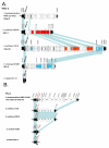
A. Schematic representation of V. parahaemolyticus island-1 (VPaI-1) and the homologous tRNA-Met insertion site in V. vulnificus strains YJ016 and CMCP6, V. cholerae N16961, and V. fischeri ES114. Block arrows represent annotated open reading frames (ORFs). Numbers above block arrows indicate ORF number on the sequenced genome strain. Black filled in ORFs represent core chromosomal genes. Open arrows represent genomic island ORFs. Shaded arrows represent integrase genes. Vertical arrows represent homologous tRNA-Met genes within each genome. Shaded blocks indicate homologous ORFs among the Vibrio species. B. Schematic representation of VPaI-2 from V. parahaemolyticus RIMD2210633.
Figure 2
Figure 2
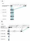
A and B. Schematic representation of V. parahaemolyticus island-3 (VPaI-3) and VPaI-4, and the homologous insertion site among the 4 sequenced Vibrio species. Block arrows represent annotated ORFs. Black filled in ORFs represent core chromosomal flanking genes. Open and shaded arrows represent genomic island ORFs. Shaded blocks indicate homologous ORFs among the Vibrio species.
Figure 3
Figure 3
A and B. Schematic representation of V. parahaemolyticus island-5 (VPaI-5) and VPAI-6, and the homologous insertion site among the 4 sequenced Vibrio species. Block arrows represent annotated ORFs. Black filled in ORFs represent core chromosomal flanking genes. Open arrows represent genomic island ORFs. Shaded blocks indicate homologous ORFs among the Vibrio species.
Figure 4
Figure 4
A. Schematic representation of VPaI-1 showing arrow heads representing primer pairs used in this study. PCR analysis of the 41 V. parahaemolyticus strains is shown representing the results from 3 primer pairs. Lanes 1 and 44 represent size markers; lanes 2 to 42 contain strains analysed in the same order as in Table 2. For overlapping PCR primer pair analysis only VPaI-1-positive strains were examined, lanes 2 to 25 contain strains analysed in the same order as in Table 3. PCR primer pairs used are indicated under each agarose gel. B. Schematic representation of tRNA-Met site in VPaI-1-negative strains. Arrow heads represent primer pair used, which bind to the 5' and 3' core chromosomal flanking genes. PCR analysis of the 17 VPaI-1-negative isolates (lanes 26 to 42) from section A. The band size is marked on the left side of the PCR image. An expected 2.9 kb product was obtained for all strains except 428/00 and 30824 (Lanes 16 and 17). Lane 1 contains marker. C. Schematic representation of tRNA-Met site in VPaI-1 negative strains 428/00 and 30824. Arrow heads represent primer pair used. Long Template Expand PCR on strains 428/00 and 30824 showed 4 kb of DNA at this insertion site. The band size is marked on the left side of the PCR image.
Figure 5
Figure 5
Southern hybridisation analysis of VPaI-1 negative strains 428/00 and 30824. PaI-1-negative strains KE9984 and KE9967, VPaI-1-positive strain RIMD2210633 and VPaI-1-negative strains 30824 and 428/00. Genomic DNA was restricted using EcoRI and the probe used was constructed from the PCR amplification using primer pair 379F and 404R with strain KE9984 as template. The expected 8 Kb and 11.4 kb band sizes in the VPaI-1 negative strains KE9984 and KE9967 are shown (Lane 1–2). The VPaI-1 positive RIMD2210633 gave the expected 11.4 Kb, 6.6 Kb and 3.1 Kb band sizes (Lane 3). The VPaI-1 negative strains 30824 and 428/00 with novel DNA inserted gave the expected band sizes of 13 Kb and 11.4 Kb.
Figure 6
Figure 6
Schematic representation of VPaI-4, VPaI-5 and VPaI-6 showing arrow heads representing overlapping and insertion site primer pairs used in this study (Table 3). Blue bars indicate regions tested for in all strains and red bars indicate regions tested for in VPaI-positive strains only. A. PCR analysis of the 24 VPaI-4-positive strains is shown. Lane 1 represents size marker; lanes 2 to 25 contain strains analysed in the same order as in Table 2. B. PCR analysis of the 24 VPaI-5-positive strains is shown. Lane 1 represents size marker; lanes 2 to 25 contain strains analysed in the same order as in Table 2. C. PCR analysis of the 24 VPaI-6-positive strains is shown. Lane 1 represents size marker; lanes 2 to 25 contain strains analysed in the same order as in Table 2.
Figure 7
Figure 7
Schematic representation of chromosomally integrated and extrachromosomal excision product of hypothetical VPaI region. Location of PCR primers used for analysis of excision products are shown as black arrows labelled PrimerR and PrimerF. Only formation of a circular intermediate or plasmid form of VPaI will allow for the amplification of a PCR product with primer pair primerF and primerR.

References

    1. Pan TM, Wang TK, Lee CL, Chien SW, Horng CB. Food-borne disease outbreaks due to bacteria in Taiwan, 1986 to 1995. J Clin Microbiol. 1997;35:1260–1262. - PMC - PubMed
    1. Yeung PS, Boor KJ. Epidemiology, pathogenesis, and prevention of foodborne Vibrio parahaemolyticus infections. Foodborne Pathog Dis. 2004;1:74–88. doi: 10.1089/153531404323143594. - DOI - PubMed
    1. Okuda J, Ishibashi M, Hayakawa E, Nishino T, Takeda Y, Mukhopadhyay AK, Garg S, Bhattacharya SK, Nair GB, Nishibuchi M. Emergence of a unique O3:K6 clone of Vibrio parahaemolyticus in Calcutta, India, and isolation of strains from the same clonal group from Southeast Asian travelers arriving in Japan. J Clin Microbiol. 1997;35:3150–3155. - PMC - PubMed
    1. DePaola A, Kaysner CA, Bowers J, Cook DW. Environmental investigations of Vibrio parahaemolyticus in oysters after outbreaks in Washington, Texas, and New York (1997 and 1998) Appl Environ Microbiol. 2000;66:4649–4654. doi: 10.1128/AEM.66.11.4649-4654.2000. - DOI - PMC - PubMed
    1. Abbott SL, Powers C, Kaysner CA, Takeda Y, Ishibashi M, Joseph SW, Janda JM. Emergence of a restricted bioserovar of Vibrio parahaemolyticus as the predominant cause of Vibrio-associated gastroenteritis on the West Coast of the United States and Mexico. J Clin Microbiol. 1989;27:2891–2893. - PMC - PubMed

Publication types

LinkOut - more resources