. 2006 May 3:4:14.
doi: 10.1186/1741-7007-4-14.

Compensation by tumor suppressor genes during retinal development in mice and humans

Affiliations

Compensation by tumor suppressor genes during retinal development in mice and humans

Stacy L Donovan et al. BMC Biol. .

Abstract

Background: The RB1 gene was the first tumor suppressor gene cloned from humans by studying genetic lesions in families with retinoblastoma. Children who inherit one defective copy of the RB1 gene have an increased susceptibility to retinoblastoma. Several years after the identification of the human RB1 gene, a targeted deletion of Rb was generated in mice. Mice with one defective copy of the Rb gene do not develop retinoblastoma. In this manuscript, we explore the different roles of the Rb family in human and mouse retinal development in order to better understand the species-specific difference in retinoblastoma susceptibility.

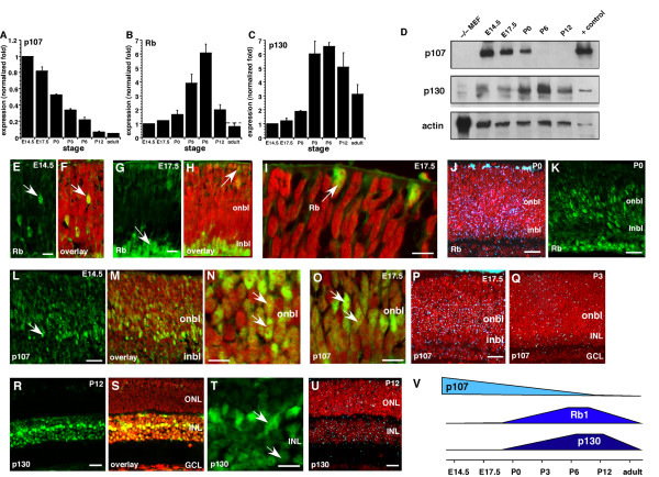
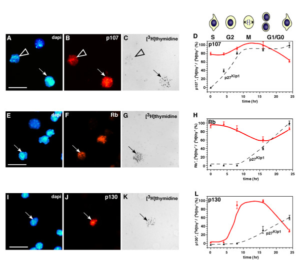
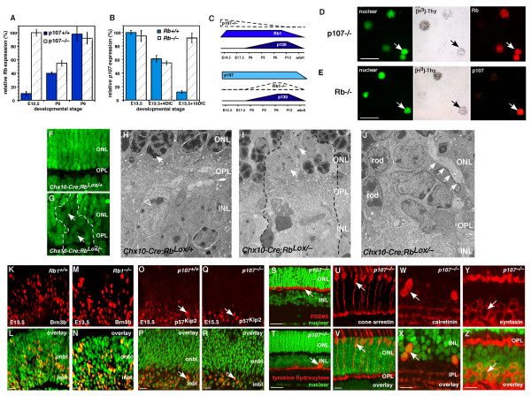
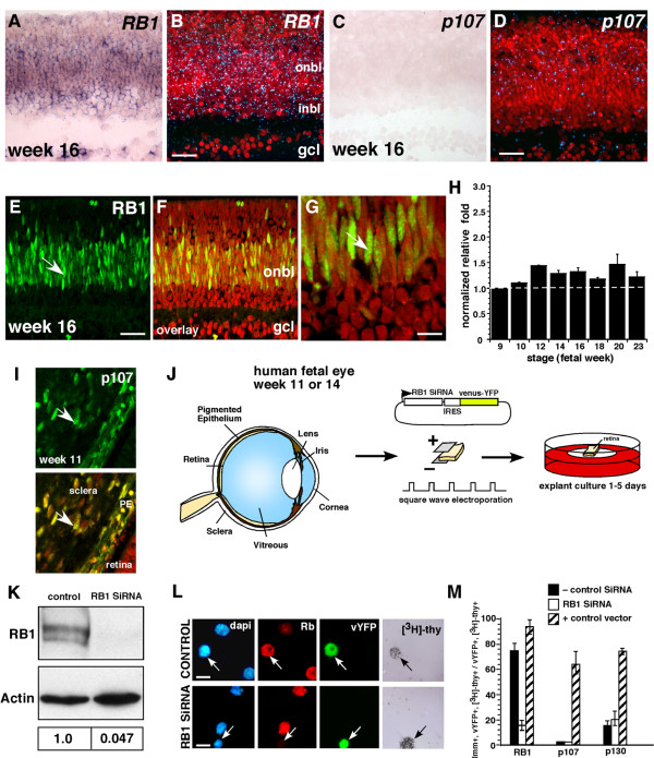
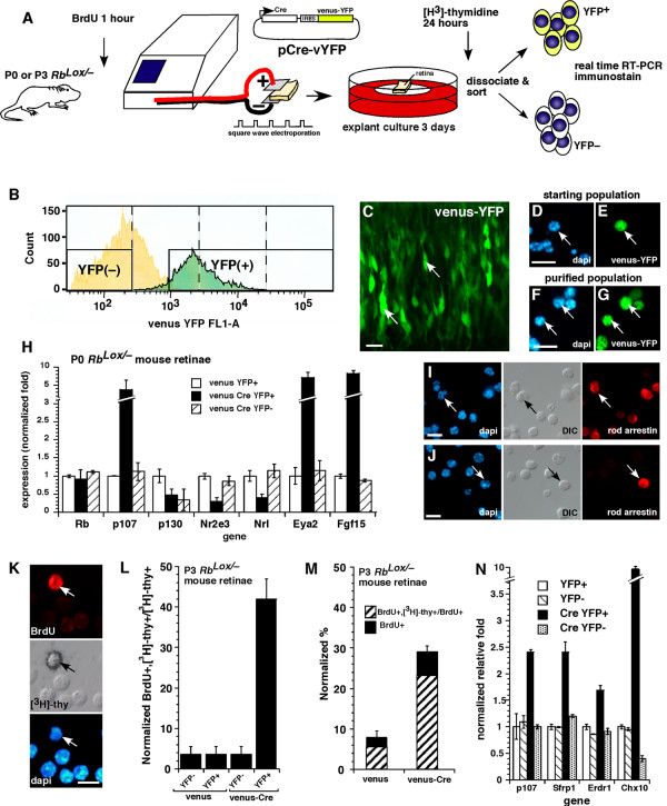
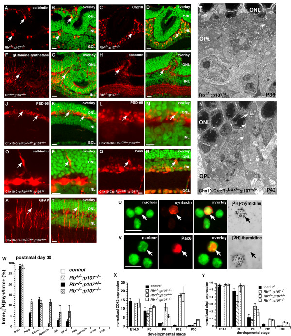
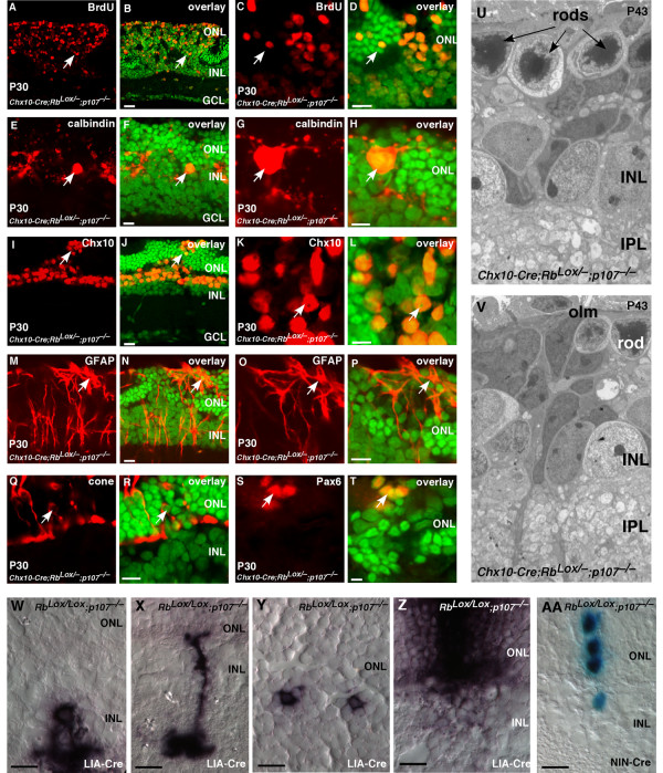
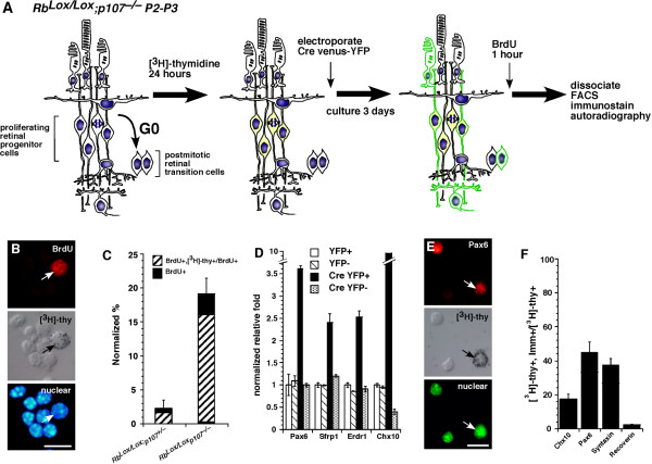
Results: We found that the Rb family of proteins (Rb, p107 and p130) are expressed in a dynamic manner during mouse retinal development. The primary Rb family member expressed in proliferating embryonic retinal progenitor cells in mice is p107, which is required for appropriate cell cycle exit during retinogenesis. The primary Rb family member expressed in proliferating postnatal retinal progenitor cells is Rb. p130 protein is expressed redundantly with Rb in postmitotic cells of the inner nuclear layer and the ganglion cell layer of the mouse retina. When Rb is inactivated in an acute or chronic manner during mouse retinal development, p107 is upregulated in a compensatory manner. Similarly, when p107 is inactivated in the mouse retina, Rb is upregulated. No changes in p130 expression were seen when p107, Rb or both were inactivated in the developing mouse retina. In the human retina, RB1 was the primary family member expressed throughout development. There was very little if any p107 expressed in the developing human retina. In contrast to the developing mouse retina, when RB1 was acutely inactivated in the developing human fetal retina, p107 was not upregulated in a compensatory manner.

Conclusion: We propose that intrinsic genetic compensation between Rb and p107 prevents retinoblastoma in Rb- or p107-deficient mice, but this compensation does not occur in humans. Together, these data suggest a model that explains why humans are susceptible to retinoblastoma following RB1 loss, but mice require both Rb and p107 gene inactivation.


Figures

Figure 1
Figure 1
Dynamic expression of the Rb family during mouse retinal development. (A-C) Real-time PCR analysis of p107, Rb and p130 was done at seven stages of mouse retinal development. Data sets of three retinas per stage were analyzed twice, normalized to Gapdh and averaged. The standard deviations were within 5% of the mean. (D) Immunoblot analysis was done at five stages of mouse retinal development. For negative controls, MEF lysates prepared from knockout embryos were used. Positive controls were taken from 293T cells ectopically expressing the full-length cDNAs. Data were normalized to actin. (E-I) Immunofluorescent detection of Rb (green) in E14.5 and E17.5 retinas was overlaid on the nuclear counterstain (red). (J) Radioactive in situ hybridization analysis of Rb (blue silver grains) levels at P0 demonstrated broad expression in the onbl and inbl. This finding was consistent with immunofluorescent detection (green) of the protein (K). (L-O) Immunofluorescent detection of p107 (green) in E14.5 and E17.5 retinas was overlaid on the nuclear counterstain (red). (P) Radioactive in situ hybridization for p107 (blue silver grains) at E17.5 demonstrated broad expression in the onbl and little expression at P3 (Q). (R-T) Immunofluorescence of p130 at P12 (green) was overlaid on the nuclear counterstain (red). (U) Expression of p130 mRNA at P12 was consistent with the protein expression data. (V) Summary of the dynamic expression of the Rb family during retinal development. Abbreviations: GCL, ganglion cell layer; inbl, inner neuroblastic layer; INL, inner nuclear layer; MEF, mouse embryonic fibroblast; onbl, outer neuroblastic layer; ONL, outer nuclear layer. Scale bars: E, G, I, N, O and T, 10 μm; J, K, L, M, P, R and S, 25 μm.
Figure 2
Figure 2
Expression of the Rb family during the cell cycle. (A-D) The E14.5 retinal explants were maintain in culture in the presence of [3H]thy for 1 h, washed and then maintained in culture for different periods. Dissociated retinas (dapi in A) were then immunostained for p107 (red in B) and overlaid with autoradiographic emulsion to detect the [3H]thy (C). The proportion of p107+ cells was then scored (250 cells in duplicate), and normalized data were plotted (D). The peak level of p107 was detected after 8 h of exposure, which coincided with the G2 and M phases of the cell cycle, and declined at 24 h, which coincided with the G1/G0 phase. (E-H) Similar experiments were done using P0 retinal explants and in P2 retinal explants (I-L). (H) The proportion of [3H]thy-labeled Rb+ cells from P0 retinal explants was high during 0 to 4 h of exposure (S, G2) and subsequently declined. (L) The level of [3H]thy-labeled p130+ cells in P2 retinal explants was low during the first 4 h (S, G2), peaked at 8 and 16 h (M phase) and then declined during G1/G0. Similar data for p27 are plotted as an internal reference for cell cycle phase estimates. Scale bars: A, E and I, 10 μm.
Figure 3
Figure 3
Reciprocal compensation after Rb or p107 inactivation. (A) Real-time PCR analysis of Rb expression in retinas from three developmental stages of p107-deficient or wild-type mice. At E13.5, Rb was upregulated. Analysis was done in triplicate samples analyzed in duplicate. (B) In a similar experiment, p107 expression was analyzed in Rb-deficient retinas. Due to the embryonic lethality of deleting Rb, cultured retinas were used for the later stages; p107 was upregulated in a compensatory manner. (C) A summary of the changes in Rb family gene expression after Rb or p107 inactivation. (D) E13.5 p107-deficient retinas were labeled with [3H]thy for 4 h and immunostained for Rb expression (red). (E) In a similar experiment, cultured Rb-deficient retinas were labeled for p107 expression. (F, G) In the absence of Rb in Chx10-Cre;RbLox/- P12 retinas, rod photoreceptors failed to form, and chromatin failed to condense in the ONL (arrow). (H) Chromatin (arrow) in rod photoreceptors normally condenses during differentiation. (I) In the absence of Rb, rods remained immature with diffuse chromatin (arrow). (J) These defects in cell-fate specification also caused defects in synaptogenesis of horizontal cells (arrows); with processes extending apically into the ONL. (K-N) Wild-type and Rb-deficient E13.5 retinas were immunostained with an antibody against Brn3b (red), a ganglion cell marker. Brn3b was distributed across the retina (green nuclear stain overlay) and was indistinguishable in the presence or absence of Rb. (O-R) Wild-type and p107-deficient E15.5 retinas were immunostained for p57Kip2 (red), which was expressed in a subset of retinal progenitor cells and newly postmitotic amacrine cells. (S) PSD95 (red) was expressed in the OPL and was normally distributed in the absence of p107. (T) Amacrine cells stained with tyrosine hydroxylase were also distributed normally. (U, V) Cone photoreceptors, (W, X) calretinin+ amacrine cells and (Y, Z) amacrine cells and horizontal cells were distributed normally in the p107-knockout retinas. Abbreviations: DIC, days in culture; INL, inner nuclear layer; IPL, inner plexiform layer; ONL, outer nuclear layer; OPL, outer plexiform layer. Scale bars: B, D, F, H, L, N, P, T and U, 10 μm; I and J, 25 μm.
Figure 4
Figure 4
Acute inactivation of RB1 in human fetal retinas. (A-D) Representative in situ hybridization of fetal week 16 human retinas with DIG-labeled probes (A, C) and radioactively labeled probes (B, D) shows that RB1 is the major RB family member expressed in proliferating retinal progenitor cells during development. (E-G) Immunofluorescent detection of RB1 (green) in fetal week 16 human retinas confirmed that the protein is expressed in the same pattern as the mRNA. (H) Real-time RT-PCR analysis using TaqMan® probes for RB1 demonstrated that the expression level does not dramatically change over the course of retinal histogenesis. Data sets were analyzed twice, normalized to Gapdh expression and averaged. (I) The sclera and pigmented epithelium were positive controls for p107 expression. (J) To inactivate RB1 in human fetal retinas, primary tissue was square-wave electroporated with a plasmid that encoded an siRNA to RB1 and a venus-YFP reporter gene. Retinas were then maintained in culture for several days and analyzed for compensation by p107. (K) COS cells transfected with the RB1 SiRNA vector shown in (J) showed a 21-fold reduction in the level of RB1 protein. Densitometry of normalized values for RB1 is shown in the lower portion of panel (K). (L, M) The venus-YFP+ retinal cells that were also [3H]thy+ downregulated RB1 but did not upregulate p107. The negative control SiRNA shown in (M) is the Gapdh SiRNA, but other nonspecific siRNAs gave similar results. The positive control samples are retinas that were square-wave electroporated with a plasmid expressing RB1, p107 or p130 and processed side-by-side with the SiRNA samples. Abbreviations: GCL, ganglion cell layer; inbl, inner neuroblastic layer; onbl, outer neuroblastic layer; PE, pigmented epithelium. Scale bars: G and L, 10 μm; B, D and E, 25 μm.
Figure 5
Figure 5
Acute inactivation of Rb in the developing mouse retina. (A-G) In vivo or in vitro square-wave electroporation and purification of cells with acute Rb inactivation. (C) A retina after electroporation is shown. (D-G) Dissociated cells before and after FACS purification are shown. (H) Real-time PCR analysis of YFP+ purified cells after acute Rb inactivation; each sample was analyzed in duplicate and normalized to Gapdh and Gpi1. (I, J) Immunostaining of purified cells after Rb inactivation. (K-N) To determine if retinal progenitor cells continue to divide after acute Rb inactivation, we scored the proportion of [3H]thy-labeled BrdU+ cells. An example of a double-positive cell is shown in (K). (L) A significant proportion of progenitor cells was sensitive to deregulated proliferation after acute Rb inactivation, as indicated by the increase in double-positive cells. Data are normalized to account for the fraction of cells labeled with BrdU during a 1-h pulse. (M) The proportion of retinal progenitor cells that continue to divide after acute Rb inactivation is shown in the venus-Cre, YFP+ column. (N) Real-time PCR analysis of cell samples used in (L) and (M) revealed a significant increase in the expression of p107 and retinal progenitor cell markers Sfrp1, Erdr1 and Chx10 after acute Rb inactivation. Scale bars: C, D, F, I and J, 10 μm.
Figure 6
Figure 6
Haploinsufficiency of p107 in the developing mouse retina. Immunostaining of P30 Rb+/-; p107-/- retinas in the region of the retinal dysplasia. (A, B) Antibodies against calbindin were used to identify horizontal cells; (C, D) those against Chx10, bipolar cells; (F, G) those against glutamine synthetase, Müller glia; and (H, I) those against bassoon, synapses in the OPL and IPL. (E) EM analysis of the P30 Rb+/-; p107-/- retinas showing the OPL with normal organization of synaptic connections in contrast to the Rb-deficient retinas. (J-T) Immunostaining of P14 Chx10-Cre;RbLox/-; p107+/- retinas in the region of Cre-mediated Rb inactivation using antibodies to OPL synapses (PSD-95, J-M), horizontal cells (calbindin, O, P), amacrine/progenitor cells (Pax6, Q, R), and reactive Müller glia (GFAP, S, T). (N) EM analysis of the P14 Chx10-Cre;RbLox/-; p107+/- retinas showing the OPL with disrupted synaptic connections and an apical horizontal process (arrows). (U-Y) To determine if there are any ectopically dividing cells in Rb+/-; p107-/-, Rb-/-; p107+/- or Rb-/-; p107-/- retinas at E14.5, P0, P6, P12 and P30, we labeled retinas with [3H]thy and BrdU for 1 h and then dissociated and immunostained the cells with antibodies against 12 markers of different retinal cell types. BrdU and [3H]thy always colocalized. (W) The only markers that colocalized with [3H]thy were amacrine/progenitor cell markers Pax6 and syntaxin and bipolar/progenitor cell marker Chx10. There were also some reactive Müller glia, as indicated by GFAP, that incorporated [3H]thy. Ectopic proliferation was more prevalent in the double-knockout retinas than in the other genotypes. Each bar is the average of triplicate samples scored in duplicate. (X, Y) Real-time PCR analysis demonstrated that throughout development there were more progenitor cell markers such as Cdk2 and Sfrp1 expressed in the double-knockout and Rb-/-; p107+/- retinas. Abbreviations: GCL, ganglion cell layer; INL, inner nuclear layer; ONL, outer nuclear layer; OPL, outer plexiform layer. Scale bars: B, D, G, I, K, M, P, Q, T, U and V, 10 μm.
Figure 7
Figure 7
Retinal progenitor cells continue to proliferate throughout development in the absence of Rb and p107. (A-T) Immunostaining of P30 Chx10-Cre;RbLox/-; p107-/- retinas by using antibodies against (A-D) BrdU to identify ectopically dividing cells; (E-H) calbindin, horizontal cells and Chx10, bipolar/progenitor cells; (M-P) GFAP, reactive Müller glia; (Q, R) cone arrestin, cone photoreceptors and (S, T) Pax6, amacrine/progenitor cells. (U, V) Electron micrographs showing that most retinal structures are disrupted in the absence of Rb and p107 and that continued proliferation eventually leads to retinoblastoma. (W-Z) Newborn RbLox/Lox; p107-/- mice were injected with a retrovirus expressing Cre (LIA-Cre), and 3 weeks later, the retinas were sectioned and stained for alkaline phosphatase expression to identify the neurons that had lost Rb and p107. Amacrine cells (W) and bipolar cells (X) formed normally in the absence of Rb and p107. Rod photoreceptors (Y) failed to form normally. There were also several clones that spanned the INL or INL and ONL, which was indicative of hyperproliferation (Z). Similar experiments were carried out in E17.5 retinal explants by using a Cre-expressing retrovirus that also expresses nuclear LacZ (AA). These clones can readily be scored for cell number to determine if loss of Rb and p107 leads to deregulated proliferation. Abbreviations: GCL, ganglion cell layer; INL, inner nuclear layer; olm, outer limiting membrane; ONL, outer nuclear layer. Scale bars: B, D, F, H, J, L, N, P, R, T and W-AA, 10 μm.
Figure 8
Figure 8
Retinal progenitor cells are sensitive to deregulated proliferation after acute inactivation of Rb and p107. (A) To determine if retinal progenitor cells are sensitive to deregulated proliferation after acute Rb inactivation in a p107-deficient genetic background, we labeled RbLox/Lox; p107-/- P0 or P3 retinas for 24 h with [3H]thy and then electroporated them with a Cre-venus-YFP plasmid. After 3 days in culture, the retinas were labeled for 1 h with BrdU and dissociated, and the YFP+ and YFP- cells were purified by FACS. (B-F) The cells were then analyzed by immunostaining, single-cell scoring and real-time PCR. (B, C) Colocalization of BrdU and [3H]thy demonstrated that most cells that continued to proliferate after Rb inactivation were retinal progenitor cells. (D) Real-time RT-PCR analysis confirmed that the proliferating cells expressed retinal progenitor cell markers. (E, F) Immunostaining and colocalization with [3H]thy further confirmed the retinal progenitor cell characteristics of these cells. Scale bars: B and E, 10 μm.

Similar articles

Cited by

References

    1. Donovan SL, Dyer MA. Regulation of proliferation during central nervous system development. Semin Cell Dev Biol. 2005;16:407–421. doi: 10.1016/j.semcdb.2005.02.012. - DOI - PubMed
    1. Dyer MA, Cepko CL. Regulating proliferation during retinal development. Nat Rev Neurosci. 2001;2:333–342. doi: 10.1038/35072555. - DOI - PubMed
    1. Clarke AR, Maandag ER, van Roon M, van der Lugt NM, van der Valk M, Hooper ML, Berns A, te Riele H. Requirement for a functional Rb-1 gene in murine development. Nature. 1992;359:328–330. doi: 10.1038/359328a0. - DOI - PubMed
    1. Jacks T, Fazeli A, Schmitt EM, Bronson RT, Goodell MA, Weinberg RA. Effects of an Rb mutation in the mouse. Nature. 1992;359:295–300. doi: 10.1038/359295a0. - DOI - PubMed
    1. Lee EY, Chang CY, Hu N, Wang YC, Lai CC, Herrup K, Lee WH, Bradley A. Mice deficient for Rb are nonviable and show defects in neurogenesis and haematopoiesis. Nature. 1992;359:288–294. doi: 10.1038/359288a0. - DOI - PubMed

Publication types

Substances

LinkOut - more resources