. 2006 May 16;103(20):7895-900.
doi: 10.1073/pnas.0511232103. Epub 2006 May 3.

Inhibition of an equilibrative nucleoside transporter by cannabidiol: a mechanism of cannabinoid immunosuppression

Affiliations

Inhibition of an equilibrative nucleoside transporter by cannabidiol: a mechanism of cannabinoid immunosuppression

Erica J Carrier et al. Proc Natl Acad Sci U S A. .

Abstract

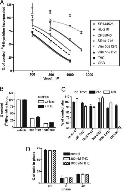
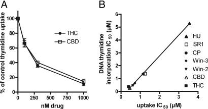
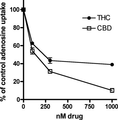
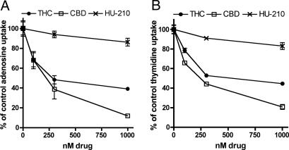
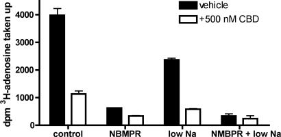
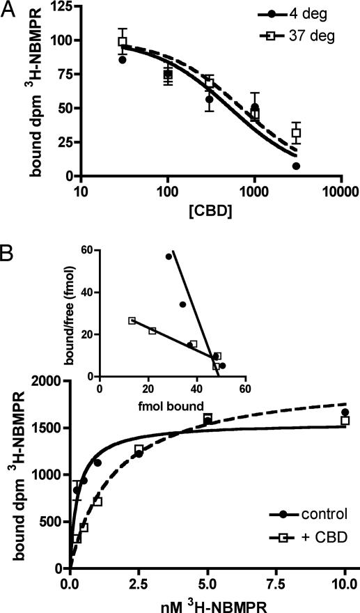
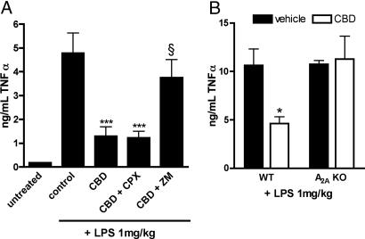
The plant-derived cannabinoids delta9-tetrahydrocannabinol (THC) and cannabidiol (CBD) both have immunosuppressive effects; although some effects of THC are mediated by the CB2 receptor, CB2 binds CBD weakly. In examining the effects of THC and CBD on microglial proliferation, we found that these compounds potently inhibit [3H]thymidine incorporation into a murine microglial cell line with no effect on cell cycle. Treatment with THC and CBD decreased [3H]thymidine uptake into microglia, with IC50 values that match inhibition of [3H]thymidine incorporation into DNA. CBD and, less potently, THC decreased uptake of [3H]adenosine to a similar extent as [3H]thymidine in both murine microglia and RAW264.7 macrophages. Binding studies confirm that CBD binds to the equilibrative nucleoside transporter 1 with a Ki < 250 nM. Because adenosine agonists have antiinflammatory effects, and because uptake of adenosine is a primary mechanism of terminating adenosine signaling, we tested the hypothesis that CBD is immunosuppressive because it enhances endogenous adenosine signaling. In vivo treatment with a low dose of CBD decreases TNFalpha production in lipopolysaccharide-treated mice; this effect is reversed with an A2A adenosine receptor antagonist and abolished in A2A receptor knockout mice. These studies demonstrate that CBD has the ability to enhance adenosine signaling through inhibition of uptake and provide a non-cannabinoid receptor mechanism by which CBD can decrease inflammation.


Conflict of interest statement

Conflict of interest statement: No conflicts declared.

Figures

Fig. 1.
Fig. 1.
Cannabinoids inhibit [3H]thymidine incorporation into EOC-20 microglia with no effect on cell cycle or MTT reduction. (A) [3H]Thymidine incorporation assay. Cannabinoids were present during the 4-h [3H]thymidine incubation. Results are expressed as a percent of control, and vertical lines represent SEM (n = 4); the single-site competition equation was used to determine IC50. (B) PTx treatment during [3H]thymidine incorporation assay. Cells were treated with 100 ng/ml PTx during the 24-h stimulation with LMCM; THC or vehicle was added for the final 4 h. Indicated concentrations are in nM. Results are shown as a percentage of control proliferation; respective controls are shown. Treatment with PTx alone resulted in ≈70% of normal proliferation (n = 6). (C) MTT assay. EOC-20 microglia were treated for 24 or 48 h with vehicle, THC, or CBD before assaying for reduction of MTT tetrazolium salt. Indicated concentrations are in nM; shown are the means and SEM of the resulting absorbance at 562 nm. (D) Cell cycle analysis by FACS after 4 h of treatment with THC. To exclude fragmented or fused cells, cells per cell cycle stage were calculated as a percentage of total gated cells (n = 2).
Fig. 2.
Fig. 2.
CBD and THC inhibit uptake of [3H]thymidine into EOC-20 microglia. (A) EOC-2 cells were pretreated with cannabinoid for 30 min at 37°C, and uptake of 0.5 μCi [3H]thymidine was assayed for a period of 1 min. Nonspecific 3H counts, determined in the presence of 1 mM thymidine, were subtracted from each data point. Results are expressed as a percentage of vehicle-treated control, and vertical lines represent SEM (n = 3). (B) Comparison of cannabinoid IC50 required to inhibit DNA [3H]thymidine incorporation over 4 h, and [3H]thymidine uptake over 1 min. Nonspecific uptake was not subtracted. The linear regression line is shown; r2 = 0.995. HU, HU-210; SR1, SR144528; CP, CP55940; Win-3, Win 55212-3; Win-2, Win 55212-2.
Fig. 3.
Fig. 3.
CBD and THC inhibit [3H]adenosine uptake in EOC-20 microglia. Cells were pretreated with cannabinoid for 30 min at 37°C, and uptake of 0.5 μCi [3H]adenosine over a period of 1 min was assayed. Nonspecific uptake, determined in the presence of 1 mM adenosine, was subtracted from each data point. Results are expressed as a percentage of vehicle-treated control, and vertical lines represent SEM (n = 3).
Fig. 4.
Fig. 4.
Plant-derived cannabinoids inhibit adenosine and thymidine uptake in RAW264.7 macrophages. Cells were pretreated with cannabinoid for 30 min at 37°C, and uptake of radiolabeled adenosine (A) or thymidine (B) was assayed over 1 min. Nonspecific uptake, determined in the presence of 1 mM unlabeled nucleotide, was subtracted from each data point. Results are expressed as a percentage of vehicle-treated control; vertical lines represent SEM (n = 3).
Fig. 5.
Fig. 5.
Adenosine uptake in EOC-20 cells is mediated by ENT transporters. To demonstrate NBMPR sensitivity, uptake assays were carried out in microglia as described. For sodium-free experiments, the NaCl in normal BSS buffer was replaced with N-methyl-glucamine. Cannabinoids or 100 nM NBMPR was added 30 min before the addition of 3H nucleoside; uptake was measured for 1 min. Nonspecific uptake was subtracted from the total uptake. Error bars reflect SEM (n = 3).
Fig. 6.
Fig. 6.
CBD is a competitive inhibitor at the ENT1 transporter in EOC-20 cells. (A) Cells were preincubated for 30 min with indicated concentrations of CBD before incubation with 1 nM [3H]NBMPR for 30 min at 4°C or 37°C. Nonspecific binding, determined in the presence of 10 μM nitrobenzylthioguanosine, was subtracted; the combined results of three experiments are shown. IC50 values, derived from the solution of a single-site competition equation in individual experiments (each n = 3), were used to calculate the dissociation constant for CBD. (B) Kinetics of [3H]NBMPR binding. EOC-20 cells were preincubated with vehicle or 500 nM CBD before [3H]NBMPR was added for a 30-min incubation at 37°C. Nonspecific binding was subtracted. Shown is one representative experiment of three; the lines represent the solution of the one-site binding equation (n = 3). A Scatchard plot of the data is shown (Inset).
Fig. 7.
Fig. 7.
CBD decreases TNFα via activation of A2A adenosine receptors. (A) Male ICR mice were pretreated for 1 h before LPS injection with a single dose of CBD (1 mg/kg, i.p.) or vehicle. Thirty minutes later, mice were given vehicle, 8-cyclopentyl-1,3-dipropylxanthine (CPX, 3 mg/kg, i.p), or ZM 241385 (ZM, 10 mg/kg, i.p.). Mice were treated with LPS (1 mg/kg, i.v.) 1 h after CBD injection. One hour after LPS treatment, mice were killed, and serum was collected. TNFα levels were determined by ELISA (n = 6). ∗∗∗, P < 0.001 compared with vehicle control (one-way ANOVA followed by Bonferroni's posttest); §, P > 0.05 compared with control and P < 0.05 compared with CBD alone (one-way ANOVA followed by Bonferroni's posttest). (B) Wild-type or A2A-null (KO) C57BL/6 mice were pretreated for 1 h with 1 mg/kg CBD or vehicle. Mice were treated with LPS, serum was collected after 1 h, and TNFα was determined by ELISA (n = 4). ∗, P = 0.014 compared with vehicle-treated by unpaired t test.

References

    1. Croxford J. L., Yamamura T. J. Neuroimmunol. 2005;166:3–18. - PubMed
    1. Wade D. T., Makela P., Robson P., House H., Bateman C. Mult. Scler. 2004;10:434–441. - PubMed
    1. Buckley N. E., McCoy K. L., Mezey E., Bonner T., Zimmer A., Felder C. C., Glass M. Eur. J. Pharmacol. 2000;396:141–149. - PubMed
    1. Mechoulam R., Parker L. A., Gallily R. J. Clin. Pharmacol. 2002;42:11S–19S. - PubMed
    1. Belgrave B. E., Bird K. D., Chesher G. B., Jackson D. M., Lubbe K. E., Starmer G. A., Teo R. K. Psychopharmacology (Berlin) 1979;64:243–246. - PubMed

Publication types

MeSH terms