Identification of a novel 74-kiloDalton immunodominant antigen of Pythium insidiosum recognized by sera from human patients with pythiosis
- PMID: 16672392
- PMCID: PMC1479217
- DOI: 10.1128/JCM.44.5.1674-1680.2006
Identification of a novel 74-kiloDalton immunodominant antigen of Pythium insidiosum recognized by sera from human patients with pythiosis
Abstract
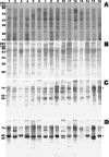
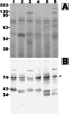
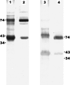
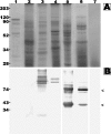
The oomycetous, fungus-like, aquatic organism Pythium insidiosum is the etiologic agent of pythiosis, a life-threatening infectious disease of humans and animals that has been increasingly reported from tropical, subtropical, and temperate countries. Human pythiosis is endemic in Thailand, and most patients present with arteritis, leading to limb amputation and/or death, or cornea ulcer, leading to enucleation. Diagnosis of pythiosis is time-consuming and difficult. Radical surgery is the main treatment for pythiosis because conventional antifungal drugs are ineffective. The aims of this study were to evaluate the use of Western blotting for diagnosis of human pythiosis, to identify specific immunodominant antigens of P. insidiosum, and to increase understanding of humoral immune responses against the pathogen. We performed Western blot analysis on 16 P. insidiosum isolates using 12 pythiosis serum samples. These specimens were derived from human patients with pythiosis who had different forms of infection and lived in different geographic areas throughout Thailand. We have identified a 74-kDa immunodominant antigen in all P. insidiosum isolates tested. The 74-kDa antigen was also recognized by sera from all patients with pythiosis but not by control sera from healthy individuals, patients with thalassemia, and patients with various infectious diseases, indicating that Western blot analysis could facilitate diagnosis of pythiosis. Therefore, the 74-kDa antigen is a potential target for developing rapid serodiagnostic tests as well as a therapeutic vaccine for pythiosis. These advances could lead to early diagnosis and effective treatment, crucial factors for better prognosis for patients with pythiosis.
Figures


Similar articles
-
The 74-kilodalton immunodominant antigen of the pathogenic oomycete Pythium insidiosum is a putative exo-1,3-beta-glucanase.Clin Vaccine Immunol. 2010 Aug;17(8):1203-10. doi: 10.1128/CVI.00515-09. Epub 2010 Mar 17. Clin Vaccine Immunol. 2010. PMID: 20237199 Free PMC article.
-
Seroprevalence of anti--Pythium insidiosum antibodies in the Thai population.Med Mycol. 2019 Apr 1;57(3):284-290. doi: 10.1093/mmy/myy030. Med Mycol. 2019. PMID: 29846667
-
An initial survey of 150 horses from Thailand for anti-Pythium insidiosum antibodies.J Mycol Med. 2021 Mar;31(1):101085. doi: 10.1016/j.mycmed.2020.101085. Epub 2020 Nov 17. J Mycol Med. 2021. PMID: 33259982
-
Human pythiosis, a rare cause of arteritis: case report and literature review.Semin Arthritis Rheum. 2003 Dec;33(3):204-14. doi: 10.1016/j.semarthrit.2003.09.003. Semin Arthritis Rheum. 2003. PMID: 14671729 Review.
-
History and Perspective of Immunotherapy for Pythiosis.Vaccines (Basel). 2021 Sep 26;9(10):1080. doi: 10.3390/vaccines9101080. Vaccines (Basel). 2021. PMID: 34696188 Free PMC article. Review.
Cited by
-
Combat-Related Pythium aphanidermatum Invasive Wound Infection: Case Report and Discussion of Utility of Molecular Diagnostics.J Clin Microbiol. 2015 Jun;53(6):1968-75. doi: 10.1128/JCM.00410-15. Epub 2015 Apr 1. J Clin Microbiol. 2015. PMID: 25832301 Free PMC article.
-
Development of an immunochromatographic test for rapid serodiagnosis of human pythiosis.Clin Vaccine Immunol. 2009 Apr;16(4):506-9. doi: 10.1128/CVI.00276-08. Epub 2009 Feb 18. Clin Vaccine Immunol. 2009. PMID: 19225072 Free PMC article.
-
Pythium aphanidermatum infection following combat trauma.J Clin Microbiol. 2011 Oct;49(10):3710-3. doi: 10.1128/JCM.01209-11. Epub 2011 Aug 3. J Clin Microbiol. 2011. PMID: 21813724 Free PMC article.
-
Biochemical and genetic analyses of the oomycete Pythium insidiosum provide new insights into clinical identification and urease-based evolution of metabolism-related traits.PeerJ. 2018 Jun 5;6:e4821. doi: 10.7717/peerj.4821. eCollection 2018. PeerJ. 2018. PMID: 29888122 Free PMC article.
-
The Immunoreactive Exo-1,3-β-Glucanase from the Pathogenic Oomycete Pythium insidiosum Is Temperature Regulated and Exhibits Glycoside Hydrolase Activity.PLoS One. 2015 Aug 11;10(8):e0135239. doi: 10.1371/journal.pone.0135239. eCollection 2015. PLoS One. 2015. PMID: 26263509 Free PMC article.
References
-
- Bollag, D., M. Rozycki, and S. Edelstein. 1996. Protein concentration determination, p. 62-67. In D. Bollag, M. Rozycki, and S. Edelstein (ed.), Protein methods, 2nd ed. Wiley-Liss, Inc., New York, N.Y.
-
- Chetchotisakd, P., C. Pairojkul, O. Porntaveevudhi, B. Sathapatayavongs, P. Mairiang, K. Nuntirooj, B. Patjanasoontorn, O. T. Saew, A. K. Chaiprasert, and M. R. Haswell-Elkins. 1992. Human pythiosis in Srinagarind Hospital: one year's experience. J. Med. Assoc. Thai. 75:248-254. - PubMed
-
- Dixon, D. M., A. Casadevall, B. Klein, L. Mendoza, L. Travassos, and G. S. Deepe, Jr. 1998. Development of vaccines and their use in the prevention of fungal infections. Med. Mycol. 36:57-67. - PubMed
-
- Grooters, A. M., and M. K. Gee. 2002. Development of a nested polymerase chain reaction assay for the detection and identification of Pythium insidiosum. J. Vet. Intern. Med. 16:147-152. - PubMed
Publication types
MeSH terms
Substances
LinkOut - more resources
Full Text Sources
Other Literature Sources

