. 2006 May;188(10):3631-44.
doi: 10.1128/JB.188.10.3631-3644.2006.

The ornibactin biosynthesis and transport genes of Burkholderia cenocepacia are regulated by an extracytoplasmic function sigma factor which is a part of the Fur regulon

Affiliations

The ornibactin biosynthesis and transport genes of Burkholderia cenocepacia are regulated by an extracytoplasmic function sigma factor which is a part of the Fur regulon

Kirsty Agnoli et al. J Bacteriol. 2006 May.

Abstract

Burkholderia cenocepacia mutants that fail to produce the siderophore ornibactin were obtained following mutagenesis with mini-Tn5Tp. These mutants were shown to be growth restricted under conditions of iron depletion. In eight of the mutants, the transposon had integrated into one of two genes, orbI and orbJ, encoding nonribosomal peptide synthetases. In the other mutant, the transposon had inserted into an open reading frame, orbS, located upstream from orbI. The polypeptide product of orbS exhibits a high degree of similarity to the Pseudomonas aeruginosa extracytoplasmic function (ECF) sigma factor PvdS but possesses an N-terminal extension of approximately 29 amino acids that is not present in PvdS. Three predicted OrbS-dependent promoters were identified within the ornibactin gene cluster, based on their similarity to PvdS-dependent promoters. The iron-regulated activity of these promoters was shown to require OrbS. Transcription of the orbS gene was found to be under the control of an iron-regulated sigma(70)-dependent promoter. This promoter, but not the OrbS-dependent promoters, was shown to be a target for repression by the global regulator Fur. Our results demonstrate that production of ornibactin by B. cenocepacia in response to iron starvation requires transcription of an operon that is dependent on the Fur-regulated ECF sigma factor gene orbS. A mechanism is also proposed for the biosynthesis of ornibactin.


Figures

FIG. 1.
FIG. 1.
Structure and biosynthesis of ornibactin. A. Structure of the ferric-ornibactin complex showing chelation of the metal ion by three bidentate ligands provided by the derivatized Nδ-amino groups of the N- and C-terminal ornithine residues and by the hydroxyaspartate residue (adapted from reference 74). B. Model for the biosynthesis of ornibactin by the two predicted NRPSs, OrbI and OrbJ. Each NRPS (represented by the two large rectangles) is composed of adenylation (A), peptidyl carrier (P), and condensation (C) domains. Each peptide chain elongation unit is indicated with a horizontal bar above the corresponding NRPS. OrbI also contains a predicted epimerase domain (E) to convert l-hydroxyaspartate to the d form. Phosphopantetheine (P-pant) “arms,” to which the activated amino acids are covalently attached by a thioester bond, are shown as vertical zig-zag motifs. Curved arrows represent peptide bond formation by nucleophilic displacement of one amino acid from the P-pant arm by the amino group of the next amino acid in the peptide chain (for clarity, the growing peptide chain is not shown attached to each P-pant arm). The amino acid precursors are shown in their modified forms, although it is not clear at what step the N5-amino group of the N-terminal ornithine is derivatized with a β-hydroxy acid. See text for details.
FIG. 2.
FIG. 2.
Analysis of siderophore production by ornibactin mutants. A. Siderophore production by ornibactin mutants streaked on CAS agar. Plates were incubated at 37°C for 16 h. Orange haloes are due to production of siderophore (the parental strain, KLF1, produces only ornibactin). B. Ornibactin production by mutant OM3 and its complemented derivative. Ornibactin production was analyzed by IEF of culture supernatants followed by overlaying the gel with an agarose gel containing CAS reagent. Ornibactin migrates as a single spot near the cathode. Lane 1, KLF1; lane 2, OM3; lane 3, OM3 containing pBBR1MCS; lane 4, OM3 containing pBBR1MCS-orbS. C. Complementation of OM3 by orbS provided in trans. OM3 containing pBBR1MCS (pBBR), pBBR1MCS-orbS (pBBR-orbS), and pBBR1MCS-orbS derivatives in which one of the three putative orbS translation initiation codons (i1, i2, and i3 [see Fig. 5B, below]) was replaced by the indicated codon were streaked onto a CAS agar plate and incubated at 37°C overnight.
FIG. 3.
FIG. 3.
Transcriptional organization of the ornibactin gene cluster of B. cenocepacia. A. Genetic map of the ornibactin operon. Genes are depicted as block arrows, with proposed biosynthetic genes shown in gray, transport and utilization genes in white, and the sigma factor gene, orbS, shown in black. Flanking genes thought to be unrelated to ornibactin biosynthesis and utilization are hatched. Promoters for the ornibactin genes are indicated by small triangles (black fill for the orbS promoter and gray fill for the OrbS-dependent promoters). Sites of mini-Tn5Tp insertion for each Orb-negative mutant are shown by large black triangles, and the direction of transcription of the dfr (Tpr) gene is indicated by horizontal block arrows. Transcripts predicted from RT-PCR are shown by horizontal line arrows, with the 5′ ends indicated by a vertical bar (the broken line means the 3′ extent of the transcript has not been determined). The genes are located on chromosome 1 and have been assigned locus designations BCAL1688 to BCAL1702 (orbS to orbL, respectively) in the preliminary genome sequence annotation of strain J2315 (www.sanger.ac.uk/Projects/B_cenocepacia/). HP, hypothetical protein (BCAL1686 and BCAL1687); CHP, conserved hypothetical protein (BCAL1703). B. RT-PCR analysis of transcripts derived from the ornibactin gene cluster. Reverse transcriptase-generated cDNA obtained from ornibactin operon transcripts was amplified by PCR using pairs of primers flanking each gene junction. Amplification products were electrophoresed in a 1.6% agarose gel. As examples, analysis of transcripts overlapping the following junctions are shown: lanes 1 and 2, orbI-orbJ; lanes 3 and 4, orbA-pvdF; lanes 5 and 6, orbE-orbI; lanes 7 and 8, orbK-pvdA; lanes 10 and 11, orbH-orbG; lanes 12 and 13, orbS-orbH; lanes 14 and 15, orbF-orbB (in each case, the first lane shows the RT-PCR result for RNA obtained from iron-sufficient cells and the second lane shows the product obtained from iron-restricted cells). Corresponding primers are shown in Table S1 in the supplemental material. Lane 9, DNA size markers.
FIG. 4.
FIG. 4.
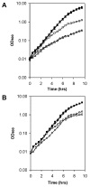
Effect of iron limitation on growth of ornibactin mutants. A. Effects of iron restriction on growth of transposon mutants OM1 (orbI::mini-Tn5Tp) and OM3 (orbS::mini-Tn5Tp). Cells were grown with aeration at 37°C in M9-glucose medium containing Casamino Acids (0.1%). Squares, parent strain (KLF1); circles, OM1; triangles, OM3. Filled symbols, medium supplemented with ferric chloride (50 μM); open symbols, medium supplemented with 2,2′-dipyridyl (100 μM). B. Effects of iron restriction on growth of the orbS mutant strain 715j-orbS::Tp. Cells were grown as described for panel A. Squares, parent strain (715j); circles, 715j-orbS::Tp. Filled symbols, medium supplemented with ferric chloride (50 μM); open symbols, medium supplemented with 2,2′-dipyridyl (100 μM).
FIG. 5.
FIG. 5.
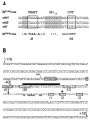
DNA sequences of the ornibactin operon promoters. A. DNA sequence of OrbS-dependent promoters. Proposed −35 and −10 regions are enclosed in boxes. G · C-rich sequences flanking the −35 region are highlighted with gray shading. The consensus sequence for the three OrbS-dependent promoters is shown below (n = any base). For comparison, the consensus PvdS-dependent promoter sequence is shown above. B. Sequence of the orbS promoter. The −35 and −10 sequences are enclosed in white boxes. Sequences located further upstream that bear close similarity to the −35 and −10 regions of the consensus σ70-dependent promoter are enclosed in gray boxes. Candidate translation initiation codons for orbS are also enclosed in boxes and labeled as i1 to i3. The true start codon (i2) was determined genetically and is indicated by a black star (see text for details). Bases within the Shine-Dalgarno sequence that are complementary to the 3′ end of B. cenocepacia 16S rRNA are in bold and underlined. The predicted Fur repressor binding site is indicated by a horizontal bar. Vertical arrows indicate the upstream endpoints of promoter deletion derivatives used in this study, except for the arrow at position +181, which was the downstream endpoint used in all the promoter derivatives. Distances shown are with respect to the proposed transcription start site (indicated by a white star). Position −178 corresponds to 209 bp upstream of the (true) translation initiation codon. The sequence was derived from strain 715j and differs from that of the sequenced strain, J2315, at only two positions (underlined).
FIG. 6.
FIG. 6.
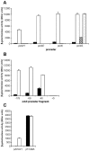
Regulation of ornibactin operon promoter activity. A. Effects of iron and orbS status on regulation of ornibactin operon promoters. β-Galactosidase activities were measured in B. cenocepacia 715j and 715j-orbS::Tp harboring pKAGd4 derivatives containing the orbH, orbE, orbI, and orbS promoters. Black bars and white bars represent the activities of the indicated promoters in 715j grown under iron-replete and iron-restricted conditions, respectively. Hatched bars and stippled bars represent the promoter activities in the orbS mutant strain grown under iron-replete and iron-restricted conditions, respectively. B. Identification of the orbS promoter. β-Galactosidase activities were measured in B. cenocepacia 715j harboring pKAGd4 containing the full-length orbS promoter (upstream endpoint at −178 relative to the predicted transcription start site) and derivatives with upstream endpoints at −69, −40, and +5 (the downstream endpoint is at +181 in all cases). Black bars and white bars represent the activities of the indicated promoters in cells grown under iron-replete and iron-restricted conditions, respectively. C. Effect of inactivation of the fur gene on regulation of the orbS promoter in E. coli. β-Galactosidase activities were measured in the E. coli Δlac Δfur::kan strain, QC1732, harboring pKAGd4ΔAp containing the full-length orbS promoter together with either pTrc99A or pAHA21 (pTrc99A-fur) (IPTG was omitted from the medium due to leaky transcription of the fur gene from the trc promoter [42]). Black bars and white bars represent the activities of the indicated promoters in cells containing pAHA21 grown under iron-restricted and iron-replete conditions, respectively. Hatched bars and stippled bars represent the promoter activities in cells containing pTrc99A grown under iron-restricted and iron-replete conditions, respectively. All assays were performed on triplicate cultures, and error bars represent the standard deviations.
FIG. 7.
FIG. 7.
Analysis of Fur binding to the ornibactin operon promoters. (A and B) Analysis of Fur binding in vivo by FURTA. E. coli H1717 harboring p3ZFBS, pBluescript II KS, or pBluescript II KS containing different orbS promoter fragments (as indicated) was streaked onto MacConkey-lactose agar supplemented with ampicillin and 40 μM Fe(NH4)2(SO4)2 and incubated overnight at 37°C. (A) Fur titration by the orbS promoter. The upstream deletion endpoint of each orbS promoter derivative is indicated (downstream endpoints are at position +181 relative to the predicted transcription start site). (B) Fur titration assay with OrbS-dependent promoters. Note that plasmid pBS-porbE also contains the divergently oriented orbI promoter. (C to E) Analysis of Fur binding in vitro by electrophoretic mobility shift assay. Radiolabeled DNA fragments containing ornibactin operon promoters were incubated with purified E. coli Fur at the indicated concentrations and electrophoresed through a 6% polyacrylamide gel. (C) porbS DNA fragment with upstream endpoint at −40; (D) porbS DNA fragment with upstream endpoint at −69; (E) effect of Fur on mobility of fragments containing porbS (−69 and +5 upstream endpoints, as indicated), porbH, and a fragment containing the divergently arranged orbE and orbI promoters. All orbS promoter fragments have a downstream endpoint at +181 relative to the predicted transcription start site.

References

    1. Aaron, S. D., W. Ferris, D. A. Henry, D. P. Speert, and N. E. Macdonald. 2000. Multiple combination bactericidal antibiotic testing for patients with cystic fibrosis infected with Burkholderia cepacia. Am. J. Respir. Crit. Care Med. 161:1206-1212. - PubMed
    1. Alexeyev, M. F. 1999. The pKNOCK series of broad-host-range mobilisable suicide vectors for gene knockout and targeted DNA insertion into the chromosome of gram-negative bacteria. BioTechniques 26:824-828. - PubMed
    1. Alting-Mees, M. A., and J. M. Short. 1989. pBluescript II: gene mapping vectors. Nucleic Acids Res. 17:9494. - PMC - PubMed
    1. Amman, E., B. Ochs, and K.-J. Abel. 1988. Tightly regulated tac promoter vectors useful for the expression of unfused and fused proteins in Escherichia coli. Gene 69:301-315. - PubMed
    1. Baichoo, N., and J. D. Helmann. 2002. Recognition of DNA by Fur: a reinterpretation of the Fur box consensus sequence. J. Bacteriol. 184:5826-5832. - PMC - PubMed

Publication types

LinkOut - more resources